I. INTRODUCTION
Recently, electronic papers have attracted a great deal of interest for use in information displays due to their distinctive advantages over the conventional paper, such as low power consumption, memorization, portability, and reliability to replace conventional papers. Reference Hou, Bian, Chen and Le1–Reference Li, Le, Hou, Chen and Shen3 Electronic paper technology research mainly divided into electrophoretic display (EPD), cholesterol liquid crystal technology (CLCD), electricity wet technology, color powders display, and microelectromechanical (MEMS), etc. Among the various approaches in the electronic display technology, in-plane electrophoretic particle displays and microencapsulated electrophoretic particle displays (EPDs) offered the advantage of lower manufacturing cost, since they could be fabricated by a roll-to-roll method. EPD showed images by the movement of charged dispersed particles in the dielectric medium when a suitable voltage was applied. Reference Fang, Wang, Xiao and Li4–Reference Park, Park, Choi, Han, Kwon and Choi7 In both case, the chargeability of the particles was a key factor in determining the speed or efficiency of the EPDs. Thus, the properties of electrophoretic particles would be the important factor determining the image quality. Enhancement of image quality required very small particle sizes which had a narrow size distribution and higher surface charge of particles for precise image control and faster response to the driving applied voltage. Reference Kim, Kim, Ahn, Kang and Suh8
Furthermore, the composite particles based on organic pigments were believed to be more suitable for the fabrication of EPD for their superior merits, such as high light stability, excellent chemical durability, the intense optical absorption, and wide varieties. Reference Meng, Tang, Peng and Ren9,Reference Yuan, Zhou, Wu and You10 In recent years, a lot of efforts have been made to study organic pigments. For example, Fu Reference Liu, He, Liu, Chen and Le11 encapsulated a nano-scaled azo Pigment Yellow 13 (PY13) with poly(styrene-maleic acid) (PSMA) by free-radical precipitation polymerization, getting satisfactory particles with their high dispersion and thermal stability. Unfortunately, the morphology of composite particles was irregular by this time-consuming and complicated procedure. Han Reference Han, Li, Feng and Zhang12 encapsulated a Pigment Yellow 181 (PY181) with polyethylene (PE) by dispersion polymerization method and a Pigment Yellow 110 (PY110) by polystyrene (PS) with mini-emulsion polymerization method, respectively. At present, the contrast ratio of yellow-white was reported to be 1.48 on our previous research of PY110, which needed yet to be improved.
In this article, Pigment Yellow 110 (PY110) and Span80 were suspended into the ethanol by dispersion method to obtain PY110-S, which was subsequently coated with styrene (St) through a mini-emulsion polymerization procedure. As well as C.I. Pigment Red 146 (P.R.146) composite particles encapsulated by polystyrene (PS) were prepared by mini-emulsion polymerization method. The composite particles with high surface charge density and narrow charge distribution were obtained, which were then successfully incorporated in an EPD cell. The contrast ratio of the EPD cell was 1.46. To improve the contrast ratio of yellow-white electrophoretic fluid, we gained that yellow particles by mixing PS/PY110-S with PS/P.R.146 and chose Isopar L:C2Cl4 = 1:1 as dispersion medium, because its density was close to the densities of particles. Furthermore, we obtained mixed modified particles, which had better properties and their contrast ratio was 1.58.
II. EXPERIMENTAL
A. Materials
Isopar L was offered from Huishuo Chemicals Co. Ltd. (Shanghai, China). Pigment Yellow 110 was purchased from Clariant Tianjin Co. Ltd. (Tianjin, China). Styrene (St), sodium dodecyl benzene sulfonate (SDBS), potassium peroxydisulfate (KPS), and cetanol were obtained from Damao Chemicals Co. Ltd., Guangfu Chemicals Co. Ltd., and Standard Technology Co. Ltd., respectively. Hyper-dispersant CH-13B and CH-22 were obtained from Sanzheng Polymer Co. Ltd. (Shanghai, China). Sorbitan monooleate (Span 80) and Sorbitan trioleate (Span 85) were purchased from Donghuan Union Chemical Plant (Beijing, China). All chemicals were in reagent grade and used as purchased without further purification.
B. Preparation of PY110-S
0.5 g PY110, 16% (the weight ratio of Span 80/PY110) Span 80 and 100 mL ethanol were poured into three-necked flask, the mixture was dispersed for 2 h at room temperature. Then the mixture was separated and dried.
C. Preparation of PS/PY110-S by miniemulsion polymerization
0.7 g PY110-S was dispersed in 10 g styrene in a beaker via an ultrasonic method for 20 min. The mixture was added to a big beaker together with 1.9 g SDBS, 0.2 g cetanol, 300 mL water, and 0.4 g KPS. The resulting mixture was ultrasonicated for 30 min to get a miniemulsion and then poured into a 500 mL three-necked round bottom flask. At 70 °C, the reaction was continued for 10 h, with the stirring speed at 400 rpm. After the polymerization process, the reaction mixture was washed with deionized water and ethanol for several times, centrifuged and dried at room temperature. Then, modified yellow pigment was obtained.
D. Preparation of PS/P.R.146 by miniemulsion polymerization
0.5 g P.R.146 was dispersed in 9.0 g styrene in a beaker via an ultrasonic method for 20 min. The mixture was added to a big beaker together with 2.0 g SDBS, 0.4 g cetanol, 300 mL water, and 0.4 g KPS. The resultant mixture was ultrasonicated for 30 min to get a miniemulsion and then poured into a 500 mL three-necked round bottom flask. At 70 °C, the reaction was continued for 10 h, with the stirring speed at 400 rpm. After the polymerization process, the reaction mixture was washed with deionized water and ethanol for several times, centrifuged and dried at room temperature. Then, modified red pigment was obtained.
E. Preparation of yellow-white dual color EPD cells
The yellow electrophoretic fluid was prepared by ball milling method. 0.2 g PS/PY110-S particles as yellow electrophoretic particles, 10 mL Isopar L:C2Cl4 = 1:1 as dispersion medium, 0.2 g CH-13B and 0.05 g CH-22 as hyper-dispersants, and 0.05 g anhydrosorbitol ester of oleic acid (span 85) as stabilizer were added into 100 mL sample bottle. The mixture was milled for 48 h with 50 g ZrO2 bead as milling medium at a stirring speed of 2000 rpm. Then, the yellow electrophoretic fluid (suspension 1) was obtained.
The white electrophoretic fluid (suspension 2) was prepared by the same procedure, which was consisted of 2.0 g home-made TiO2/KH-570/PMMA Reference Bai, Meng and Feng13 as electrophoretic particles, 10 mL Isopar L:C2Cl4 = 1:1 as dispersion medium, 0.25 g Span85 as emulsifier, 0.15 g CH-5 as stabilizer, and 0.1 g T151 as charge control additive. Reference Han, Li, Feng and Zhang12
The mixture that was consisted of suspension 1 and suspension 2 in a weight ratio of 1:1 was sonicated for 20 min. The resultant yellow-white dual-particle electrophoretic fluid was filled in the space of two ITO-covered glasses in about 100 μm width.
F. Preparation of mixed yellow-white dual color EPD cells
The mixed yellow electrophoretic fluid was prepared by ball milling method. 0.195 g PS/PY110-S particles as yellow electrophoretic particles and 0.005 g PS/P.R.146 particles as red electrophoretic particles, 10 mL Isopar L:C2Cl4 = 1:1 as dispersion medium, 0.20 g CH-13B and 0.05 g CH-22 as hyper-dispersants, and 0.05 g anhydrosorbitol ester of oleic acid (Span 85) as stabilizer were added into 100 mL sample bottle. The mixture was milled for 48 h with 50 g ZrO2 bead as milling medium at a stirring speed of 2000 rpm. Then, the mixed yellow electrophoretic fluid (suspension 3) was obtained.
The mixture that was consisted of suspension 3 and suspension 2 in a weight ratio of 1.5:1 was sonicated for 20 min. The resultant yellow-white dual-particle electrophoretic fluid was filled in the space of two ITO-covered glasses in about 100 μm width.
G. Characterization
The size of particles in the electrophoretic suspension was determined by zeta potential and submicron particle size analyzer (Delsa Nano C, Beckmann Coulter Inc., Brea, California). Moreover, chemical component of the product particles was identified by Fourier transform infrared spectroscopy (FT-IR, Tensor 27, German). Their surface morphologies and shapes were observed by scanning electron microscopy (SEM, S4800, Hitachi, Japan) after Pt coating with ion sputtering for 180 s and transmission electron microscopy (TEM, JEM-2001F, Jeol, Japan). To further elucidate the characteristic findings, the UV–vis spectra of the dispersibility of particles were exhibited.
III. RESULTS AND DISCUSSION
Surfactant produces were adsorbed in the surface of pigment particles by ion pair, hydrogen bond and intermolecular force and formed orientational adsorption. Thus it could change the surface properties of the particles, improve the dispersion effect and dispersion stability.
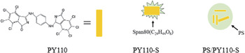
PY110 particles, Span 80 and 100 mL ethanol were dispersed for 2 h to obtain PY110-S, which was subsequently coated with styrene (St) through a mini-emulsion polymerization procedure. Based on the SEM images, TEM images and the FT-IR spectra of PY110, PY110-S, and PS/PY110-S, the structure diagrams of PY110-S and PS/PY110-S are shown in Fig. 1. Reference Han, Zhang, Li, Sun, Shao and Feng14
Structure diagrams of PY110, PY110-S and PS/PY110-S.

A. Characterization of electrophoretic particle structure
Figure 2 shows the influence of Span 80 on dispersibility of PY110. The dispersion effect of PY110 in the water was improved, and the dispersion stability of PY110 was improved from 52.02% to 96.86%, when the mass ratio of Span 80 was 16%. And the dispersion stability could maintain about 15 h, because nonionic surfactant had strong hydrophilic oxygen functional groups. And its molecular chain contained some polar groups which could adsorb suspend solid particles in water and form the bridge with large flocculate from particle surface. When two pigment particles moved toward each other, the interaction occurred between the two adsorption layers in the case the pigment surface spacing was less than half of the thickness of adsorption layer, and entropy decreased to produce entropy repulsion.
The influence of Span80 on dispersibility of PY110: (a) the influence of mass ratio of Span80, (b) the influence of time.

Figure 3 shows the FT-IR spectra of PY110, PY110-S, St and PS/PY110-S. In the spectrum of PY110-S, the peak at 2923 cm−1 and 2852 cm−1 of PY110-S were attributed to the vibrations of the C–H bond belonging to Span 80. Through comparing the IR of St and PS, we found that the differences were the peak of 2850, 2922 and 907 cm−1. 2923 and 2852 cm−1 were attributed to the vibrations of the C–H bond belonging to PS, and 907 cm−1 was attributed to the vibrations of the C–H bond of C=C bond belonging to St.
FT-IR spectra of PS/PY110-S, St, PY110-S, and PY110.

In the spectrum of PS/PY110-S, 3025 and 3059 cm−1 were attributed to the stretching vibrations of the C–H bond in phenyl ring, the peak of 907 cm−1 disappeared, the peak intensity of 2922 and 2850 cm−1 increased obviously, and it indicated that St generated the PS and more PS were coated on the surface of PS/PY110-S. Therefore, a conclusion was inferred that PY110-S was composed of PY110 with Span 80 serving as a dispersant, and PS/PY110-S was consisted of PY110, Span 80 and PS.
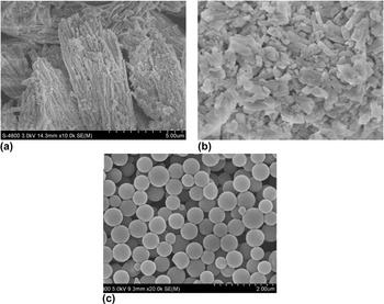
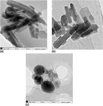
Figure 4 shows the SEM images of the unmodified pigments and corresponding modified particles. Figure 5 shows the TEM images of the same ones. The modification of PY110 by Span 80, the well dispersed PY110-S particles were obtained, as shown in Figs. 4(b) and 5(b). It was reasoned that the well dispersed particles were resulted from the covering of the pigment surface with Span 80, when the mass ratio of the Span 80 on PY110 was applicable (16%). Then through miniemulsion polymerization procedure and adjusting the concentration of emulsifier and monomer, regular spherical composite particles containing PY110-S particles with a narrow size distribution were formed and the average particle size was about 456.5 nm, as shown in Figs. 4(c) and Fig. 5(c). Meanwhile, it was shown in Fig. 5(b), the TEM of PY110-S were rectangular. In Fig. 5(c), we could see the core–shell structure of electrophoretic particles and PS was the spherical shell; PY110-S particles were rectangular cores. It indicated PY110-S particles were coated by PS, which improved the solvent resistance and electrophoretic migration rate of PY110 in electrophoretic liquid. Thus PS/PY110-S was suitable for EDP.
SEM images of (a) PY110, (b) PY110-S, and (c) PS/PY110-S.

TEM images of (a) PY110, (b) PY110-S and (c) PS/PY110-S.

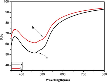
Figure 6 shows the differences of their optical properties. The same peak locations of the crude PY110 and corresponding PS/PY110-S indicated that the composite particles remained the same hues as the PY110. Reflectance peaks of the PS/PY110-S particles were intensified and broadened comparing to that of PY110, which made the PS/PY110-S particles look more albescent than the crude ones. The two kinds of particles were put into filter bag and soaked for 24 h in tetrachloroethylene. We found that the solvent resistance of composite particles had obviously been improved. Meanwhile, we measured transmittance (T s), and the T s of PY110 was 51.57%, as well as the T s of PS/PY110-S was 61.23%. This was because an interfacial reflection process was promoted in a favorable way when the PY110 nanoparticles were coated by PS, resulting in the enhancement of reflective strength. Modified materials insulated organic pigments and solvent, and made organic solvent not come into contact with the organic phase of particles. So it could improve the solvent resistance and extend the life of the organic pigment.
UV–vis spectra and solvent resistance: (a) PY110, (b) PS/PY110-S.

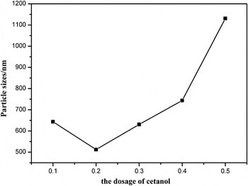
The influence of the dosage of cetanol on the particle sizes of PS/PY110-S was shown in Fig. 7. Assistant surfactant can change the water–oil balance and surface active of surfactant, adjust the polarity of the oil and water and participate in the formation of micelle. Cetanol is water-soluble, which can reduce the polarity of water and make it have better compatibility with monomer. The particle size was first decreased from 643.8 nm to 512.3 nm, and then increased to 1131.1 nm, when the dosage of cetanol was changed from 0.1 g to 0.5 g. Therefore, 0.2 g was determined to be the appropriate dosage.
The influence of the dosage of cetanol on the particle sizes.

The influence of the dosage of SDBS on the particle size of PS/PY110-S particles was shown in Fig. 8. When the dosage of SDBS was changed from 1.5 g to 2.1 g, the particle size decreased from 620.6 nm to 434.3 nm, and then increased to 669.6 nm, as well as the polydispersity index decreased from 0.28 to 0.10, and then increased. Because when the dosage of SDBS was 1.5 g, the concentration of surfactant was so low that it couldn't make monomer droplets containing pigment particles be fully dispersed, which made the composite particle size bigger. Increased the dosage of SDBS could make the formation of new microemulsion continuously and maintain the stability of the microemulsion. When we continued increasing the dosage of SDBS, the pro-oil base group of surfactant would spontaneously form micelle, which couldn't play a role in dispersion of pigment particles.
The influence of the dosage of cetanol on the particle sizes.

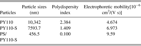
The particle sizes and electrophoretic mobility of composite particles were shown in Table I. Compared with PY110, particle size of PY110-S is reduced to 7593.7 nm and their polydispersity index relatively is reduced to 1.409. The particle sizes of PS/PY110-S reduced to 456.5 nm, and their polydispersity index is relatively reduced to 0.100, which indicated that the modified particles had better dispersion stability. As for the electrophoretic mobility, modified particles still showed better results. The electrophoretic mobility of PY110-S increased to 6.973 × 10⁻⁶ cm2/(V s) and electrophoretic mobility of PS/PY110-S increased to 9.59 × 10⁻⁶ cm2/(V s).
The particle size and electrophoretic mobility of composite particles.
B. The electrophoretic response behavior of the dual-particle electrophoretic fluid
The color contrast of the composite particles in Isopar L:C2Cl4 = 1:1 was verified as shown in Fig. 9. According to the basic color laws of three primary colors elements, yellow pigment in doping red pigment made integral color more orange. It was more advantageous to improve the contrast of yellow-white EPD device.
Color contrast of the composite particles in Isopar L:C2Cl4 = 1:1, (a) the yellow; (b) the mixed yellow.

Next, the electrophoretic movement of yellow-white particles in a simple electrophoretic display test-cell with 25 × 15 × 0.1 mm was investigated, and the contrast ratio was also determined in this test-cell. Figure 10(a) shows the yellow state and white state images in the yellow-white EPD device. The yellow particles bore positive charges, while white particles were negatively charged. When 20 V/mm DC voltage was applied, yellow and white particles moved towards the opposite direction. Having changed the direction of voltage, the display state also changed. Meanwhile, the pigments could always remain 2 h and the test-cell could keep images well when applied voltage. The contrast ratio of yellow state and white state was 1.46. Figure 10(b) shows the mixed yellow state and white state images in the mixed yellow-white EPD device. The contrast ratio of yellow state and white state was 1.58.
EPD images of electrophoretic fluid with dual-particles at 20 V DC voltage: (a) the yellow-white; (b) the mixed yellow-white.

IV. CONCLUSIONS
In summary, the composite PS/PY110-S particles via double modification of original PY110 were prepared and characterized with a simple method. The well dispersed PY110-S particles with uniform size (about 100 nm) were obtained via the coating of the pigment surface with Span 80, when the mass ratio of Span 80 on PY110 was applicable (16%). The yellow electrophoretic slurry achieved the highest zeta potential of −60 to −70 mV and the smallest particle size of 456.5 nm after various condition experiments. The contrast ratio of the fabricated yellow-white dual color EPD cell was found to be 1.46. The composite PS/PY110-S particles were mixed with PS/P.R.146 and the contrast ratio of the fabricated mixed yellow-white dual color EPD cell was found to be 1.58.
ACKNOWLEDGMENTS
This work is supported by the Key project of the Natural Science Foundation of China for international academic exchanges under the contract No. 51020105010. The support from National Key Technologies R&D Program (21476162) is also appreciated.