Implications
Piglet mortality during parturition and lactation is a major problem in pig farming worldwide. Increasing the survival of piglets will significantly enhance animal welfare and improve the ethical sustainability and economy of swine production. In order to increase piglet survival, more information is needed on the causes of piglet mortality in different production systems. This large-scale, postmortem examination study in Norwegian piglet-producing herds with loose-housed sows, provides further relevant information on this topic, indicating that trauma and starvation are important causes of death in preweaned piglets.
Introduction
A high piglet mortality during parturition and lactation is a major problem in pig farming worldwide. Preweaning mortality rates of live-born piglets in European countries range from 10% to 20% (Muns et al., Reference Muns, Nuntapaitoon and Tummaruk2016). Total preweaning mortality in piglets, including stillbirths, is estimated to be between 18.8% and 25% (Damm et al., Reference Damm, Pedersen, Heiskanen and Nielsen2005). In Europe, between 4% and 12% of piglets are stillborn (Edwards, Reference Edwards2002; KilBride et al., Reference KilBride, Mendl, Statham, Held, Harris, Cooper and Green2012; Strange et al., Reference Strange, Ask and Nielsen2013). The majority of piglets that die before weaning, die within the 1st week of life (Varley, Reference Varley1992), with stillbirth, starvation and crushing by the sow considered to be the main causes (Pedersen et al., Reference Pedersen, Berg, Jørgensen and Andersen2011). In a Swedish study, stillbirth, starvation and crushing accounted for 6.5%, 5.0% and 4.1% of piglet mortality from all piglets born, respectively (Westin et al., Reference Westin, Holmgren, Hultgren, Ortman, Linder and Algers2015). These percentages vary among studies, depending on housing systems (crates v. loose housing), management, weaning age and definitions used regarding mortality rates (KilBride et al., Reference KilBride, Mendl, Statham, Held, Harris, Cooper and Green2012).
Pig husbandry has changed considerably over the last 20 years, and the focus on animal welfare has greatly increased. In 2003, Norwegian legislation prohibited crated sows in the farrowing pen. In 1986, Norwegian sows produced, on average, 10.1 live-born piglets per litter, 1.1 piglets were stillborn (9.8%) and 8.5 piglets were weaned. The mean weaning age was 44 days (Grøndalen et al., Reference Grøndalen, Liven, Djønne, Gjestvang, Jæger and Utlev1986). In Norway today, national data can be obtained from The National Efficiency Control Database in Norway, called Ingris, which contains production results and is administrated by Norsvin (Norwegian Pig Breeding Association) and Animalia (Norwegian Meat and Poultry Research Centre). According to Ingris, in 2014, Norwegian sows produced, on average, 13.2 live-born piglets and 1.1 stillborn piglets (8.4%) per litter, and weaned, on average, 11.3 piglets per litter at day 33; the preweaning mortality of live-born piglets was 14.2% (Ingris, 2014). The preweaning mortality of total-born piglets that same year was 21%. Information on the exact causes of preweaning mortality is needed in order to be able to reduce piglet losses. A number of studies of piglet mortality have relied on diagnoses made by farmers. However, up to 40% of such diagnoses have been incorrect (Edwards et al., Reference Edwards, Smith, Fordyce and MacMenemy1994). In order to understand the etiology behind most piglet deaths, a thorough postmortem examination is necessary. The aim of the present study was to use postmortem examinations to determine the etiology and prevalence of preweaning mortality in piglets in Norwegian loose-housed sow herds.
Material and methods
Study population
The data presented in this paper were obtained as part of a larger study investigating both management and sow behavior in association with preweaning mortality. Details have been described previously (Rosvold et al., Reference Rosvold, Kielland, Ocepek, Framstad, Fredriksen, Andersen-Ranberg, Næss and Andersen2017). Initially, 52 herds were included based on the following criteria: firstly, farms where sows were kept loose during parturition, farms with regular records of production results to Ingris, and secondly, breed (LY: sows of Norwegian Landrace×Swedish Yorkshire). For the present study, convenience sampling was used, with samples collected from a subset of 14 herds within 3 h driving distance of the pathology laboratory. The study period was from September 2013 until May 2014. All herds were located in the southeastern part of Norway. From 347 sows with 5254 total-born piglets, of which 4924 were live-born, 4026 survived and 1228 piglets were reported dead at weaning. Altogether, 1216 dead piglets were collected, and, of these, 1200 were necropsied. The mean parity of the sows was 2.4 (±1.6 SD), ranging from 1 to 10. Farmers were requested to adhere to usual management routines during this study.
Farm characteristics
All herds included in the study used batch management systems. The number of sows per batch ranged from 20 to 60 sows, with 49% of the herds with a batch size of 20 to 25 sows. The interval between batches was mostly seven weeks (65% of the herds). In each herd, gilts and sows were loose-housed in groups before they were moved individually into standard farrowing pens without crates, mean size of 7.7 m2 (±1.1 SD, including a separate piglet creep area). Sows and gilts were, on average, moved to individual farrowing pens 8 days (±4 SD) before expected parturition, ranging from 3 to 18 days. The mean parturition duration in the herds was 4.9 h, ranging from 1 to 12.75 h. The temperature in the lactation rooms was 18°C to 19°C in all herds. Farrowings were not hormonally synchronized. Farmed animals in Norway only receive drugs administered by a veterinarian. Parturition was attended. At 1 week of age, the male piglets where castrated using local anesthesia and systemic analgesia. Teeth clipping and tail docking was not performed as this is prohibited by law in Norway. Mean weaning age in the herds was 32 days (±3.9 SD). All herds used the same vaccination regime; that is, vaccinating against Escherichia coli, porcine parvovirus, and Erysipelothrix rhusiopathiae. The sows were fed a commercial lactation diet containing 9.29 MJ NE/kg feed and 8.26 g lysine/kg feed. All sows were provided with small amounts of hay/sawdust before farrowing, and all sows had ad libitum access to water.
Data collection
Farmers wrapped each dead piglet individually in a plastic bag, and marked the bag with a preprinted sticker in which they were instructed to add the following information: Herd id, sow id, date of parturition, date of death and tentative cause of death as suspected by the farmer. For the piglets that died on the day of parturition, the time of death was recorded as lactation day 0 (LD 0). For piglets that died the day after parturition, time of death was LD 1, and so on until LD 32. Before collection, dead piglets were stored in a cool place in the barn, avoiding freezing, as that would negatively affect the quality of the necropsy. Dead piglets were collected from each herd twice weekly (Sunday and Wednesday), stored in a cooler overnight, and the necropsy was performed the next day. Sow-level data were recorded by the farmer on a separate form. Information recorded was sow id, number of live-born piglets, number of stillborn piglets, duration of parturition (time between first and last piglet born), whether the farmer had intervened during parturition, and/or if oxytocin was used. Production results from 2014 for the 14 herds were extracted from Ingris (Ingris, 2014). The total number of piglets born, which is synonymous with the term ‘total born’, was defined as the number of live-born plus the number of stillborn piglets per sow. As a part of the necropsy, piglets where weighed (kg) and measured (cm). Body length was measured from os occipitale to the root of the tail. Body mass index (BMI) was calculated using BW (kg) and the square of body length (m2): BMI=BW (kg)/the square of body length (m2).
Postmortem examinations
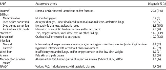
All piglets found dead or euthanized before weaning, and traceble back to the farm, were necropsied (n=1200). The necropsy included general external and internal inspection at the Norwegian Veterinary Institute. In order to categorize each piglet as having one primary cause of death, pathoanatomical diagnoses (PAD) were based on the postmortem examination (Engblom et al., Reference Engblom, Eliasson-Selling, Lundeheim, Belák, Andersson and Dalin2008). Each piglet was assigned to one of the nine primary PAD categories (Table 1). When several PAD categories were identified in an individual piglet, the PAD that was considered most likely to be the cause of death was used. For example, a piglet with anemia and broken ribs would be categorized as death due to trauma. When the primary cause of death was not possible to determine, the piglets were given the PAD No clear PAD. The category stillborn, was subdivided into four different groups; mummification, dead before parturition, dead during parturition and dead due to aspiration of amniotic fluids with or without meconium.
Causes of death and pathoanatomical diagnoses (PAD) based on postmortem examination of piglets and prevalence among the necropsied piglets (n=1200)

NPAD=no clear PAD.
1 PAD is ranged according to the mortality percentage of the 1200 piglets.
2 When the farmer reported that the piglet had been euthanized and the skull was crushed, these piglets were then categorized as euthanized. Without reports from the farmer, but with obvious signs of crushed skull, these were also categorized as euthanized.
3 Included seven piglets with (n): esophageal obstruction (2) atresia ani (2), drowning (1) or no aorta present (2).
4 NPAD: the majority of piglets in this group had several of the PADs, but determining which PAD that was the primary cause of death was not possible.
The main criteria for each PAD are defined in Table 1. Detailed postmortem findings, such as subcutaneous edema (SOD) and skin lesions, are defined below. The definition of SOD is a non-inflammatory accumulation of fluid in subcutaneous tissue. Skin lesions were defined as present when either discoloration, or swelling, or ulcerations were observed. When signs of infection were present, affected organs(s) were analyzed by standard microbiological techniques. The results were used to confirm/refute a PAD. The results from the microbiological examinations of 156 samples from 98 piglets were categorized as follows; (N) normal flora or no growth; (C) contamination or non-specific mixed growth; (U) unspecific growth and dependent on other findings; (P) pathogenic bacteria.
Statistics
Descriptive statistics were conducted using the statistical software STATA (Stata SE/10 for Windows; Stata Corp., College Station, TX, USA). When calculating the preweaning mortality among total-born piglets, the data set included all piglets born within the corresponding batch and herd (n=5254). Preweaning mortality were calculated for both total-born piglets (n=5254) and live-born piglets (n=4924). The prevalence of each PAD was calculated at three levels; (a) among total-born piglets, (b) among live-born piglets and (c) among the necropsied piglets. The prevalence of each detailed postmortem finding, that is, SOD, skin lesions, circulation failure, etc., was calculated for each individual PAD. Any association between skin lesions and cause of death due to an infection (excluding the stillborn), was investigated using χ 2 test on contingency tables. κ was used to evaluate the agreement between the farmers’ tentative diagnoses of the cause of death and the cause of death according to PAD. κ Values between 0.21 and 0.40 are considered as a fair agreement, 0.41 and 0.60 indicates moderate agreement and 0.61 and 0.80 equals substantial agreement (Viera and Garreth, Reference Viera and Garreth2005). In the analyses, statistical significance was considered at P<0.05.
Missing data
Among the reported dead piglets (n=1228), 12 were not collected for necropsy and the origins of 16 could not be identified and therefore these were not included in the postmortem analyses. Nevertheless, in order to obtain the correct mortality percentage for all piglets in the 14 herds, these 28 piglets were included when the preweaning mortality of the total number of piglets was calculated. In 63 piglets, the date of death had not been recorded and the time of death could not be calculated. Length (cm) was not recorded for 14 piglets, eight due to mummification. The start and end of parturition, that is, the time between first and last piglets being born, was recorded during 178 parturitions. A tentative diagnosis regarding the cause of death was obtained from the farmers for 980 piglets. In the comparison of the farmers’ and pathologists’ diagnoses, 220 piglets that did not receive an ‘on-farm diagnosis’ were excluded.
Results
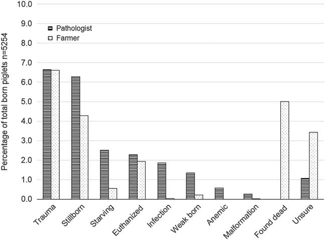
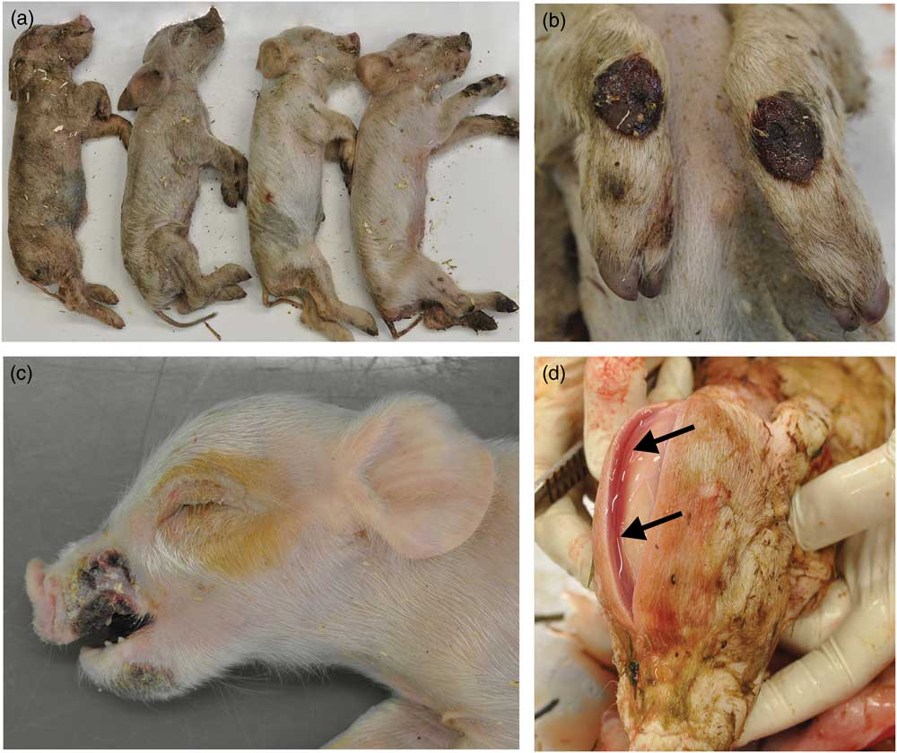
Total preweaning mortality was 23.4% (1228/5254), with 6.3% (330/5254) of the piglets diagnosed as stillborn. The piglets were categorized into nine different PAD (Table 2), and Figure 1 displays the prevalences of different causes of death among total-born piglets included in the study. Preweaning mortality of live-born piglets was 18.2% (898/4924). The most prevalent cause of death according to PAD among all live-born piglets (n=4924), was trauma (7.1%, 349/4924). Starvation was diagnosed in 2.7% (132/4924) of the piglets (Figure 2a), and 3.0% were euthanized (120/4924). Piglets dying of an infection accounted for 2.0% (98/4924) of deaths, and being weak born for 1.5% (71/4924).
Preweaning mortality of all piglets born (n=5254) assigned to nine causes of death categories, based on pathoanatomical diagnoses (PAD) (black and white stripes) and the cause of death given by the farmer (black dots). The category ‘Found Dead’ had no other explanation and the cause of death was not diagnosed by the farmer.

Photographs from the postmortem examination of piglets. (a) Four 3 to 6 lactation days (LD) old piglets. These piglets died from starvation and dehydration. (b) 1 week-old piglet, front legs. The piglet had severe skin ulcerations with granulation tissue formation on the cranial aspects of the carpal joints. (c) 1 week-old piglet. The piglet had bilateral extensive skin ulcerations on the snout and a focal skin erosion on the lower jaw. (d) 1 day-old piglet. The piglet had pronounced blood-tinted subcutaneous edema (arrows) on the forehead.

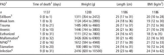
Descriptive data (mean (min. to max.)) at time of death (days), weight (g), length (cm) and body mass index (BMI; kg/m2) at death of 1200 piglets, categorized according to the pathoanatomical diagnosis (PAD)

NPAD=no clear PAD.
1 PAD is ranged according to the mean age when the piglets were found dead.
2 Time of death in lactation days.
3 Including piglets that died before and during parturition, piglets with aspired amniotic fluids and mummified piglets.
4 Included seven piglets with (n): esophageal obstruction (2) atresia ani (2), drowning (1) or no aorta present (2).
5 NPAD: the majority of piglets in this group had several of the PADs, but determining which PAD that was the primary cause of death was not possible.
6 Included both piglets with a general infection and those with enteritis.
Postmortem examination (n=1200)
The PAD of the primary cause of death in each piglet are shown in Table 1. Among the necropsied piglets (n=1200), 29% died due to trauma (n=349), 26.8% of piglets were categorized as stillborn (n=330), 11% died of starvation (n=132) and 8% died from an infection. Among the piglets diagnosed as stillborn (n=330), 45.5% (150/330) were normal mature fetuses that died during parturition, and 27.3% (90/330) were normal mature fetuses with aspired amniotic fluids, that is 72.8% died intrapartum. The remaining stillborn piglets died before parturition and were not fully grown (24.8%, 82/330) or were mummified (0.6%, 8/330). Excluding stillborn from the 1200 necropsied piglets, 39.9% (347/870) of the piglets were categorized as having died due to trauma.
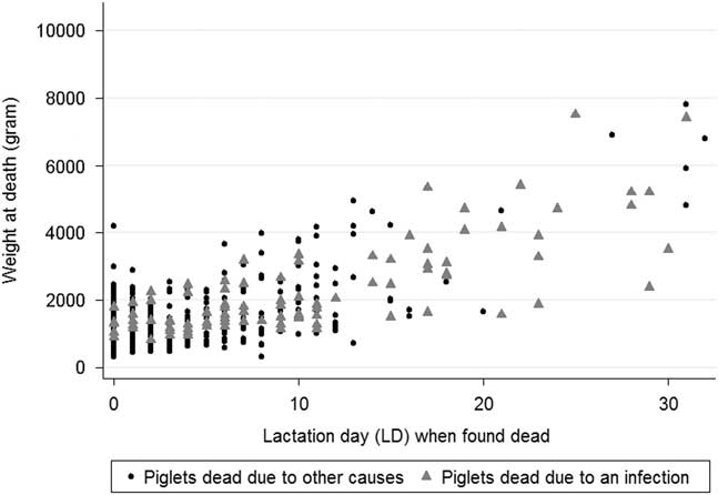
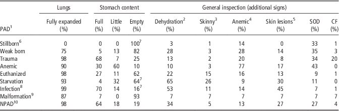
For all necropsied piglets with known time of death (n=829), and excluding those with the PAD stillborn, 30.7% died at LD 0, 18.9% died at LD 1, 15.7% died at LD 2, 7.1% died at LD3, and 27.6% died between LD 4 and LD 32. The mean time of death for live-born piglets that died was LD 3.5 (ranging from LD 0 to LD 32). The association between time of death (LD) and BW (g) measured during the necropsy is shown in Figure 3. The lowest mean BWs were found in piglets that died of starvation, were weak-born or were euthanized (Table 2). The mean BMI-values for starving and euthanized piglets were 16 and 18 kg/m2, respectively. Piglets that died of an infection were among the heaviest piglets (Figure 3) and had a high BMI. Of all live-born piglets that died, 17.3% had skin lesions, with 59.0% of these lesions on the front legs, mainly carpus (39.1%: Figure 2b). A considerable number of piglets had lesions on both carpus and tarsus (23.0%), and some had deep skin ulcerations (Figure 2c). There was a significant association between macroscopic local skin lesions and death due to an infection (P<0.001), with piglets that had died due to an infection were more likely to have skin lesions than not. When exploring the detailed findings of the necropsy for the most prevalent PAD (trauma, n=349), it was apparent that these piglets were among the heavier piglets that died (Table 2). Most of the piglets that died of trauma did so within LD 3 (82.2%). The remaining 17.8% died between LD 4 and LD 21. Most of these piglets had full stomachs (68%), but 25% had empty stomachs and 7% had only a small amount of milk in their stomachs (Table 3). Additionally, 20% of piglets that died of trauma had signs of anemia, 33% had signs of SOD (Figure 2d), and 13.2% had signs of dehydration. Other detailed descriptions from the necropsies for each PAD are provided in Table 3. Unusual findings, such as hyperkeratosis of the pars esophagea were demonstrated in the ventricles of four piglets. Other infrequent findings were: esophagus obstruction, atresia ani, one piglet had drowned in the feeding trough, and another piglet had no aorta.
Time of death (lactation days (LD)) and weight (g) recorded during the necropsy of all the necropsied piglets (n=1200). Piglets that died of an infection is highlighted with a gray triangle (n=98), the remaining piglets are marked with small black dots (n=1102)

Pathoanatomical diagnosis (PAD) and detailed postmortem findings of 1200 piglets

SOD=subcutaneous edema; CF=circulation failure (congestion of internal organs, mainly lungs and liver); NPAD=no clear PAD.
1 PAD is ranged according to the mean age when the piglets were found dead and the numbers are row % according to categories.
2 Piglets with sunken eyes and reduced skin elasticity.
3 Piglets with little subcutaneous fat and reduces muscle mass.
4 Piglets with pale skin and pale organs.
5 Skin lesions present, regardless of location.
6 Including piglets that died before and during parturition, mummified and also piglets with aspired amnion fluids.
7 Hyperkeratosis of the pars esophagea was demonstrated in the ventricle in four piglets; two piglets were stillborn, one died of starvation and one died of an infection.
8 Included both piglets with a general infection and those with enteritis.
9 Included seven piglets with esophagus obstruction, atresia ani, drowning, lacking aorta.
10 NPAD: the majority of piglets in this group had several of the PADs, but determining which PAD that was the primary cause of death was not possible.
Microbiological analyses
Of the microbiological analyses from 8% of all necropsied piglets, 47.4% of the samples were from the small (39.7%) or large (7.7%) intestines. Among the samples analyzed, 44.3% of the bacteriological findings were categorized as N, 31.4% as C, 12.8% as U and 11.5% as P. Among the samples in category P and U, pathogenic bacteria were identified in 18 (47.4%). The most prevalent species was E. coli (n=13, 34.2%); with four isolates of the type E. coli F4+ (intestine) and one hemolytic E. coli (small intestine). The other pathogenic bacteria found were identified as Staphylococcus hyicus (skin, spleen and lungs; n=5), Staphylococcus aureus (heart and spleen; n=3), Streptococcus sp. (joint; n=1) and Trueperella pyogenes (joint; n=1).
Farmers’ diagnoses
Farmers provided a tentative diagnosis on the cause of death for 980 (81.6%) of the 1200 necropsied piglets. Comparison of the farmers’ diagnoses with the PAD, indicates Fair agreement (34%, P<0.001). The highest agreement found was for piglets that had died due to crushing (farmers’ diagnosis)/trauma (PAD), Figure 1. Among the piglets assigned the tentative diagnosis ‘found dead’ by the farmer (Figure 1), 35% were in the PAD category Stillborn. Among piglets assigned the PAD category ‘Starvation’, the farmer used the word ‘starvation’ for 13.6% of the piglets Three percent of the total-born piglets were euthanised. The most frequent reasons for euthanasia given by the farmer were; starvation (34.3%, 36/105), weak born (22.9%, 24/105,), being small (9.5%, 10/105).
Discussion
Comparison of these results with those from a similar study of piglet mortality in Norwegian herds published over 30 years ago (Grøndalen et al., Reference Grøndalen, Liven, Djønne, Gjestvang, Jæger and Utlev1986), indicates that the preweaning mortality of piglets has been reduced in Norway over this period. Whereas the former study reported 24.7% total loss of piglets, in the present study, the total piglet loss was 23.4%. The sows were housed differently in these two studies, with crates used in the former and loose housing in the current study. The genetics, nutrition, and management of piglets have also changed greatly during the intervening period between these studies. Studies from other countries have also shown comparable figures of piglet mortality in different housing systems (Weber et al., Reference Weber, Keil, Fehr and Horat2009; KilBride et al., Reference KilBride, Mendl, Statham, Held, Harris, Cooper and Green2012). Interestingly, the average percentage of preweaning mortality of live born reported to Ingris from the 14 study herds during the period from June 2013 to June 2014 was 18.2% (Ingris, 2014). However, the mortality recorded in our study was higher, and our figures were also higher than the country average of 21% that was reported during the same period. These discrepancies indicate problems in the registration system and possible under-reporting of piglet mortality at the national level. Under-reporting may be partly due to farmers not including mummified and malformed piglets in their report to the national registry. It is also known that farmers may euthanize very small piglets without counting these as being born alive, and this can influence numbers in the national database. A well-functioning recording system is essential for modern pig production, and our data suggest that validation of the current Norwegian database, Ingris, could be advantageous, both for breeding purposes and for animal welfare assessments. The number of stillborn piglets was 2% lower than the number in the national database, Ingris, which should be representative of the Norwegian pig population. The numbers of stillborn were also lower than those reported in a study of loose-housed herds in England (KilBride et al., Reference KilBride, Mendl, Statham, Held, Harris, Cooper and Green2012). Participant bias may be one reason for the number of stillborn being lower than expected, as the study may have selected for participation of particularly concerned farmers. The study also demanded considerable efforts from each farmer during parturitions, and this may have influenced the number of piglets that were saved as the farmers were more likely to be present during parturition. Results from a larger study performed in the same herds as the present study (Rosvold et al., Reference Rosvold, Kielland, Ocepek, Framstad, Fredriksen, Andersen-Ranberg, Næss and Andersen2017), showed that farmer presence during parturition decreased the preweaning mortality.
Of all the piglets necropsied, the percentage of stillborn piglets was reduced from that reported in 1986 (Grøndalen et al., Reference Grøndalen, Liven, Djønne, Gjestvang, Jæger and Utlev1986; 37.5%) until today (26.8%). This suggests that management procedures, feeding, and/or genetics have improved with respect to reduction of stillbirths. Comparing the distribution between the categories of stillborn, the majority of the piglets were fully grown and died during parturition. This is in accordance with a study on stillbirths preformed in the Netherlands (Leenhouwers et al., Reference Leenhouwers, van der Lende and Knol1999). Piglets born late during the parturition process, and/or with a broken umbilical cord, have been suggested as explanations for 71% of stillbirths in a previous study in Norwegian piglets (Rootwelt et al., Reference Rootwelt, Reksen, Farstad and Framstad2012). Our finding that 14% of stillborn piglets had signs of anemia, could suggest that a broken umbilical cord is a possible cause of death. Interestingly, we found that 33% of stillborn piglets had clear signs of SOD; this could be due to cardiac failure caused by asphyxia, as cited in a review (Alonso-Spilsbury et al., Reference Alonso-Spilsbury, Mota-Rojas, Villanueva-Garcia, Martinez-Burnes, Orozco, Ramirez-Necoechea, Mayagoitia and Trujillo2005). However, SOD may also be a normal finding as newborns may have some degree of edema due to a large amount of total body fluid (80%) (Fanaroff and Martin, Reference Fanaroff and Martin2002). The postmortem examinations showed that most live-born piglets that die before weaning, die of trauma (7.1%). This is comparable with other studies with loose-housed sows (4.5% to 9.8%; Weber et al., Reference Weber, Keil, Fehr and Horat2009). However, other studies have reported a slightly higher risk of crushing in comparable housing systems (KilBride et al., Reference KilBride, Mendl, Statham, Held, Harris, Cooper and Green2012; Hales et al., Reference Hales, Moustsen, Nielsen and Hansen2013). However, the overall mortality in crate systems and loose-housing systems is reported to be similar, and sow welfare is greatly improved in loose-housing systems (KilBride et al., Reference KilBride, Mendl, Statham, Held, Harris, Cooper and Green2012; Hales et al., Reference Hales, Moustsen, Nielsen and Hansen2013). In addition, trauma (caused by crushing) is highly associated with mothering style (Andersen et al., Reference Andersen, Berg and Bøe2005); lateral lying style and nest-building activities are strongly associated with the risk of crushing (Pedersen et al., Reference Pedersen, Jørgensen, Heiskanen and Damm2006). Planned future studies will explore how mothering style is associated with each cause of death.
In 1986, Grøndalen et al. (Reference Grøndalen, Liven, Djønne, Gjestvang, Jæger and Utlev1986) reported that one of the most common causes of preweaning mortality of total-born was dehydration, with a prevalence of 2.9%. We assume that the dehydrated piglets reported in 1986, would have been categorized as starved in our study (2.5%), as 65% of the piglets that were given the PAD ‘starvation’ also had signs of dehydration.
Preweaning mortality among live-born piglets due to bacterial infection occurred in 2.0% of preweaning deaths in these 14 pig farms, indicating that bacterial infections are currently not an important factor in preweaning mortality in Norway. The proportion of infections among necropsied piglets has been reduced in Norway, from 25% in 1986 (Grøndalen et al., Reference Grøndalen, Liven, Djønne, Gjestvang, Jæger and Utlev1986) to 8.0% today. One reason may be that vaccination of sows is more common today than 30 years ago. Another reason may be the common use of all-in-all-out management today, which includes thorough cleaning of farrowing pens between batches. In a Swedish experimental study, 24% of all necropsied piglets had an infection (Westin et al., Reference Westin, Holmgren, Hultgren, Ortman, Linder and Algers2015), most of which was enteritis (60%). In our study, there was a significant association between skin lesions and death due to an infection in the piglets. Skin lesions may provide an entrance route for pathogenic bacteria and thereby increase the risk of death in piglets. Piglets that died of an infections, died closer to weaning. Late losses are more costly in terms of management and labor, and may also be considered as having a more severe impact on animal welfare as infections often cause pain and discomfort over a longer time period.
Our findings were in accordance with a review stating that most of the total preweaning mortality occurs during the first 72 h (Muns et al., Reference Muns, Nuntapaitoon and Tummaruk2016). However, most studies report higher piglet mortality during the 1st days of life than we observed in our study (Pedersen et al., Reference Pedersen, Berg, Jørgensen and Andersen2011; KilBride et al., Reference KilBride, Mendl, Statham, Held, Harris, Cooper and Green2012; Muns et al., Reference Muns, Nuntapaitoon and Tummaruk2016). Our findings suggest that in Norwegian pig farms, the survival rate of piglets could be improved by farmers directing greater efforts towards detection of starving piglets and preventing skin lesions from occurring. Starving and euthanized piglets had the lowest mean BWs and BMI in our study, both close to 17 kg/m2. Kielland et al. (Reference Kielland, Rootwelt, Reksen and Framstad2015) found that piglets with BMI below 17 kg/m2 had significantly lower Immunoglobulin G (IgG) concentrations in the blood at LD 1. In addition, a lower BMI is associated with a relatively increased body surface, which increases heat loss (Quesnel et al., Reference Quesnel, Farmer and Devillers2012). Thus, achieving a higher BMI should be a goal in piglet production, both to reduce piglet mortality by adequate intake of colostrum, and to prevent heat loss. Achieving adequate milk intake by piglets is important, especially with the demand for larger litter sizes resulting in greater competition around suckling. Not surprisingly, the diagnosis crushing/trauma showed the highest agreement between farmer and pathologist, as external and/or internal lacerations and/or fractures are easily observed macroscopically. Piglets that died due to starvation were more challenging for the farmer to diagnose correctly, and agreement was only 13.6%. Increasing farmers’ knowledge on how to identify a starving piglet, improve their milk supply, and manage these piglets, remains a work in progress, especially for high-producing sows with large litters.
Conclusion
Our study shows that there are still challenges in pig farming regarding preweaning mortality in piglets due to trauma and starvation. Many of the stillborn piglets were fully developed, a finding indicating that there is a potential to reduce the number of stillborn. Most piglets that died in the preweaning period, died of trauma. Surprisingly, this included large and well-fed piglets. The second most prevalent cause of pre-weaning mortality was starvation. Improved monitoring may reveal piglets with low BMI, and additional nutrition may contribute to increase the survival rate.
Acknowledgments
The authors would like to thank the farmers participating in this work-intensive study. We are also grateful for the funding provided by the Agricultural Agreement Research Fund and the Foundation for Research Levy on Agricultural Products.