Introduction
The La Meseta Formation (Eocene), Antarctic Peninsula, is a highly fossiliferous unit in terms of the quantity and diversity of remains. The invertebrate taxa include bivalves, gastropods (Stilwell & Zinsmeister Reference Stilwell and Zinsmeister1992, Bitner Reference Bitner1996), bryozoan colonies (Hara Reference Hara2001), crinoids (Rasmussen Reference Rasmussen1979, Baumiller & Gaździcki Reference Baumiller and Gaździcki1996), ophiuroids (Aronson et al. Reference Aronson, Blake and Oji1997, Blake & Aronson Reference Blake and Aronson1998), and echinoids (McKinney et al. Reference McKinney, McNamara and Wiedman1988). Within the vertebrates, fishes (Jerzmańska Reference Jerzmańska1988, Jerzmańska & Świdnicki Reference Jerzmańska and Świdnicki1992), whales (Wiman Reference Wiman1905, Borsuk-Białynicka Reference Borsuk-Bia̡ynicka1988, Fordyce Reference Fordyce1989), a sparnotheriodontid mammal (Vizcaino et al. Reference Vizcaino, Bond, Reguero and Pascual1997), a ratite bird (Tambussi et al. Reference Tambussi, Noriega, Gazdzicki, Tatur, Reguero and Vizcaino1994) and penguins (Myrcha et al. Reference Myrcha, Jadwiszczak, Tambussi, Noriega, Gazdzicki, Tatur and Del Valle2002, Jadwiszczak Reference Jadwiszczak2006a, Hospitaleche & Reguero Reference Hospitaleche and Reguero2010 and references cited therein) have been described.
Penguins (Aves, Sphenisciformes) are particularly abundant in this unit. Even though knowledge of this group has increased greatly in the past few years, there are still many unanswered questions, especially regarding penguin anatomy and functional morphology. Due to the fact that the systematics of Sphenisciformes relies on the morphology and proportions of the tarsometatarsi, and sometimes humeri, few Antarctic studies have been performed regarding other skeletal elements. Only in a few cases elements other than the tarsometatarsi and humeri have been described (Hospitaleche & Reguero Reference Hospitaleche and Reguero2010), and analysed in detail (Hospitaleche & Di Carlo Reference Hospitaleche and Di Carlo2010, see also Jadwiszczak Reference Jadwiszczak2006a, Reference Jadwiszczak2006b).
The first Antarctic penguin skull to be described was a very fragmentary bill assigned by Olson (Reference Olson1985) to ?Palaeeudyptes sp. Other isolated remains including cranial elements have been described (Myrcha et al. Reference Myrcha, Tatur and Del Valle1990) and more recently re-studied (Jadwiszczak Reference Jadwiszczak2003, Reference Jadwiszczak2006a). Additional remains from the Submeseta Allomember were described and treated as problematic specimens (Jadwiszczak Reference Jadwiszczak2006a) due to their poor preservation and lack of comparable material. In spite of this, Jadwiszczak's valuable contribution has provided the basis for advancing the knowledge of morphological aspects that might be useful in systematic and palaeobiological studies. According to their general morphology and size, some of these remains have been assigned to Palaeeudyptes gunnari (Wiman, Reference Wiman1905) (Jadwiszczak Reference Jadwiszczak2006a), although previous studies assigned them to Anthropornis sp. or Palaeeudyptes sp. Jadwiszczak (Reference Jadwiszczak2000, Reference Jadwiszczak2003) and to ?Palaeeudyptes sp., probably P. klekowskii Myrcha, Tatur & del Valle, 1990 (Jadwiszczak Reference Jadwiszczak2006a, see also Myrcha et al. Reference Myrcha, Tatur and Del Valle1990). Other specimens studied by Jadwiszczak (Reference Jadwiszczak2006a) were assigned to P. gunnari or Archaeospheniscus wimani (Marples, 1953) or were too fragmentary for identification according to Jadwiszczak (Reference Jadwiszczak2006a). Only one of them corresponds to an incomplete and deformed neurocranium and constitutes until today the only reference to a Palaeogene Antarctic cranium (Jadwiszczak Reference Jadwiszczak2006a). All of them come from the Submeseta Allomember.
More recently, new findings from La Meseta Formation were described and at least three species were identified on the basis of size differences (Ksepka & Bertelli Reference Ksepka and Bertelli2006), although the fragmentary state of the material does not allow their systematic assignment.
The above seems to indicate that the preservation of craniums and mandibles is improbable. They are easily destroyed and only small fragments can be recovered from the sediments.
In this context of patchy information, even minor skull characteristics that can be detected will be useful for: 1) increasing the knowledge of the anatomy of penguin skulls, in particular of Sphenisciformes from Antarctica, 2) elucidating, as much as the material allows and within the limitations imposed by its preservation state, their trophic habits, and 3) analysing the faunal changes detectable from the specimens studied here.
In the present contribution, several cranial and mandibular penguin remains from La Meseta Formation (Eocene) of the Antarctic Peninsula are studied, including the oldest penguin neurocranium from Antarctica. A comparative description including modern and fossil species is given in order to identify the presence of different morphotypes in this penguin assemblage.
Abbreviations
DPV (División Paleontología Vertebrados del Museo de La Plata, Argentina), IAA (Instituto Antártico Argentino), MEF-PV (Museo Paleontológico Egidio Feruglio, Argentina), MLP (Museo de La Plata, Argentina), RNP (Rae Natalie Prosser, The R. Natalie P. Goodall Foundation, Argentina), RV (University of California at Riverside, USA), UCPM (University of California Museum of Paleontology, USA).
Geological and geographical setting
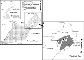
The La Meseta Formation (Rinaldi et al. Reference Rinaldi, Massabie, Morelli, Rosenman and Del Valle1978, Elliot & Trautman Reference Elliot and Trautman1982, Marenssi et al. Reference Marenssi, Santillana and Rinaldi1998a) crops out on Isla Marambio (Seymour Island) and Cockburn Island, close to the northern tip of the Antarctic Peninsula (Fig. 1). This unit has been assigned to the Eocene and is composed of sandstones and mudstones with interbedded shell rich conglomerates, organized into six erosionally based internal units. They are from base to top: Valle de Las Focas, Acantilados, Campamento, Cucullaea I, Cucullaea II and Submeseta Allomembers (Telms 1–7 according to Sadler Reference Sadler1988). These units were deposited during the Eocene in deltaic, estuarine and shallow marine settings, mostly within a NW–SE trending valley (Marenssi et al. Reference Marenssi, Santillana and Rinaldi1998a, Reference Marenssi, Santillana and Rinaldi1998b).
a. Map showing the location of Isla Marambio (Seymour Island), Antarctic Peninsula. b. Sketch map of the northern part of Isla Marambio showing the distribution of the Submeseta Allomember and the fossil penguin bearing localities cited in the text.

In particular, penguins are one of the most abundant groups, represented throughout the sequence by 14 species (Tambussi et al. Reference Tambussi, Hospitaleche, Reguero and Marenssi2006, see also Jadwiszczak Reference Jadwiszczak2006a). The oldest penguin from La Meseta Formation appears in the Valle de las Focas Allomember or the Acantilados Allomember (Telm 1-2 according to Jadwiszczak Reference Jadwiszczak2006b) and is represented by an indeterminate genus and species. One species was collected in the Campamento Allomember, whereas the diversity increases in the highest levels of the formation. At least eight species are known from the Cucullaea I Allomember, six species from Cucullaea II, and in the Submeseta Allomember, at the top of the sequence, 14 species are present (see however Jadwiszczak Reference Jadwiszczak2006a, Reference Jadwiszczak2006b, Reference Jadwiszczak2008, Reference Jadwiszczak2010).
Twelve facies were described by Marenssi et al. (Reference Marenssi, Santillana and Rinaldi1998a), three of which are present in the La Meseta Formation. Facies Association I extends from the Valle de Las Focas, through the Acantilados and Campamento allomembers. The Formation represents valley-confined deposition in progradational/agradational tide dominated and wave influenced delta front/delta plain environments at the beginning of the infill of the incised valley. Energy increased as the delta built up to sea level. The incision of third order surfaces in the upper part of the Acantilados Allomember indicates the change from a wave reworked delta front to a tide dominated delta plain environment. Only a few fragments of penguin bills have been described from these levels (Jadwiszczak Reference Jadwiszczak2006b).
Facies Association II is the intermediate element, and includes the Cucullaea I, Cucullaea II and the lower part of the Submeseta Allomembers. A diverse and abundant macrofauna has been found here, corresponding to a valley confined estuary mouth to inner estuary complex. Tidal channels and mixed flats, tidal inlets and deltas, and washover and beach environments represent the interfingering of high and low energy environments (Marenssi et al. Reference Marenssi, Santillana and Rinaldi1998a). Within the vertebrates, several penguin remains including MLP 96-I-6-48, MLP 92-II-2-115a, MLP 92-II-2-108, MLP 92-II-2-250 and MLP 92-II-2-203 of the material here described, come from this facies.
The uppermost Facies Association III is characterized by a more uniform sandy lithology that represents non-confined tide and storm influenced nearshore environments. A sea level rise is suggested on the top of the Submeseta Allomember. Thin shell beds, gravel beds and clay levels are intercalated (Marenssi et al. Reference Marenssi, Santillana and Rinaldi1998a). Most of the remains described in this contribution, including the neurocranium, come from this level.
Systematic palaeontology
Aves
Sphenisciformes
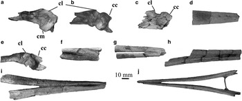
a. Dorsal view of articular region (MLP 96-I-6-48). b. Dorsal view of articular region (MLP 92-II-2-115a). c. Dorsal view of articular region (MLP 91-II-4-223). d. Lateral view of symphysis (MLP 96-I-6-48). e. Dorsal view of articular region (MLP 92-II-2-108). f. Medial view of right ramus mandibularis (MLP 78-X-26-143). g. Ventral view of fragmentary mandible (MLP 78-X-26-144). h. Dorsal view of rami mandibularis (MLP 92-II-2-195). i. Ventral view of rami mandibulae (MLP 91-II-4-221). j. Dorsal view of rami mandibulae (MLP 78-X-26-2). cl = cotyla lateralis, cm = cotyla medialis, cc = cotyla caudalis. Scale bar = 10 mm.

Material: MLP 96-I-6-48 (several fragments of a mandible, Fig. 2a–d).
Provenance: Cucullaea I Allomember (Telm 5).
Locality: IAA 1/90.
Description: The anterior end of the pars symphysialis is broken. Its ventral surface is straighter than in living species. The height of the rami mandibulae increases in anterior–posterior direction. Although the material is fragmentary and lacks most of the ramus mandibularis, the symphysis shows latero-medial compression as opposed to 91-II-4-221 and 78-X-26-2 (mentioned below), both of which show a stronger curvature on the lateral side.
The articular region is narrower cranio-caudally than in living penguins. Its processus mandibularis medialis extends posteriorly; its end is slightly damaged, only the tip of the process is broken. The margin of the cotyla medialis is sub-quadrangular and extends further medially, more than in UCMP 321057 described by Ksepka & Bertelli (Reference Ksepka and Bertelli2006). The tuberculum pseudotemporale is well developed as in those described by Ksepka & Bertelli (Reference Ksepka and Bertelli2006), and sharper than in modern taxa (also developed in Ksepka & Bertelli Reference Ksepka and Bertelli2006). The crista transversa fossa is elevated and sharp, similar to that of Spheniscus magellanicus (Forster, 1781) and Pygoscelis, whereas in Eudyptes chrysocome (Forster, 1781) this crest is less pronounced. The processus retroarticularis seems not to be developed, although the material is badly damaged in this region. The cotyla caudalis and the cotyla lateralis are merged, whereas they are separated by a crest in modern penguins. They form a fossa deeper than in the modern penguins. The attachments of the membrana postmeatica and ligamentum occipitomandibulare form a rounded tubercle as in modern species. A well-marked sulcus intercotylaris, deeper than in modern and Miocene species, occurs between the cotyla lateralis and the cotyla medialis. The fossa articularis quadratica has no distinguishing features. The posterior end of the fossa aditus canalis mandibulae is more rounded than in living and Miocene species.
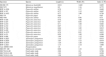
The articular region is similar in shape to Paraptenodytes antarcticus (Moreno & Mercerat, 1891). The proportion of the articular surface (length from beginning of processus lateralis to end of processus caudalis if possible or to the most caudal end of the processus lateralis/width from the most medial point of the cotyla medialis to the most lateral point of the cotyla lateralis) is similar to that of Pygoscelis and also to Paraptenodytes antarcticus, although MLP 96-I-6-48 is slightly wider than the latter (see Table I).
Measurements (mm) taken on the fossa articularis quadratica of the materials under study and the comparison specimens.

Material: MLP 92-II-2-115a (articular region of the right ramus mandibularis, Fig. 2b).
Provenance: Cucullaea I Allomember (Telm 5).
Locality: IAA 1/90.
Description: It is less robust, 2.95% smaller in length and 20.4% in width than MLP 96-I-6-48 (see Table I), although the processus mandibularis medialis is similarly extended in both. The edge of the cotyla medialis is more rounded and less extended medially than in MLP 96-I-6-48, Madrynornis, and the living species of Spheniscus and Eudyptes, while it is similar to that of Pygoscelis.
The tuberculum pseudotemporale is very well developed, much more than in the materials described by Ksepka & Bertelli (Reference Ksepka and Bertelli2006) and even more prominent than in MLP 96-I-6-48. The crista trasversal fossa is less elevated with respect to the articular surface than in MLP 96-I-6-48, but more than Madrynornis and the modern species. The processus retroarticularis seems not to be developed, although the material is badly damaged in this region, showing irregular edges that might indicate a missing end. The cotyla caudalis and the cotyla lateralis are clearly separated, as in the living penguins and Spheniscus megaramphus Stucchi, Urbina & Giraldo, 2003. The cotyla lateralis is well separated from the rest of the articular surface, rounded in shape and concave. The attachments of the membrana postmeatica and ligamentum occipitomandibulare do not show any particular feature. The sulcus intercotylaris between the cotyla lateralis and the cotyla medialis is almost half the length of that of MLP 96-I-6-48 and one third narrower than that of MLP 92-II-2-108, it is around the same size than Pygoscelis antarctica (Forster, 1781) and Aptenodyptes patagonicus Miller, 1778. The foramen located anterior to the cotyla lateralis, described by Ksepka & Bertelli (Reference Ksepka and Bertelli2006) in Antarctic materials, is present here. The fossa articularis quadratica is shallower than in MLP 96-I-6-48 and Pygoscelis papua Forster, 1781, but similar to that of the other modern species. The posterior end of the fossa aditus canalis mandibulae is rounded. The proportion of the articular surface is similar to that of Aptenodyptes patagonicus and MLP 92-II-2-108 (see below).
Material: MLP 92-II-2-108 (articular region of the right rama mandibularis, Fig. 2e).
Provenance: Cucullaea I Allomember (Telm 5).
Locality: DPV 6/84.
Description: The proportion of the articular surface is around 6% smaller than MLP 92-II-2-115a (see Table I) but very similar in shape, even though the specimen is not as well preserved as the former. No significant differences were observed, however the tuberculum pseudotemporale seems to be less developed, as in UCMP 321057 (Ksepka & Bertelli Reference Ksepka and Bertelli2006). Concerning the proportion of the articular surface, this specimen is similar to A. patagonica and to MLP 92-II-2-115a, as stated before.
Material: MLP 91-II-4-223 (articular region of ramus mandibulae, Fig. 2c).
Provenance: Submeseta Allomember (Telm 7).
Locality: DPV 15/84.
Description: This specimen is eroded and badly damaged. Only the articular surface is preserved, but not its processes or its most ventro-caudal region. As in MLP 92-II-2-108 and MLP 92-II-2-115a, the cotyla caudalis is divided from the cotyla lateralis, which is concave and very well developed.
Material: MLP 94-III-15-409 (articular region of left mandible).
Provenance: Submeseta Allomember (Telm 7).
Locality: DPV 13/84.
Description: The fragment is badly damaged, which does not permit comparison of features. The only one visible is the foramen already observed in MLP 92-II-2-115 and described by Ksepka & Bertelli (Reference Ksepka and Bertelli2006).
Material: MLP 78-X-26-2 (rami mandibulae, Fig. 2i)
Provenance: Submeseta Allomember (Telm 7).
Locality: DPV 14/84.
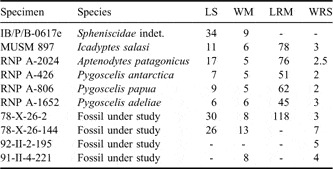
Description: This ramus mandibularis is 118 mm length (see Table II), it is 40 mm longer than that of the modern species, and similar in length to the one studied by Jadwiszczak (Reference Jadwiszczak2006a), Waimanu (Slack et al. Reference Slack, Jones, Ando, Harrison, Fordyce, Arnason and Penny2006) and Icadyptes salasi Clarke, 2007. Both rami mandibulae are joined through the symphysis, lacking their articular region. The symphysis is more than twice the length of mandibles of living representatives (see Table II) and the tip is not curved. Each ramus mandibularis is stout and slender. Its height is nearly constant (about 10 mm) along its entire length, scarcely increasing gradually towards the caudal end, a feature also present in UCMP 321057 (Ksepka & Bertelli Reference Ksepka and Bertelli2006), Perudyptes devriesi Clarke, 2007 (Clarke et al. Reference Clarke, Ksepka, Stucchi, Urbina, Giannini, Bertelli, Narváez and Boyd2007), Spheniscus megaramphus (Stucchi et al. Reference Stucchi, Urbina and Giraldo2003) and Icadyptes salasi (Ksepka et al. Reference Ksepka, Clarke, Devries and Urbina2008). The lateral side of the mandible is concave, whereas it is flat in modern and Neogene species. The ventral outline is straight, as in all Palaeocene and Eocene penguins. The vascular foramina covering the external surface of the tip of the mandible are present as in all penguins (see Ksepka et al. Reference Ksepka, Clarke, Devries and Urbina2008).
Measurements (mm) taken on the mandibles under study and the comparison specimens. LS = length of symphysis. WM = width of mandible at the union of both rami mandibulae. LRM = length of the ramus mandibularis from the tip to the fenestra rostro mandibularis. WRS = width of the ramus mandibularis right at the caudal end of the symphysis.

Material: MLP 78-X-26-143 (fragment of right ramus mandibulae, Fig. 2f) and MLP 78-X-26-144 (fragment of mandible, Fig. 2g).
Provenance: Submeseta Allomember (Telm 7).
Locality: DPV 14/84.
Description: The symphysis of the 78-X-26-144 is similar in length (26 mm) but more robust than that of MLP 78-X-26-2 (30 mm). The rest of the features resemble the former in terms of the concave lateral surfaces and the straightness of the entire ramus. MLP 78-X-26-143 is very fragmentary and shows the end of the insertion of the ramphoteca on its medial surface.
Material: MLP 93-X-1-68 (fragment of right rami mandibulae, without articular region or symphysis).
Provenance: Submeseta Allomember (Telm 7).
Locality: DPV 14/84.
Description: The specimen is deformed, therefore measurements could not be taken. The inner surface shows the insertion of the ramphoteca.
Material: MLP 92-II-2-203 (fragment of right ramus mandibulae, without the articular region or symphysis).
Provenance: Cucullaea II Allomember (Telm 6).
Locality: IAA 1/93.
Description: The material is badly damaged and does not present particular characters.
Material: Several fragments of mandibles: MLP 92-II-2-195 (right and left ramus mandibulae joined by sediment, but without the articular region or symphysis, Fig. 2(h), MLP 92-II-2-197 (fragment of left? ramus mandibularis), MLP 92-II-2-198 (small fragment of the left ramus madibularis), MLP 92-II-2-199 (fragment of the left ramus madibularis), MLP 92-II-2-200 (fragment of the ramus madibularis), MLP 92-II-2-201 (fragment of the right ramus madibularis).
Provenance: Submeseta Allomember (Telm 7).
Locality: DPV 14/84.
Description: Most of the remains are very fragmentary; MLP 92-II-2-195 is the most complete. It is larger in size, considering the width of the ramus mandibularis (Table II), than MLP 78-X-26-2, Icadyptes and extant penguins. However, it is slender and stout as the other remains described here.
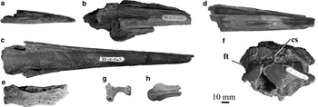
Material: MLP 93-X-1-67 (whole bill with missing distalmost tip, Fig. 3c).
a. Lateral view of bill (MLP 92-II-4-202). b. Dorsal view of proximal portion of bill (MLP 93-X-1-115). c. Dorsal view of bill (MLP 93-X-1-67). d. Dorsal view of rami mandibularis without the tip or proximal portion (MLP 93-X-1-91). e. Dorsal view of interorbital region (MLP 78-X-26-158). f. Dorsal view of posterior portion of skull (MLP 84-II-1-10). g. Lateral view of left quadrate (MLP 94-III-15-413). h. Dorsal view of interorbital region (MLP 92-II-2-250). ft = fossa temporalis, cs = crista nuchalis sagittalis. Scale bar = 10 mm.

Provenance: Submeseta Allomember (Telm 7).
Locality: DPV 14/84.
Description: The bill is 179 mm long and slender, gradually narrowing toward the tip. As in Icadyptes, there is no indication of a downturned tip, whereas the end is slightly curved in the materials described by Jadwiszczak (Reference Jadwiszczak2006b) and even more recurved or decurved in modern and Miocene penguins.
The nares extend through most of the length of the bill and are wider posteriorly. In dorsal view they are well differentiable as in the living Spheniscus, Eudyptes, Eudyptula and Pygoscelis. The nasal-premaxillar suture seems not obliterated like in modern and Miocene species, and unlike Perudyptes.
Material: MLP 91-II-4-202 (distal end of bill, Fig. 3a).
Provenance: Submeseta Allomember (Telm 7).
Locality: DPV 16/84.
Description: Distal end of bill similar to MLP 93-X-1-67 described above. The length is 70 mm, the most proximal section is 15 mm width, larger than that described by Jadwiszczak Reference Jadwiszczak2006a (see Table II).The dorsal outline is rounded, but the cross section is sub-triangular. Tomial edges are straight. Laterally the dorsal and ventral surfaces form an angle of approximately 10°.
Material: MLP 93-X-1-115 (proximal portion of bill, Fig. 3b).
Provenance: Submeseta Allomember (Telm 7).
Locality: DPV 13/84.
Description: Similar in size to MLP 93-X-1-67, the nasal fossae present the same configuration as well.
Material: MLP 93-X-1-91 (fragment of bill without the tip or proximal portion, Fig. 3d).
Provenance: Submeseta Allomember (Telm 7).
Locality: DPV 14/84.
Description: Similar in size to MLP 93-X-1-67. Particular features are not observable because of the degree of deformation.
Material: MLP 91-II-4-221 (fragment of ramus mandibularis with the symphysis broken and without the articular region, Fig. 2i).
Provenance: Submeseta Allomember (Telm 7).
Locality: DPV 15/84.
Description: Right and left rami mandibulae joined by symphysis as well as by sediment. The fossil remain lacks the articular region and the tip of the mandible as well.
On lateral view vascular foramina are present. The ramus appears to be long and slender similar to MLP 78-X-26-2. Measurements cannot be taken because the mandible is not complete. Although MLP 91-II-4-221 resembles MLP 78-X-26-2 in shape, it seems remarkably larger than the latter, but smaller than MLP 96-I-6-48 and MLP 78-X-26-4. MLP 91-II-4-221 is similar to MLP 78-X-26-2 in that they both show a marked curvature on the lateral side of the ramus mandibulae, while this is straight in MLP 96-I-6-48.
Material: MLP 84-II-1-10 (posterior portion of skull, Fig. 3f).
Provenance: Submeseta Allomember (Telm 7).
Locality: DPV 14/84.
Description: This skull is slightly larger than those of living Spheniscus and UCPM 321265 (Ksepka & Bertelli Reference Ksepka and Bertelli2006) and preserves part of the calvaria, occipital region, nuchal crests, temporal fossa, posterior end of the left fossa glandulae nasalis and part of the neurocranium. It is more globose than those of Icadyptes and the Antarctic UCPM 321265, which is slender and more elongated antero-posteriorly.
The fossa temporalis is well developed, reaching the sagittal nuchal crest dorsally, as in Paraptenodytes and UCMP 321265 (Ksepka & Bertelli Reference Ksepka and Bertelli2006). The dorsal end of this fossa is wider antero-posteriorly than in fossil and living species of Spheniscus. In the other modern species compared, the fossa ends in a triangular shape and does not reach the sagittal crest. The fossa temporalis is similar in size and shape to that of the Peruvian fossil Perudyptes, and to the Antarctic skull UCPM 321265.
The temporal crest is almost perpendicular to the sagittal crest, while in most penguins this crest is curved toward the anterior region. The sagittal crest is prominent as in Paraptenodytes and comparatively shorter.
The occipital region is trapezoidal as in Paraptenodytes, while it is more quadrangular in modern species and Madrynornis. The occipital condyle is robust and rounded as in the Miocene species, Pygoscelis adeliae (Hombron & Jacquinot), P. papua and Eudyptes; it is oblong in Spheniscus, P. papua and Aptenodytes. The occipital condyle is smaller than that of UCMP 321265 (Ksepka & Bertelli Reference Ksepka and Bertelli2006), but it is wider than high in both. The subcondylar fossa is deeper and more defined than in Paraptenodytes antarcticus and even more so than in UCPM 321265.
The tubercles of the lamina parasphenoidalis are stronger than those of the living and Miocene species, and elongated as in Paraptenodytes.
Material: MLP 94-III-15-413 (left quadrate, Fig. 2p).
Provenance: Submeseta Allomember (Telm 7).
Locality: DPV 13/84.
Description: The processus orbitalis is broken. The quadrate forms a more concave angle between the otic and squamosal capitula of the condylus caudalis than in Spheniscus, Pygoscelis, and similar to Aptenodytes and Eudyptes. The tubercle for attachment of the adductor mandibulae externus described by Clarke et al. (Reference Clarke, Ksepka, Stucchi, Urbina, Giannini, Bertelli, Narváez and Boyd2007) in Icadyptes, and also present in Miocene and modern species, is absent in this material.
The condylus lateralis is proportionally smaller than in living species and similar to that of Paraptenodytes and Madrynornis, the compared Miocene species. A wide sulcus separates the otic and squamosal capitula, as in all penguins. The condylus caudalis is flat and less pronounced than in Icadyptes and similar to that of modern and Miocene species.
In palatal view, the pterygoid condyle is not so rounded as in Madrynornis, Paraptenodytes and modern penguins. This condyle is located more antero-medially than the cotyla medialis as well, as in Miocene and modern penguins. However, the sharp tip of the pterygoid condyle points medially as opposed to the extant and Miocene penguins whose cotyla medialis points antero-medially. The quadratojugal articulation is deep and rounded, similar to that of all penguins.
Material: MLP 78-X-26-158 (fragment of skull including interorbital region, Fig. 3e).
Provenance: La Meseta Formation (upper levels).
Locality: DPV 2/84.
Description: The preserved fragment includes both frontals and a portion of the sulcus glandulae nasalis. The interorbital region is 90% wider than in Madrynornis and Pygoscelis (these taxa present stick-like frontals), similar to that of Spheniscus, Aptenodytes, and Paraptenodytes and 10% narrower than in 321223 (Ksepka & Bertelli Reference Ksepka and Bertelli2006).
The sulcus glandulae nasalis is narrower towards the anterior end, as in Spheniscus and Paraptenodytes. The supraorbital edge is also absent in these three taxa. The lateral edges of the fossa glandulae nasalis curve ventrally, as in UCMP 321223 (Ksepka & Bertelli Reference Ksepka and Bertelli2006).
Material: MLP 92-II-2-250 (fragment of skull including interorbital region, Fig. 3h).
Provenance: Cucullaea I Allomember (Telm 5).
Locality: IAA 1/90.
Description: It is less than half the size of MLP 78-X-26-158 and exhibits basically the same features.
Discussion and conclusions
Little background information exists about the functional morphology of Antarctic penguins. In the oldest species, improved diving capability may have been linked to the development of stronger bones and probably muscular structures enabling them to endure greater forces operating in water (see Hospitaleche & Di Carlo Reference Hospitaleche and Di Carlo2010). Such robust structures would not have been optimal for speed swimming. This idea was explored by Jenkins (Reference Jenkins1985), who based on the morphology of the flippers, considered that Anthropornis nordenskjoeldi Wiman, Reference Wiman1905, one of the “giant penguins”, would have been a slow swimmer. Even more, it was suggested that its long neck probably favoured the capture of motile prey (fishes) rather than krill and small squids (Tambussi et al. Reference Tambussi, Hospitaleche, Reguero and Marenssi2006).
Although the available cranial materials are too fragmentary to make a morphometric analysis, some considerations about their functional morphology can be advanced. A set of skull characters were evaluated in living penguins and showed promise as good indicators of trophic preferences and life habits (see Acosta Hospitaleche & Tambussi Reference Acosta Hospitaleche and Tambussi2006 and references therein).
With respect to the neurocranium, only two previously reported Antarctic specimens are available for comparisons (Jadwiszczak Reference Jadwiszczak2006a, Ksepka & Bertelli Reference Ksepka and Bertelli2006). MLP 84-II-1-10 studied here comes from the Submeseta Allomember in the upper levels of the unit. The configuration of the temporal fossa suggests a wide and well-developed temporal portion of the adductor mandibulae externus muscle. This muscle depresses the mandible and protracts the upper jaw. The extensive attachment area could indicate great forces acting to close the jaws (see Acosta Hospitaleche & Tambussi Reference Acosta Hospitaleche and Tambussi2006). Although the temporal fossae are shallow, the presence of a long sagittal nuchal crest supports the idea of powerful muscles acting in this area. This morphology suggests a piscivorous habit.
On the other hand, most of the previously described mandibular remains from Antarctica come from the Submeseta Allomember (see Jadwiszczak Reference Jadwiszczak2006a, Ksepka & Bertelli Reference Ksepka and Bertelli2006). The articular regions here described were mainly from the lower and medium beds of the La Meseta Formation. One specimen (MLP 96-I-6-48) shows a peculiarity with respect to the cotyla caudalis and lateralis, which does not present a clear separation between them as opposed to the other articular regions, which show two different cotylae developed.
In addition, the processus retroarticularis, which is present in all extant penguins, was not observed in any of the material here described, in some cases due to lack of preservation, but in others it does not seem to had been developed.
Although fragmentary, these specimens always exhibit the same morphology, consisting of elongated and dagger-like bills. This morphology is closely associated to the capture of large prey such as fish or squids. This morphology is widely developed in medium and large Palaeogene species. Elongate and powerful bills have been also described for the Peruvian Perudyptes, Icadyptes and Inkayacu (Clarke et al. Reference Clarke, Ksepka, Stucchi, Urbina, Giannini, Bertelli, Narváez and Boyd2007, Reference Clarke, Ksepka, Salas-Gismondi, Altamirano, Shawkey, D'Alba, Vinther, DeVries and Baby2010), and the two New Zealand species of Waimanu (Slack et al. Reference Slack, Jones, Ando, Harrison, Fordyce, Arnason and Penny2006). The single exception is a fragmentary rostrum maxillare (IB/P/B–0617e) studied by Jadwiszczak (Reference Jadwiszczak2006b) that comes from the Acantilados Allomember. This upper jaw is basally wide and strongly narrowing toward the tip in dorsal view, a particular shape that implies an important advance in the knowledge of the Antarctic penguins. It is the first evidence to suggest that not all the Eocene Antarctic penguins had long slender bills (Jadwiszczak Reference Jadwiszczak2006b).
The specimens MLP 96-I-6-48 and 92-II-2-203 here described, exhumed from the lower and middle units of the La Meseta Formation, are medium to large in size and partially resemble the shape observed in Miocene species. It means a mandible shape more similar to extant penguins than to those of the Eocene species. The former also shows a singular morphology in the articular region (see above). In contrast, the mandibles from the upper levels of the sequence are long and slender, similar to those classically used in giant penguin reconstructions, and probably belonged to taxa specialized in catching fish. These two morphotypes support the hypothesis proposed by Jadwiszczak (Reference Jadwiszczak2003) about the co-existence of penguins with different trophic habits, and explain a possible strategy to avoid interspecific competition. Although the evidence is fragmentary, a niche partitioning like this is expected when the number of sympatric species is high, as in the case of the penguin assemblage of the late Eocene of Antarctica. Future research involving articulated skeletons or at least associated remains will be very useful to clarify these ideas.
Acknowledgments
We thank the Agencia Nacional de Promoción Científica y Tecnológica and the Consejo Nacional de Investigaciones Científicas y Técnicas for financial support. Materials were collected by Juan José Moly and Marcelo Reguero from Museo de La Plata. We would like to thank Dr Natalie Goodall who kindly lent us comparative material, Leonel Acosta who prepared the fossils, Dr Cecilia Morgan who improved the English grammar and Dr Marcelo Reguero for help. We also wish to thank the reviewers Dr Herculano Alvarenga and Dr Piotr Jadwiszczak, and the editor for useful suggestions.