




No CrossRef data available.
Article contents
Selection of peptides recognized by human antibodies against the surface of Plasmodium falciparum-infected erythrocytes
Published online by Cambridge University Press: 13 December 2004
Abstract
In an attempt to identify mimotopes of the surface antigens of P. falciparum-infected erythrocytes (iRBC), antibodies were eluted from iRBC that had been treated with a pool of sera from malaria-infected individuals (IHS), and were used to screen a phage display library (PDL). After repeated panning of the PDL on immobilized antibodies, phage that selectively bound to IHS were accumulated. Of 23 randomly chosen clones that were sequenced, 13 individual sequences were detected at varying frequencies and 3 of the 13 sequences had homology with membrane proteins known to exist on iRBC. The majority of phage clones (7 out of 8 clones) selected after the 4th panning bound selectively to IgG in IHS. Specific binding of the selected phage to IgG in IHS was also confirmed using 24 IHS and 11 sera from uninfected individuals. One phage clone was the most frequently found in the sequenced clones after the 4th panning, and the binding of this clone to IgG in all IHS was greater than in any serum from uninfected individuals. A rabbit antiserum against the peptide expressed on the clone specifically recognized the surface of iRBC and resulted in iRBC haemolysis.
Information
- Type
- Research Article
- Information
- Copyright
- © 2004 Cambridge University Press
INTRODUCTION
Development and use of chemotherapeutics and insecticides have been important factors in controlling many vector-borne infectious diseases, including malaria. However, the emergence of drug-resistant malaria parasites and insecticide-resistance in the vector, the Anopheles mosquito, has increased the number of malaria infections in the world, resulting in millions of deaths each year (Trigg & Kondrachine, 1998). In addition, most people in malaria-endemic areas suffer not only from this devastating disease but also from poverty and lack of appropriate health care systems. Therefore, the development of malaria vaccines is now considered to be an urgent need for the control of malaria infections. Most of the efforts in developing malaria vaccines have been made against the life-threatening species, Plasmodium falciparum.
Since irradiated sporozoites were found to elicit protective immunity against a malaria infection approximately 30 years ago (Clyde et al. 1973; Cochrane et al. 1976), various malaria-parasite-derived proteins have been tested for their ability to elicit protective immunity with the goal of developing a practical vaccine (reviewed by Moore, Surgey & Cadwgan, 2002). When tested in animal models, promising results have been obtained using some proteins from the invasive stages of malaria parasites, such as the circumsporozoite surface protein and the merozoite surface proteins. However, the effectiveness of such vaccine candidates in human trials were, so far, less than satisfactory for practical use i.e. partial effectiveness in a large-scale human trial or short-lasting immunity (Moore et al. 2002). As one of the ways to overcome the difficulties in inducing universal and effective immunity, DNA-based multi-component vaccines are now being developed, and one such vaccine has been tested in human volunteers (Moore et al. 2002). As suggested by Doolan & Hoffman (2002), it should be possible to develop a potent DNA-based vaccine when multiple antigens from all blood stages are included in the construct. However, at present, the surface antigens of erythrocytes infected with mature-stage parasites have not been considered as a component of the vaccine despite the fact that the erythrocytic stages remain in the bloodstream and are exposed to the host immune system for the longest period of time (48–72 h). Indeed, the establishment of a protective immune response against these stages has been suggested by the following observations. (1) Adults who survive multiple malaria infection develop naturally acquired immunity that limits the level of parasitaemia and clinical symptoms caused by the erythrocytic-stage parasites (Baird, 1998; Rhee et al. 2001). (2) Parasitaemia in infected children was suppressed by passive transfer of antibodies from adults with protective immunity against malaria (Kumar, Epstein & Richie, 2002; Sabchareon et al. 1991).
It has been demonstrated that the plasma membrane of the erythrocyte undergoes dramatic changes when P. falciparum parasites mature therein. Such changes include the appearance of protrusions (called knobs) (Aikawa et al. 1983; Nagao, Kaneko & Dvorak, 2000), a decrease in cholesterol and sphingomyelin content (Maguire & Sherman, 1990), externalization of aminophospholipids (Eda & Sherman, 2002; Maguire, Prudhomme & Sherman, 1991), insertion of parasite-derived proteins (Baruch, Rogerson & Cooke, 2002b; Beeson & Brown, 2002; Heddini, 2002; Smith et al. 2001), and modification of host erythrocyte-membrane proteins (Sherman, Crandall & Smith, 1992). Of these, a parasite-derived protein, P. falciparum erythrocyte membrane protein-1 (PfEMP-1), and an erythrocyte protein, band 3, have been shown to be recognized by antibodies produced in P. falciparum-infected individuals (Baruch et al. 2002b; Beeson & Brown, 2002; Crandall, Guthrie & Sherman, 1995; Heddini, 2002; Smith et al. 2001). PfEMP-1, a family of proteins encoded by about 60 copies of var genes (Gardner et al. 2002), acquires extensive antigenic diversity by genetic recombination (Freitas-Junior et al. 2000). At the trophozoite and schizont stages, PfEMP-1 molecules are transported to the surface of iRBC and contribute to antigenic variation (Baruch et al. 2002b; Beeson et al. 2002; Heddini, 2002; Smith et al. 2001). Moreover, it has been shown that PfEMP-1 plays a pivotal role in the pathogenesis of malaria by mediating the sequestration of iRBC in the deep tissues leading to cerebral and placental malaria (Baruch et al. 2002b; Beeson et al. 2002; Heddini, 2002; Smith et al. 2001). PfEMP-1 proteins are composed of adhesive domains responsible for iRBC binding to various endothelial and placental receptors. For instance, domains DBLβ, CIDR-1, and DBLγ were shown to bind ICAM-1, CD36 and chondroitin sulfate A, respectively (Baruch et al. 2002b; Beeson et al. 2002; Heddini, 2002; Smith et al. 2001). In spite of its antigenic diversity, PfEMP-1 has been studied as a candidate for a malaria vaccine. This is based on the findings that a recombinant protein of CIDR-1 domain could induce monoclonal antibodies that cross-reacted with other variants (Gamain, Miller & Baruch, 2001) and that the immunization of Aotus monkeys with CIDR-1 protein could induce a high level of protection when challenged with a homologous strain of P. falciparum (Baruch et al. 2002a). However, the protection by CIDR-1 was not observed when the immunized monkey was challenged with a different strain. Therefore, further investigations are required for this well-studied surface antigen on iRBC before it can be used as a component of a DNA-based multivalent malaria vaccine.
To date, the CIDR-1 domain of PfEMP-1 is the only epitope tested for development of a vaccine based on iRBC surface antigens. Therefore it is of great importance to identify novel epitopes on the iRBC. In the last decade, phage display library (PDL), a library of phage expressing random peptides on their surface, has been used successfully for the identification of epitopes by panning the library on various antibodies. Here we have attempted to identify surface epitopes or mimotopes of iRBC surface antigens utilizing PDL, and antibodies eluted from the surface of iRBC that had been treated with a pool of sera from malaria-infected individuals (IHS).
MATERIALS AND METHODS
Materials
Bacto tryptone and Bacto agar were obtained from Becton Dickinson and Co. (Sparks, MD). Yeast extract and DNA preparation kit, QIAprep Spin M13 kit, were obtained from DIFCO laboratories (Detroit, MI) and QIAGEN Inc. (Valencia, CA), respectively. Streptavidin-labelled Alexa488, NeutrAvidin-labelled microspheres (ϕ1 μm) were purchased from Molecular Probes Inc. (Eugene, OR) and HRP-conjugated goat anti-human IgG antibody was purchased from Bio-Rad Laboratories (Hercules, CA). Biotin-labelled goat anti-human IgG antibody, biotin-labelled anti-rabbit IgG antibody, and Protease Inhibitor Cocktail were obtained from Calbiochem, EMD Biosciences Inc. (San Diego, CA). Other chemicals were obtained from Sigma Chemicals Co. (St Louis, MO). Synthesis of the peptide identified in this work and production of polyclonal antibody against the peptide conjugated with keyhole limpet haemocyanin was carried out by New England Peptide (Fitchburg, MA). A pool of sera from P. falciparum-infected individuals (IHS) in Africa was provided by Dr William E. Collins (Centers for Disease Control and Prevention, Atlanta, GA) and serum samples from P. falciparum-infected individuals in Thailand were provided by Dr Nicholas J. White (Mahidol University, Bangkok, Thailand). Sera of uninfected individuals (NHS) were obtained from healthy volunteers at the University of California at Riverside (Riverside, CA).
Parasites
Two P. falciparum cloned lines, FCR-3 (knobby, CD36-preferring line), and CS2 (knobby, CSA-preferring line) were continuously cultured in O+ human erythrocytes as described by (Trager & Jensen, 1976). The binding characteristics of FCR-3 and CS2-infected iRBC were confirmed using CD36-expressing CHO cells and CSA-overexpressing CHO cells as described previously (Eda et al. 1999). Cultures of parasites were synchronized at the mature stage by gelatin flotation (Pasvol et al. 1978) and at the ring stage by lysis of mature-stage iRBC using a 5% sorbitol solution (Lambros & Vanderberg, 1979).
Isolation of human antibodies bound to the surface of iRBC
iRBC infected with mature FCR-3 parasites (trophozoites and schizonts) were concentrated by gelatin floatation (Pasvol et al. 1978) resulting in parasitaemias of 70%. The concentrated iRBC were incubated at room temperature for 2 h with 1[ratio ]10 diluted NHS or a pool of IHS from P. falciparum-infected individuals in Africa in the presence of 10% bovine serum albumin (BSA). Unbound materials were removed by washing the cells with 5 ml of PBS 3 times and bound materials were eluted at room temperature for 2 min with saline-glycine buffer (0·1 M glycine, pH 3·0 and 0·1 M NaCl). We conducted Western blotting using the antibody eluted from NHS- or IHS-treated iRBC and found that antibody from IHS-treated iRBC specifically recognized a band with a molecular weight of about 240 kDa and the same band was detected by an anti-PfEMP-1 antibody (data not shown), showing that the eluted materials contained antibodies recognizing the surface molecules of iRBC. The eluate was neutralized by adding 1[ratio ]50 volume of 0·5 M Tris-base solution and was stored at −20 °C until use.
Panning the phage display peptide library and phage binding assays
PDL and Escherichia coli strain, K91Kan, used in this work were kind gifts from Dr Renata Pasqualini, (University of Texas, M. D. Anderson Cancer Center, TX) and the details of library construction and protocols have been described previously (Scott & Smith, 1990; Koivunen et al. 1993). Phage in the library express 7-mer random peptides at the extracellular N-terminal of coat proteins, pIII, and the random peptides are constrained by disulfide bonding of two cysteine residues at the flanking region of the peptides. The complexity of the library was approximately 2×10⁸. Human antibodies eluted from the surface of iRBC were diluted 1[ratio ]50 in PBS and immobilized on the well of a plastic 96-well plate by incubating at 4 °C overnight, and the well was blocked at room temperature for 2 h with PBS-0·2% Tween (PBST) containing carrier proteins, such as BSA. Phage suspended in PBST-2% BSA containing 1[ratio ]50 diluted eluate from NHS-treated iRBC was incubated at room temperature for 2 h with the immobilized antibodies. Unbound phage were removed by washing 5 times with PBST, and bound phage were eluted by incubation at room temperature for 2 min with saline-glycine buffer (pH 3·0). Log-phase E. coli were infected by phage in the eluate by incubation at room temperature for 30 min in Luria-Bertani (LB) broth containing 100 μg/ml kanamycin. Infected bacteria were selected by culturing them in the presence of 20 μg/ml of tetracycline at 37 °C for 1 h. The titre of the infected E. coli suspension was checked by culturing the infected bacteria on tetracycline-containing agar plates. The remaining suspension of phage-infected bacteria was incubated in LB medium containing 20 μg/ml of tetracycline at 37 °C overnight and phage were isolated from the pellet by polyethylene glycol (PEG) precipitation. The titre of amplified phage was determined as described above and the phage were suspended in PBST containing carrier proteins and subjected to the following panning.
The panning process was repeated 4 times to select phage that bind selectively to IgG in IHS. For the 1st panning, 6×10⁹ TU of phage were used and 2×10⁸ TU of phage were used for the 2nd, 3rd, and 4th panning (Table 1). To avoid collecting phage that non-specifically bind to carrier proteins, different proteins were used for each panning. For the 1st and 4th panning, BSA (2 w/v% in PBST) was used as a carrier protein. Superblock (PIERCE, Rockford, IL, 10 v/v% in PBST) and BlockAid (10 v/v% in PBS) were used for the 2nd and 3rd panning, respectively. Antibodies eluted from NHS-treated iRBC were used in the all pannings to avoid selection of mimotopes of surface antigens of nRBC and, for the 4th panning, phage were incubated with immobilized antibodies in the presence of 10% normal serum to remove phage binding to the constant region of antibodies and serum proteins. After the 4th panning, colonies of phage-infected E. coli were randomly selected, amplified and used for assessing the binding specificity of each phage clone against immobilized antibodies. The phage binding assays were conducted in the same fashion as the panning procedure described above; however, anti-human IgG polyclonal antibody was used to capture IgG molecules specifically in plastic wells. We immobilized goat anti-human IgG antibody in a 96-well plate using 0·6 μg of antibody per well and, after removal of unbound antibody by washing and blocking with 2% BSA, the wells were incubated with 30 μl of 1[ratio ]50 diluted NHS and IHS that contain excess amount (approx. 6 μg) of human IgG. Therefore, the amount of bound human IgG in each well should be limited by the amount of immobilized anti-human IgG antibodies on the wells and thus it is expected that the same amount of human IgG from NHS and IHS was immobilized in each well.
Table 1. Panning of phage display library on antibodies isolated from the surface of iRBC (See Materials and Methods section for details.)

DNA sequencing
Phage clones were randomly selected after the 4th panning and amplified for DNA sequencing. DNA of selected phage was purified using QIAprep Spin M13 kit according to the manufacturer's instructions. To determine the sequence of the random peptide on the selected phage, partial sequencing was carried out using BigDyeR Terminator v3.0 cycle Sequencing Kit (Applied Biosystems Inc., Foster City, CA) and 5′ primer (5′-CCCTCATAGTTAGCGTAACG-3′). The PCR products were analysed on a 3730 DNA analyser (Applied Biosystems Inc.). All DNA sequencing was carried out at the UCR Genomics Institute.
Anti-peptide antibody binding assay
Five μl of packed iRBC (FCR-3 strain, trophozoite or schizont stages, 10% parasitaemia) were incubated at room temperature for 1 h with anti-peptide antiserum diluted 1[ratio ]2 in RPMI-HEPES and unbound materials were removed by washing the cells 3 times with PBS-0·01% BSA. After the last centrifugation, the cell pellet was resuspended in RPMI-HEPES containing 1% BSA and was subjected to a binding assay against beads coated with anti-rabbit IgG polyclonal antibody. Coating of beads with anti-rabbit IgG antibody was conducted as follows. Neutravidin-coated fluorescent beads (5 μl packed volume) were first blocked with 1% BSA in PBS at room temperature for 30 min and then incubated with biotin-labelled anti-rabbit IgG antibody at room temperature for 1 h. Unbound antibodies were removed by washing the beads 3 times with PBS-1% BSA. The beads were resuspended in 100 μl of 1% BSA in RPMI-HEPES and mixed with 10 μl of packed iRBC previously incubated with anti-peptide antibody. After incubation with gentle shaking, 2% glutaraldehyde solution was added to the bead suspension to make a final concentration of 0·2%. To remove unbound beads, iRBC were washed with PBS by centrifugation at 1500 g for 30 sec, and the pellet suspended in PBS. Beads bound to the surface of iRBC were observed and counted by fluorescent microscopy using a combination of a phase-contrast filter for identification of iRBC and a fluorescent filter for visualization of beads. nRBC and iRBC were distinguishable under phase-contrast microscopy and were counted until the number reached at least 100. The number of beads bound on nRBC or iRBC was also counted and bead binding was expressed as the number of beads bound on 100 RBC.
Haemolysis assay
Twenty μl of packed iRBC and uninfected RBC suspended in RPMI-HEPES were incubated at 37 °C for 1 h with 20 μl of various dilutions of anti-peptide antiserum. After adding 60 μl of PBS to the suspension of iRBC, the cells were sedimented by centrifugation at 600 g for 3 min. Fifty μl of the supernatant were collected, and the concentration of released haemoglobin was determined by measuring the absorbance at 419 nm using a microplate reader (Bio-Rad). Since haemolysis of trophozoite and schizont stage of iRBC by sorbitol solution is known to be very specific and complete (Lambros & Vanderberg, 1979), we used the haemoglobin concentration obtained by sorbitol lysis to calculate the percentage of lysed RBC.
Statistical analysis
Differences between values in Fig. 3 were examined using ANOVA (single factor) and were regarded as significant when P-values were less than 0·05.
RESULTS
Panning of PDL on antibodies isolated from the iRBC surface
Human antibodies recognizing surface molecules on iRBC were eluted from iRBC previously treated with a pool of IHS from P. falciparum-infected individuals in Africa. To isolate phage expressing peptides mimicking antigens on iRBC, a library of phage expressing random peptides (7 mer peptides constrained by disulfide bonding of two cysteine residues) was panned on immobilized antibodies eluted from iRBC. After extensive washing, bound phage were eluted with an acidic buffer and amplified in E. coli. The amplified phage were purified and subjected to panning on immobilized antibodies. As shown in Table 1, the recovery of phage after panning (the ratio of TU of bound phage to that of phage used for the panning) was increased after each panning and dramatically increased after the 4th panning. The recovery was 600-fold higher at the 4th panning when compared with that of the 1st panning, suggesting the accumulation of phage specific for binding to antibodies eluted from the iRBC surface. Further panning did not increase the recovery rate.
Binding specificity of phage against IgG in IHS
The original phage library and phage after the 3rd and 4th panning were tested for their binding activity against NHS and IHS. In the phage binding assay, IgG molecules were captured on plastic wells coated with anti-human IgG polyclonal antibodies. The binding of phage collected after the 4th panning to IgG in IHS was the highest among phage mixtures tested and was 160-fold higher than that of the original library (Fig. 1), confirming the accumulation of phage binding to IgG in IHS. Phage after the 4th panning bound 2·1-fold higher to IgG in IHS than to that in NHS (Fig. 1). This suggested that phage clones collected after the 4th panning contained clones that were binding specifically to IgG in IHS.

Fig. 1. Binding specificity of original phage display library and phage selected after panning against antibodies isolated from the iRBC surface. Phage (1×10⁸ TU) were panned on immobilized IgG from NHS (open bar)- or with IHS (closed bar)-treated iRBC and TU of bound phage was determined as described in the Materials and Methods section. Each bar represents the mean±S.D. of triplicate determinations.
Sequences of peptides on selected phage
Twenty-three clones from the phage mixture obtained by the 4th panning were randomly selected, and the sequences of random peptides expressed on the phage were determined. As shown in Table 2, the sequences of clones 1 and 2 were found in 5 and 3 clones in the 23 sequenced clones, respectively. The sequences of clones 3–6 were found twice in 23 clones, and the sequences of clones 7 and 8 were identical except for one amino acid substitution. This suggested the selection of specific phage during the panning process. Homology of identified peptidic sequences with known proteins on the surface of iRBC was determined using the BLAST program. The sequence of the clone 1 peptide had homology with an extracellular domain (DBL3γ) of the parasite-derived iRBC surface protein, PfEMP-1 (Baruch et al. 1995), and the sequences of clones 2 and 3 had homology with extracellular loops 2 and 7 of the erythrocyte membrane protein, band 3 (Zhu, Lee & Casey, 2003).

Binding specificity of selected phage to IgG in IHS
To confirm the binding specificity of isolated clones, phage of clones 1–8 were amplified and tested for their binding to IgG in NHS and IHS. All of the phage clones, except for clone 2, specifically bound to immobilized IgG in IHS as shown in Fig. 2, whereas phage in the original phage display library and phage expressing an irrelevant peptide neither showed such binding activity nor specificity against IgG in IHS. Prevalence of antibodies against the peptide of phage clones in IHS was examined using 11 NHS and 24 IHS. IHS from Thailand was used for this phage binding assay in order to identify mimotopes conserved in different geographical areas. (Note that IHS from Africa was used for the panning and phage binding assay shown in Fig. 2.) Clone 2 was excluded in this experiment, because specific binding of clone 2 phage was not found with IHS. Binding of phage of clones 1 and 3–7 to a group of IHS was higher than that to a group of NHS, and the difference between phage binding to NHS and IHS was statistically significant (P value<0·05). The original library and phage of clone 8 bound to each group of sera to almost the same extent (Fig. 3). Most interestingly, in the case of clone 1 phage, all IHS showed a higher binding of phage when compared with NHS (Fig. 3), demonstrating that all of the IHS tested contained IgG against the peptide expressed on clone 1 phage (named mimotope 1, M1). Therefore, in the following experiments, we focused on the M1 peptide as a candidate of iRBC surface mimotope.

Fig. 2. Binding specificity of original phage display library, phage expressing an irrelevant peptide, and phage clones selected after the panning against antibodies isolated from iRBC surface. Phage (1×10⁸ TU) were panned on immobilized IgG from NHS (open bar)- or with IHS (closed bar)-treated iRBC and TU of bound phage was determined as described in the Materials and Methods section. Each bar represents the mean±S.D. of triplicate determinations.

Fig. 3. Binding specificity of the original phage display library and phage clones selected after the 4th panning against IgG in NHS (11 samples, open circle) or IHS (24 samples, filled circle). Phage (1×10⁸ TU) were panned on immobilized IgG and TU of bound phage was determined as described in the Materials and Methods section. Horizontal lines in each graph represent the mean values. The numbers in each graph indicate P values calculated using ANOVA (single factor). Relative phage binding was calculated by dividing each value of phage binding (TU/well) to NHS or IHS by the mean of phage binding (TU/well) to NHS.
Binding of anti-M1 antibody to the surface of iRBC
To examine whether the M1 peptide could elicit the production of antibodies against a surface antigen of iRBC, rabbits were immunized with the M1 peptide conjugated to keyhole limpet haemocyanin, and the antisera tested for their activity to recognize the surface of iRBC infected with trophozoites/schizonts. Binding of anti-M1 antiserum to the surface of iRBC could not be detected by indirect immunofluorescent and flow cytometrical assays (data not shown), suggesting that the number of the antigens on iRBC was too low to be detected by such methods. Therefore, we elected to detect antibody binding to the surface of iRBC using fluorescent beads. iRBC were incubated with the rabbit anti-M1 antiserum and antibody binding was detected by fluorescent beads coated with anti-rabbit IgG antibody. Although non-specific binding of beads was observed when iRBC were treated with pre-immune rabbit serum (Fig. 4A), a much larger number of beads was observed on the surface of iRBC treated with rabbit anti-M1 antiserum (Fig. 4B). No significant binding was observed on uninfected erythrocytes. Significant binding of anti-M1 antibodies on iRBC was confirmed by counting the bound beads (Fig. 4C). The number of beads bound to the surface of iRBC was 3-fold higher when the cells had been treated with rabbit anti-M1 antiserum when compared to the number of beads bound to untreated iRBC or to iRBC treated with pre-immune serum. These findings suggest that the M1 peptide could induce the production of antibodies against a surface antigen of iRBC in the immunized rabbit.

Fig. 4. Binding of anti-M1-peptide antibody to iRBC (FCR-3 strain). (A, B) Photographs of fluorescent beads bound to the surface of iRBC pre-treated with pre-immune rabbit serum (A) or with immune serum raised against the M1 peptide (B). Arrowheads indicate bound fluorescent beads on iRBC and arrows indicate nRBC present in the iRBC suspension. (C) Binding of antibodies to the surface of uninfected erythrocytes (open bar) and iRBC (closed bar). Fluorescent beads were coated with anti-rabbit IgG antibody and used to detect the binding of anti-M1 antibody on iRBC as described in the Materials and Methods section. Each bar represents the mean±S.D. of triplicate determinations.
Haemolysis of iRBC by rabbit anti-M1 antiserum
Since anti-M1 rabbit antibodies recognized the surface of iRBC, we tested whether anti-M1 antiserum could result in complement-mediated lysis of iRBC. Significant haemolysis was observed when iRBC were incubated at 37 °C for 2 h with anti-M1 antiserum, whereas such haemolysis was absent when the cells were treated with pre-immune serum (Fig. 5A). The haemolysis of iRBC was observed with two phenotypically different strains of malaria parasite, i.e. FCR-3 and CS2 (Fig. 5A). Antiserum did not cause significant haemolysis when nRBC were used instead of iRBC (data not shown). Haemolysis was completely inhibited when the antiserum was heat inactivated or when the incubation was conducted in the presence of EDTA (Fig. 5B). Since it is well known that activity of complement factors can be abolished by these treatments, this result suggested that the observed haemolysis was due to complement activation of antibodies fixed to the iRBC surface.

Fig. 5. Haemolysis of iRBC by rabbit anti-M1 antiserum. (A) Pre-immune serum (open symbols) and immune serum (closed symbols) were incubated with a suspension of iRBC infected with FCR-3 strain (circle) or with CS2 strain (triangle) and the lysis of RBC was evaluated. Each value represents the mean±S.D. of triplicate determinations. (B) Lysis of iRBC infected with FCR-3 strain after incubation with rabbit pre-immune and immune serum raised against the M1 peptide was examined as in (A). Heat: heat-inactivated immune serum was used. EDTA: incubation was conducted in the presence of EDTA. Each bar represents the mean±S.D. of triplicate determinations.
DISCUSSION
In this paper, we have made an attempt to identify epitopes on the iRBC by screening a PDL on human antibodies that recognize the surface changes of the iRBC membrane. Although a priori it might be expected that panning of PDL on materials with multiple specificity, such as serum, would not result in the selection of specific phage clones, it is of interest to note that some investigators have found they could identify disease-specific epitopes/mimotopes by panning PDL on sera from patients (Bowditch et al. 1996; Santamaria et al. 2001). Santamaria et al. (2001) conducted the screening of PDL expressing random peptides with sera from human papillomavirus (HPV)-infected patients and identified peptides having homologies with an HPV protein, HPV 16 L1. Further, some of the selected phage reacted specifically with sera from HPV patients. Bowditch et al. (1996) also carried out similar screening using sera from patients with chronic immune thrombocytopaenic purpura (ITP). ITP patients are known to have autoantibodies against glycoprotein IIb/IIIa complex. The screening resulted in peptides having homology with glycoprotein IIb/IIIa and the phage expressing such peptides reacted with an anti-IIb/IIIa autoantibody. Moreover, specific selection of peptides using PDL on intact cell surfaces has also been successful both in vitro and in vivo (Arap et al. 2002; da Silva et al. 2002; Samoylova et al. 2002). Thus, PDL is a powerful tool for identifying specific peptides even when the target contains multiple components.
The advantages of the approach taken in this work are that (1) small epitopes could be directly identified using short peptide-expressing PDL, (2) the identification could be carried out without predefined information, (3) it is relatively easy to study the distribution of antibodies against identified peptides in infected subjects, and (4) even peptides mimicking non-proteinaceous epitopes may be identified (Renkonen et al. 2002). To focus only on the antigens exposed on iRBC, we have used antibodies eluted from iRBC that had been treated with IHS and have identified peptides that are specifically recognized by IHS. Three out of 13 identified peptides showed significant homology with the extracellular portion of iRBC surface proteins, namely PfEMP-1 and band 3.
The peptides expressed on phage clones 2 and 3 showed homology with extracellular loops 2 and 7 of band 3 protein, respectively (Zhu et al. 2003). Band 3 is the anion-exchange protein of erythrocytes and the polypeptide of the protein is known to transverse the phospholipid bilayer 14 times. Loops 2 and 7 of band 3 are known to be relatively short, i.e. 7 and 4 amino acids in length, when compared with other loops (10–38 amino acids in length) and are flanked by two α-helical transmembrane domains. Therefore, it is possible that the structure of loops 2 and 7 could be constrained as the peptides on the phage are constrained by disulfide bonding of two cysteine residues and that could be the reason for peptides having homology with loops 2 and 7 being selected. It is interesting to note that most of the band 3 protein is transmembranous and cytoplasmic so that only about 10% of the entire protein is exposed on the outer surface of erythrocytes. Therefore, the probability for phage peptides to have homology with such an extracellular region should be very low, supporting the idea that the peptides in loops 2 and 7 of band 3 are the epitopes on the iRBC that are mimicked by peptides of clones 2 and 3. The reactivity of the phage expressing the loop 2 homologue to NHS and IHS was higher than that of the original PDL or irrelevant phage; however, the difference in reactivity to NHS and IHS was not significant. Since we have shown that cryptic regions of band 3 (amino acid residues 547–553 and 824–829) were exposed and mediated cytoadherence of iRBC (Sherman, Eda & Winograd, 2003), and Kay (1992) have shown that the regions were the senescent antigens recognized by anti-band 3 autoantibodies, the loop 2 region of band 3 with homology with the peptide on clone 2 could also be such an autoantigen exposed both on iRBC and senescent erythrocytes. On the other hand, the phage expressing the peptide with the loop 7 homologue was selectively recognized by IHS (Fig. 3, clone 3). This result is consistent with our previous data that a peptide partly containing the sequence on the phage clone 3 (WVVKSTPAL) was recognized by IHS but not by NHS (Crandall et al. 1995). However, a peptide containing the entire sequence of the peptide on phage clone 3 was not tested in this work. How can peptides of a host erythrocyte protein be antigenic during a malaria infection? Since parts of band 3, having homology with peptides of clones 2 and 3, were reported to be buried in the phospholipid bilayer in nRBC, it is possible that regions came to be exposed as the parasite matured within the RBC. In support of this hypothesis, we recently demonstrated that a transmembrane region of band 3 (amino acid residues 534–547) was exposed to the surface of iRBC upon parasite maturation within the RBC (Winograd, Eda & Sherman, 2004). However, we have not conducted any experiments to confirm whether loops 2 and 7 are exposed on iRBC due to a conformational change in the band 3 protein and are the epitopes of the peptides on phage clones 2 and 3.
The BLAST search (search for short, nearly exact matches) of the DARHRC sequence of the M1 peptide found the homologous region (DAKHRC) in DBL3γ domain of PfEMP-1 of Dd2 strain whereas no other P. falciparum protein had such homology with the M1 sequence. Further, the homology search of the M1 peptide in the database of Homo sapiens genes found 3 function-unknown genes, KIAA 0342, microrchidia homologue, and LOC132621; however, the homologies were lower when compared with the homology of DARHRC in M1 with DAKHRC in DBL3γ. Thus, the homology between the M1 peptide and the peptide in DD2var1-DBL3γ was very specific. However, this raised a question as to why the M1 peptide was homologous with DBL3γ region of Dd2 strain. Since PfEMP-1 genes of FCR-3 strain have not been completely sequenced and it is still not clear whether only 1 PfEMP-1 gene product is exclusively expressed on the surface of iRBC; possibly the FCR-3 strain expressed a PfEMP-1 gene product having the DAKHRC sequence or its homologue on the surface of iRBC. Another possibility is that a var gene in FCR-3 encodes an amino acid sequence that shares homology with DAKHRC in three-dimensional structure and the var gene product was expressed on FCR-3-iRBC. The result of the haemolysis assay might support this possibility. Namely, the haemolysis caused by anti-M1 peptide antibody appears more efficient for the CS2 strain than FCR-3 strain. Since the strains are distinct in characteristics of cytoadherence to CD36 and CSA that is mediated by PfEMP-1, the result suggests that different PfEMP-1 expressed on FCR-3 and CS2 were the targets of the anti-M1 peptide (CTGDARHRC) antibody. However, none of these possibilities was supported by our observation that anti-M1 antibody did not detect any proteins with the same molecular weight as PfEMP-1 on Western blots (data not shown). Although this result could be due to the disruption of the three-dimensional structure of PfEMP-1 conserved in FCR-3- and Dd2-iRBC during the extraction process, the possibility that it is purely accidental that M1 showed homology with DBL3γ can not be excluded. Thus, it is uncertain whether the DBL3γ domain of PfEMP-1 (var-1 product) was the original epitope of the M1 peptide; however, despite this, the following observations suggest that the M1 peptide (or its derivatives) might be used as a component of multicomponent malaria vaccine. (1) The M1 peptide could induce the production of antibodies recognizing the surface of iRBC confirming that the peptide was mimicking an iRBC surface antigen; (2) antiserum against M1 caused haemolysis of iRBC; and (3) phage bearing the M1 peptide were exclusively recognized by IHS.
It has been shown by several groups of investigators that filamentous phage expressing a peptidic mimotope on coat proteins could induce humoral and cellular immune responses without using any external adjuvant. For example, Willis, Perham & Wraith (1993) demonstrated that immunization with phage having peptides of major antigenic determinants of P. falciparum circumsporozoites could induce humoral and cellular immune responses without the addition of immunostimulants. Also, Demangel et al. (1998) identified phage-displayed peptide, CGRVCLRC (C15), by panning PDL on a monoclonal antibody against the merozoite surface protein 1 of P. vivax (Pv42). Immunization of mice with C15-expressing phage particles induced the production of antibodies against the original antigen, Pv42, even in the absence of adjuvant. These findings suggest that identifying epitope/mimotope by biopannning of PDL on antibody and immunization with peptidic epitope-expressing phage particles may be a simpler and less expensive strategy for the development of a malaria vaccine. To this end, it would be of interest to test whether immunization with phage clone 1 or a combination of phage clones 1 and 3–7 could induce antibody production and/or epitope-specific stimulation of lymphocytes and whether such induction would result in protective immunity in a P. falciparum animal model.
In this work, we have shown that panning of PDL on antibodies eluted from the iRBC surface is an effective way to identify epitopes/mimotopes of surface antigens of iRBC, and this approach may also be useful for identifying surface antigens on other stages of malaria parasites i.e. merozoites, sporozoites, and gametocytes.
The authors wish to thank Jacques Prudhomme for expert technical assistance. We also thank Dr Renata Pasqualini for the general gift of the phage display library used in this work. This work was supported by grant, AI 43608 (to I.W.S.) from the National Institute of Allergy and Infectious Diseases, National Institutes of Health. S.E. was supported by Overseas Research Fellowship from the Japan Society for the Promotion of Sciences.
References
REFERENCES
AIKAWA, M.,
RABBEGE, J. R.,
UDEINYA, I. &
MILLER, L. H. (
1983).
Electron microscopy of knobs in Plasmodium falciparum-infected erythrocytes.
Journal of Parasitology
69,
435–437.CrossRefGoogle Scholar
ARAP, W.,
KOLONIN, M. G.,
TREPEL, M.,
LAHDENRANTA, J.,
CARDO-VILA, M.,
GIORDANO, R. J.,
MINTZ, P. J.,
ARDELT, P. U.,
YAO, V. J.,
VIDAL, C. I.,
CHEN, L.,
FLAMM, A.,
VALTANEN, H.,
WEAVIND, L. M.,
HICKS, M. E.,
POLLOCK, R. E.,
BOTZ, G. H.,
BUCANA, C. D.,
KOIVUNEN, E.,
CAHILL, D.,
TRONCOSO, P.,
BAGGERLY, K. A.,
PENTZ, R. D.,
DO, K. A.,
LOGOTHETIS, C. J. &
PASQUALINI, R. (
2002).
Steps toward mapping the human vasculature by phage display.
Nature Medicine
8,
121–127.CrossRefGoogle Scholar
BAIRD, J. K. (
1998).
Age-dependent characteristics of protection v. susceptibility to Plasmodium falciparum.
Annals of Tropical Medicine and Parasitology
92,
367–390.CrossRefGoogle Scholar
BARUCH, D. I.,
PASLOSKE, B. L.,
SINGH, H. B.,
BI, X.,
MA, X. C.,
FELDMAN, M.,
TARASCHI, T. F. &
HOWARD, R. J. (
1995).
Cloning the P. falciparum gene encoding PfEMP1, a malarial variant antigen and adherence receptor on the surface of parasitized human erythrocytes.
Cell
82,
77–87.Google Scholar
BARUCH, D. I.,
GAMAIN, B.,
BARNWELL, J. W.,
SULLIVAN, J. S.,
STOWERS, A.,
GALLAND, G. G.,
MILLER, L. H. &
COLLINS, W. E. (
2002 a).
Immunization of Aotus monkeys with a functional domain of the Plasmodium falciparum variant antigen induces protection against a lethal parasite line.
Proceedings of the National Academy of Sciences, USA
99,
3860–3865.Google Scholar
BARUCH, D. I.,
ROGERSON, S. J. &
COOKE, B. M. (
2002 b).
Asexual blood stages of malaria antigens: cytoadherence.
Chemical Immunology
80,
144–162.Google Scholar
BEESON, J. G. &
BROWN, G. V. (
2002).
Pathogenesis of Plasmodium falciparum malaria: the roles of parasite adhesion and antigenic variation.
Cellular and Molecular Life Sciences
59,
258–271.CrossRefGoogle Scholar
BOWDITCH, R. D.,
TANI, P.,
FONG, K. C. &
McMILLAN, R. (
1996).
Characterization of autoantigenic epitopes on platelet glycoprotein IIb/IIIa using random peptide libraries.
Blood
88,
4579–4584.Google Scholar
CLYDE, D. F.,
MOST, H.,
McCARTHY, V. C. &
VANDERBERG, J. P. (
1973).
Immunization of man against sporozite-induced falciparum malaria.
American Journal of the Medical Sciences
266,
169–177.CrossRefGoogle Scholar
COCHRANE, A. H.,
AIKAWA, M.,
JENG, M. &
NUSSENZWEIG, R. S. (
1976).
Antibody-induced ultrastructural changes of malarial sporozoites.
Journal of Immunology
116,
859–867.Google Scholar
CRANDALL, I.,
GUTHRIE, N. &
SHERMAN, I. W. (
1995).
Plasmodium falciparum: sera of individuals living in a malaria-endemic region recognize peptide motifs of the human erythrocyte anion transport protein.
American Journal of Tropical Medicine and Hygiene
52,
450–455.CrossRefGoogle Scholar
DA SILVA, A. Jr.,
KAWAZOE, U.,
FREITAS, F. F.,
GATTI, M. S.,
DOLDER, H.,
SCHUMACHER, R. I.,
JULIANO, M. A.,
DA SILVA, M. J. &
LEITE, A. (
2002).
Avian anticoccidial activity of a novel membrane-interactive peptide selected from phage display libraries.
Molecular and Biochemical Parasitology
120,
53–60.CrossRefGoogle Scholar
DEMANGEL, C.,
ROUYRE, S.,
ALZARI, P. M.,
NATO, F.,
LONGACRE, S.,
LAFAYE, P. &
MAZIE, J. C. (
1998).
Phage-displayed mimotopes elicit monoclonal antibodies specific for a malaria vaccine candidate.
Biological Chemistry
379,
65–70.Google Scholar
DOOLAN, D. L. &
HOFFMAN, S. L. (
2002).
Nucleic acid vaccines against malaria.
Chemical Immunology
80,
308–321.CrossRefGoogle Scholar
EDA, S.,
EDA, K.,
PRUDHOMME, J. G. &
SHERMAN, I. W. (
1999).
Inhibitory activity of human lactoferrin and its peptide on chondroitin sulfate A-, CD36-, and thrombospondin-mediated cytoadherence of Plasmodium falciparum-infected erythrocytes.
Blood
94,
326–332.Google Scholar
EDA, S. &
SHERMAN, I. W. (
2002).
Cytoadherence of malaria-infected red blood cells involves exposure of phosphatidylserine.
Cellular Physiology and Biochemistry
12,
373–384.CrossRefGoogle Scholar
FREITAS-JUNIOR, L. H.,
BOTTIUS, E.,
PIRRIT, L. A.,
DEITSCH, K. W.,
SCHEIDIG, C.,
GUINET, F.,
NEHRBASS, U.,
WELLEMS, T. E. &
SCHERF, A. (
2000).
Frequent ectopic recombination of virulence factor genes in telomeric chromosome clusters of P. falciparum.
Nature, London
407,
1018–1022.Google Scholar
GAMAIN, B.,
MILLER, L. H. &
BARUCH, D. I. (
2001).
The surface variant antigens of Plasmodium falciparum contain cross-reactive epitopes.
Proceedings of the National Academy of Sciences, USA
98,
2664–2669.CrossRefGoogle Scholar
GARDNER, M. J.,
HALL, N.,
FUNG, E.,
WHITE, O.,
BERRIMAN, M.,
HYMAN, R. W.,
CARLTON, J. M.,
PAIN, A.,
NELSON, K. E.,
BOWMAN, S.,
PAULSEN, I. T.,
JAMES, K.,
EISEN, J. A.,
RUTHERFORD, K.,
SALZBERG, S. L.,
CRAIG, A.,
KYES, S.,
CHAN, M. S.,
NENE, V.,
SHALLOM, S. J.,
SUH, B.,
PETERSON, J.,
ANGIUOLI, S.,
PERTEA, M.,
ALLEN, J.,
SELENGUT, J.,
HAFT, D.,
MATHER, M. W.,
VAIDYA, A. B.,
MARTIN, D. M.,
FAIRLAMB, A. H.,
FRAUNHOLZ, M. J.,
ROOS, D. S.,
RALPH, S. A.,
McFADDEN, G. I.,
CUMMINGS, L. M.,
SUBRAMANIAN, G. M.,
MUNGALL, C.,
VENTER, J. C.,
CARUCCI, D. J.,
HOFFMAN, S. L.,
NEWBOLD, C.,
DAVIS, R. W.,
FRASER, C. M. &
BARRELL, B. (
2002).
Genome sequence of the human malaria parasite Plasmodium falciparum.
Nature, London
419,
498–511.CrossRefGoogle Scholar
HEDDINI, A. (
2002).
Malaria pathogenesis: a jigsaw with an increasing number of pieces.
International Journal for Parasitology
32,
1587–1598.CrossRefGoogle Scholar
KAY, M. M. (
1992).
Molecular mapping of human band 3 aging antigenic sites and active amino acids using synthetic peptides.
Journal of Protein Chemistry
11,
595–602.CrossRefGoogle Scholar
KOIVUNEN, E.,
GAY, D. A. &
RUOSLAHTI, E. (
1993).
Selection of peptides binding to the alpha 5 beta 1 integrin from phage display library.
Journal of Biological Chemistry
268,
20205–20210.Google Scholar
KUMAR, S.,
EPSTEIN, J. E. &
RICHIE, T. L. (
2002).
Vaccines against asexual stage malaria parasites.
Chemical Immunology
80,
262–286.CrossRefGoogle Scholar
LAMBROS, C. &
VANDERBERG, J. P. (
1979).
Synchronization of Plasmodium falciparum erythrocytic stages in culture.
Journal of Parasitology
65,
418–420.CrossRefGoogle Scholar
MAGUIRE, P. A. &
SHERMAN, I. W. (
1990).
Phospholipid composition, cholesterol content and cholesterol exchange in Plasmodium falciparum-infected red cells.
Molecular and Biochemical Parasitology
38,
105–112.CrossRefGoogle Scholar
MAGUIRE, P. A.,
PRUDHOMME, J. &
SHERMAN, I. W. (
1991).
Alterations in erythrocyte membrane phospholipid organization due to the intracellular growth of the human malaria parasite, Plasmodium falciparum.
Parasitology
102,
179–186.CrossRefGoogle Scholar
MOORE, S. A.,
SURGEY, E. G. &
CADWGAN, A. M. (
2002).
Malaria vaccines: where are we and where are we going?
Lancet Infectious Diseases
2,
737–743.Google Scholar
NAGAO, E.,
KANEKO, O. &
DVORAK, J. A. (
2000).
Plasmodium falciparum-infected erythrocytes: qualitative and quantitative analyses of parasite-induced knobs by atomic force microscopy.
Journal of Structural Biology
130,
34–44.CrossRefGoogle Scholar
PASVOL, G.,
WILSON, R. J.,
SMALLEY, M. E. &
BROWN, J. (
1978).
Separation of viable schizont-infected red cells of Plasmodium falciparum from human blood.
Annals of Tropical Medicine and Parasitology
72,
87–88.CrossRefGoogle Scholar
RENKONEN, R.,
FUKUDA, M. N.,
PETROV, L.,
PAAVONEN, T.,
RENKONEN, J.,
HAYRY, P. &
FUKUDA, M. (
2002).
A peptide mimic of selectin ligands abolishes in vivo inflammation but has no effect on the rat heart allograft survival.
Transplantation
74,
2–6.CrossRefGoogle Scholar
RHEE, M. S.,
AKANMORI, B. D.,
WATERFALL, M. &
RILEY, E. M. (
2001).
Changes in cytokine production associated with acquired immunity to Plasmodium falciparum malaria.
Clinical and Experimental Immunology
126,
503–510.CrossRefGoogle Scholar
SABCHAREON, A.,
BURNOUF, T.,
OUATTARA, D.,
ATTANATH, P.,
BOUHAROUN-TAYOUN, H.,
CHANTAVANICH, P.,
FOUCAULT, C.,
CHONGSUPHAJAISIDDHI, T. &
DRUILHE, P. (
1991).
Parasitologic and clinical human response to immunoglobulin administration in falciparum malaria.
American Journal of Tropical Medicine and Hygiene
45,
297–308.CrossRefGoogle Scholar
SAMOYLOVA, T. I.,
AHMED, B. Y.,
VODYANOY, V.,
MORRISON, N. E.,
SAMOYLOV, A. M.,
GLOBA, L. P.,
BAKER, H. J. &
COX, N. R. (
2002).
Targeting peptides for microglia identified via phage display.
Journal of Neuroimmunology
127,
13–21.CrossRefGoogle Scholar
SANTAMARIA, H.,
MANOUTCHARIAN, K.,
ROCHA, L.,
GONZALEZ, E.,
ACERO, G.,
GOVEZENSKY, T.,
URIBE, L. I.,
OLGUIN, A.,
PANIAGUA, J. &
GEVORKIAN, G. (
2001).
Identification of peptide sequences specific for serum antibodies from human papillomavirus-infected patients using phage display libraries.
Clinical Immunology
101,
296–302.CrossRefGoogle Scholar
SCOTT, J. K. &
SMITH, G. P. (
1990).
Searching for peptide ligands with an epitope library.
Science
249,
386–390.CrossRefGoogle Scholar
SHERMAN, I. W.,
CRANDALL, I. &
SMITH, H. (
1992).
Membrane proteins involved in the adherence of Plasmodium falciparum-infected erythrocytes to the endothelium.
Biology of the Cell
74,
161–178.CrossRefGoogle Scholar
SHERMAN, I. W.,
EDA, S. &
WINOGRAD, E. (
2003).
Cytoadherence and sequestration in Plasmodium falciparum: defining the ties that bind.
Microbes and Infection
5,
897–909.CrossRefGoogle Scholar
SMITH, J. D.,
GAMAIN, B.,
BARUCH, D. I. &
KYES, S. (
2001).
Decoding the language of var genes and Plasmodium falciparum sequestration.
Trends in Parasitology
17,
538–545.CrossRefGoogle Scholar
TRAGER, W. &
JENSEN, J. B. (
1976).
Human malaria parasites in continuous culture.
Science
193,
673–675.CrossRefGoogle Scholar
TRIGG, P. &
KONDRACHINE, A. (
1998).
The current global malaria situation. In
Malaria: Parasite Biology, Pathogenesis and Protection (
ed. Sherman, I. W.), pp.
11–22.
ASM Press,
Washington, D.C.
WILLIS, A. E.,
PERHAM, R. N. &
WRAITH, D. (
1993).
Immunological properties of foreign peptides in multiple display on a filamentous bacteriophage.
Gene
128,
79–83.CrossRefGoogle Scholar
WINOGRAD, E.,
EDA, S. &
SHERMAN, I. W. (
2004).
Chemical modifications of band 3 protein affect the adhesion of Plasmodium falciparum-infected erythrocytes to CD36.
Molecular and Biochemical Parasitology
136,
243–248.CrossRefGoogle Scholar
ZHU, Q.,
LEE, D. W. &
CASEY, J. R. (
2003).
Novel topology in C-terminal region of the human plasma membrane anion exchanger, AE1.
Journal of Biological Chemistry
278,
3112–3120.CrossRefGoogle Scholar

Table 1. Panning of phage display library on antibodies isolated from the surface of iRBC

Fig. 1. Binding specificity of original phage display library and phage selected after panning against antibodies isolated from the iRBC surface. Phage (1×10⁸ TU) were panned on immobilized IgG from NHS (open bar)- or with IHS (closed bar)-treated iRBC and TU of bound phage was determined as described in the Materials and Methods section. Each bar represents the mean±S.D. of triplicate determinations.
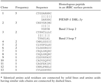
Table 2. Sequences of peptides expressed on selected phage

Fig. 2. Binding specificity of original phage display library, phage expressing an irrelevant peptide, and phage clones selected after the panning against antibodies isolated from iRBC surface. Phage (1×10⁸ TU) were panned on immobilized IgG from NHS (open bar)- or with IHS (closed bar)-treated iRBC and TU of bound phage was determined as described in the Materials and Methods section. Each bar represents the mean±S.D. of triplicate determinations.

Fig. 3. Binding specificity of the original phage display library and phage clones selected after the 4th panning against IgG in NHS (11 samples, open circle) or IHS (24 samples, filled circle). Phage (1×10⁸ TU) were panned on immobilized IgG and TU of bound phage was determined as described in the Materials and Methods section. Horizontal lines in each graph represent the mean values. The numbers in each graph indicate P values calculated using ANOVA (single factor). Relative phage binding was calculated by dividing each value of phage binding (TU/well) to NHS or IHS by the mean of phage binding (TU/well) to NHS.

Fig. 4. Binding of anti-M1-peptide antibody to iRBC (FCR-3 strain). (A, B) Photographs of fluorescent beads bound to the surface of iRBC pre-treated with pre-immune rabbit serum (A) or with immune serum raised against the M1 peptide (B). Arrowheads indicate bound fluorescent beads on iRBC and arrows indicate nRBC present in the iRBC suspension. (C) Binding of antibodies to the surface of uninfected erythrocytes (open bar) and iRBC (closed bar). Fluorescent beads were coated with anti-rabbit IgG antibody and used to detect the binding of anti-M1 antibody on iRBC as described in the Materials and Methods section. Each bar represents the mean±S.D. of triplicate determinations.
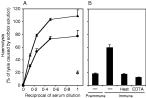
Fig. 5. Haemolysis of iRBC by rabbit anti-M1 antiserum. (A) Pre-immune serum (open symbols) and immune serum (closed symbols) were incubated with a suspension of iRBC infected with FCR-3 strain (circle) or with CS2 strain (triangle) and the lysis of RBC was evaluated. Each value represents the mean±S.D. of triplicate determinations. (B) Lysis of iRBC infected with FCR-3 strain after incubation with rabbit pre-immune and immune serum raised against the M1 peptide was examined as in (A). Heat: heat-inactivated immune serum was used. EDTA: incubation was conducted in the presence of EDTA. Each bar represents the mean±S.D. of triplicate determinations.
 You have
Access
- 11
- Cited by