Introduction
Malaria is one of the most serious infectious diseases affecting humans, causing 250 million clinical cases annually and leaving 3 billion people at risk of infection globally (World Health Organisation, 2017). Blood stage parasite infection is responsible for all clinical symptoms associated with malaria (Schofield and Grau, Reference Schofield and Grau2005), which can range from relatively mild and uncomplicated disease to severe complications including cerebral malaria, respiratory distress, hypoglycaemia, renal failure and pulmonary oedema (Miller et al., Reference Miller, Baruch, Marsh and Doumbo2002). Children under 5 years of age, with low levels of pre-existing immunity, are most vulnerable to these severe manifestations of disease. Individuals living in malaria-endemic regions acquire clinical immunity to the disease only after repeated exposure to the parasite over a number of years. This type of immunity is non-sterilizing, but can prevent clinical episodes by substantially reducing parasite burden (Marsh and Kinyanjui, Reference Marsh and Kinyanjui2006), commonly resulting in asymptomatic infection of adults. Parasite-specific antibody responses are known to play an important role in protective immunity to malaria (Cohen et al., Reference Cohen, McGREGOR and Carrington1961; Sabchareon et al., Reference Sabchareon, Burnouf, Ouattara, Attanath, Bouharoun-Tayoun, Chantavanich, Foucault, Chongsuphajaisiddhi and Druilhe1991) and engage in various effector functions during blood-stage infection including the prevention of erythrocyte invasion and sequestration (Bolad and Berzins, Reference Bolad and Berzins2000; Stanisic et al., Reference Stanisic, Richards, McCallum, Michon, King, Schoepflin, Gilson, Murphy, Anders, Mueller and Beeson2009), promotion of complement-mediated lysis, and opsonization for phagocytic parasite clearance (Schmidt et al., Reference Schmidt, Kennedy and Tham2015).
The development of long-lived, antibody-mediated immunological memory requires the establishment of germinal centre (GC) structures. Following initial interaction with antigen, naive B cells in lymphoid organs either differentiate into short-lived plasma cells or migrate deeper into the B cell follicles where they establish a GC. Within the GCs, activated B cells undertake somatic hypermutations in their Ig genes, followed by selective survival of B cells producing antibodies with higher affinity. GC establishment and function require a specific subset of T cells- named T follicular helper (TFH) cells (Crotty, Reference Crotty2011). Recent studies investigated how these processes are modulated in malaria in order to identify immunological processes which underpin the slow acquisition of immunity. Epidemiological studies revealed that the development of IgG+ memory B cells specific for Plasmodium falciparum antigens is delayed in individuals residing in areas of high seasonal malaria transmission, who are vulnerable clinical episodes of disease (Weiss et al., Reference Weiss, Traore, Kayentao, Ongoiba, Doumbo, Doumtabe, Kone, Dia, Guindo, Traore, Huang, Miura, Mircetic, Li, Baughman, Narum, Miller, Doumbo, Pierce and Crompton2010). Consistent with those findings, using a rodent infection model we previously showed that the same inflammatory cytokines that contribute to the induction of malaria disease symptoms such as IFN-γ, upregulate the expression of the transcription factor T-bet in TFH cells, which prevents their normal differentiation and capacity to provide help for the induction of plasma cells and IgG+ memory B cells (Ryg-Cornejo et al., Reference Ryg-Cornejo, Ioannidis, Ly, Chiu, Tellier, Hill, Preston, Pellegrini, Yu, Nutt, Kallies and Hansen2016a, Reference Ryg-Cornejo, Ly and Hansen2016b).
Emerging evidence started to delineate multiple layers of memory B cells, with distinct effector functions during recall responses (Dogan et al., Reference Dogan, Bertocci, Vilmont, Delbos, Mégret, Storck, Reynaud and Weill2009). Whereas most studies examining long-lived memory B cell responses to infection and vaccination have focussed on the IgG+ memory B cell compartment, IgM+ memory B cells have only recently started to receive attention. Evidence from murine studies has shown that this compartment remains stable (Good-Jacobson and Tarlinton, Reference Good-Jacobson and Tarlinton2012) and recognizes antigen with low affinity (Pape et al., Reference Pape, Taylor, Maul, Gearhart and Jenkins2011) compared to class-switched memory B cells. Unlike IgG+ memory B cells, which rapidly differentiate into antibody-secreting plasma cells upon antigen re-encounter, recent studies in humans and murine models suggest that IgM+ memory B cells have the additional capacity to establish secondary GC responses, undertake further rounds of somatic hypermutation and affinity selection to remodel their existing B cell receptor specificity, and undergo switch recombination (Taylor et al., Reference Taylor, Jenkins and Pape2012; Seifert et al., Reference Seifert, Przekopowitz, Taudien, Lollies, Ronge, Drees, Lindemann, Hillen, Engler, Singer and Küppers2015). As such, it has been proposed that IgM+ memory B cells may assist in the maintenance of long-term immunity by replenishing the IgG+ memory B cell pool when reactivated by their specific antigen.
Recent studies revealed that IgM antibody responses specific for a range of P. falciparum surface antigens are associated with protection from symptomatic malaria (Arama et al., Reference Arama, Skinner, Doumtabe, Portugal, Tran, Jain, Traore, Doumbo, Davies, Troye-Blomberg, Dolo, Felgner and Crompton2015; Boyle et al., Reference Boyle, Chan, Handayuni, Reiling, Feng, Hilton, Kurtovic, Oyong, Piera, Barber, William, Eisen, Minigo, Langer, Drew, de Labastida Rivera, Amante, Williams, Kinyanjui, Marsh, Doolan, Engwerda, Fowkes, Grigg, Mueller, McCarthy, Anstey and Beeson2019), suggesting a potential role for IgM+ memory B cells in the acquisition of clinical immunity to the infection. Only a few studies have identified IgM+ memory B cells in malaria-exposed individuals and in malaria mouse infection models (Stephens et al., Reference Stephens, Ndungu and Langhorne2009; Krishnamurty et al., Reference Krishnamurty, Thouvenel, Portugal, Keitany, Kim, Holder, Crompton, Rawlings and Pepper2016; Wendel et al., Reference Wendel, He, Qu, Wu, Hernandez, Ma, Liu, Xiao, Crompton, Pierce, Ren, Chen and Jiang2017), with those induced by murine malaria found to rapidly differentiate into plasma cells upon antigenic re-stimulation (Krishnamurty et al., Reference Krishnamurty, Thouvenel, Portugal, Keitany, Kim, Holder, Crompton, Rawlings and Pepper2016). Memory B cells isolated from malaria-exposed individuals have been shown to undertake Ig somatic hypermutations (Triller et al., Reference Triller, Scally, Costa, Pissarev, Kreschel, Bosch, Marois, Sack, Murugan, Salman, Janse, Khan, Kappe, Adegnika, Mordmüller, Levashina, Julien and Wardemann2017) and accumulate further mutations after acute infection (Wendel et al., Reference Wendel, He, Qu, Wu, Hernandez, Ma, Liu, Xiao, Crompton, Pierce, Ren, Chen and Jiang2017) raising the intriguing possibility that IgM+ memory B cells might re-enter GC reactions during secondary infection. To address that question, here we have used a murine model of severe malaria to investigate the differentiation capacity of IgM+ memory B cells during re-infection. Our results revealed that this compartment adopts a GC B cell-like phenotype upon parasite-specific antigenic re-stimulation.
Materials and methods
Mice and infections
Eight to twelve-week-old C57BL/6 mice were used for all experiments in this study. Mice were infected with 5.0 × 10⁴P. berghei ANKA parasitized red blood cells (pRBCs) via intravenous (i.v.) tail vein injection. In most experiments, mice were treated with dose chloroquine (10 mg kg−1) and pyrimethamine (10 mg kg−1) intraperitoneally (i.p.) at the onset of severe malaria symptoms on day 5 post infection (pi), followed by treatment with drinking water containing chloroquine and pyrimethamine (10 mg kg−1) for 7 days. Parasitemia was assessed from Giemsa-stained smears of tail blood. All experiments were conducted with approval from the Walter & Eliza Hall Institute Animal Ethics Committee.
Flow cytometry
Splenocytes suspensions were incubated with anti-CD16/CD32 antibody. Cells were washed twice prior to incubation with fluorescent antibodies for 30 min at 4 °C. To detect of IgM+ memory B cells, splenocytes were stained with: APC anti-CD21 (7G6, Miltenyi Biotec), FITC anti-IgM (Il/41, BD Biosciences), PE anti-IgD (11–26c.2a, BD Biosciences), PerCP Cy5.5 anti-CD19 (1D3, BD Biosciences), BV650 anti-Fas/CD95 (Jo2, BD Biosciences), PE/Cy7 anti-CD38 (90, BioLegend) and BV421 anti-CD138 (281–2, BioLegend). To detect GC B cells splenocytes were stained with the following antibodies: PerCP Cy5.5 anti-CD19 (1D3, BD Biosciences), PE anti-GL7 (GL7, BD Biosciences), FITC anti-CD38 (90, BioLegend), BV785 anti-CD45.1 (A20, BioLegend) and PE/Cy7 anti-CD45.2 (281–2, BioLegend). To detect plasma cells, splenocytes were stained with the following antibodies: PerCP Cy5.5 anti-CD19 (1D3, BD Biosciences), FITC anti-IgM (Il/41, BD Biosciences), PE anti-IgD (11–26c.2a, BD Biosciences), BV421 anti-CD138 (281–2, BioLegend), BV785 anti-CD45.1 (A20, BioLegend) and PE/Cy7 anti-CD45.2 (281–2, BioLegend). For the detection of proliferating cells or intranuclear transcription factors, cells were fixed and permeabilized with 200μL of eBioscience permeabilization buffer for 1hr at 22 °C, then washed and stained with AF594 anti-Ki67 (SOIA15), or with APC anti- Bcl-6 (GC B cells, BD Biosciences) or APC anti-Blimp-1 (Plasma cells, BD Biosciences) for another hour. After staining, cells were washed twice and analysed either by flow cytometry on a BD LSRFortessa™ X-20 cell analyser (BD Biosciences), or sorted on BD FACSAria™ III/W FACS cell sorters (BD Biosciences). Data were analysed using FlowJo v12 software (Treestar Inc.).
Adoptive transfer
Ly5.1+ C57BL/6 mice were infected with 5.0 × 10⁴P. berghei ANKA pRBCs and drug cured at day 5 pi. On day 21 pi, CD19+CD21−CD138−Fas−CD38+IgD−IgM+ cells were isolated by sorting on a BD FACS Aria III system (BD Biosciences) with BD Diva software. Isolated IgM+ memory B cells were adoptively transferred (1.0 × 10⁶/mouse) into Ly5.2+ C57BL/6 recipients, which were infected with 5.0 × 10⁴P. berghei ANKA pRBCs the following day. Spleen cells from recipient animals were collected on day 3 pi for the assessment of immune responses.
Statistical analysis
Statistical analysis was performed in Prism version 8 (GraphPad Software Inc.). The Student's t-test was used for the analysis of data. Information on replicates/error/significance is indicated in the figure legends.
Results
IgM+ memory B cells can be detected after infection with Plasmodium berghei
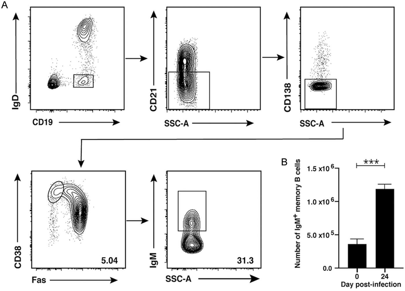
We have previously shown that P. berghei ANKA infection results in the induction of IgG+ memory B cells (Ryg-Cornejo et al., Reference Ryg-Cornejo, Ioannidis, Ly, Chiu, Tellier, Hill, Preston, Pellegrini, Yu, Nutt, Kallies and Hansen2016a, Reference Ryg-Cornejo, Ly and Hansen2016b; Ly et al., Reference Ly, Liao, Pietrzak, Ioannidis, Sidwell, Gloury, Doerfinger, Triglia, Qin, Groom, Belz, Good-Jacobson, Shi, Kallies and Hansen2019). To determine if this infection also induces IgM+ memory B cells, C57BL/6 mice were infected with 5.0 × 10⁴ pRBCs. As P. berghei ANKA infection is lethal in C57BL/6 mice, infected animals were cured with anti-malarial drugs after the onset of symptoms at day 5 pi to allow the assessment of immune responses at later time-points as described (Ryg-Cornejo et al., Reference Ryg-Cornejo, Ioannidis, Ly, Chiu, Tellier, Hill, Preston, Pellegrini, Yu, Nutt, Kallies and Hansen2016a). After full resolution of parasitemia on day 24 pi, splenocytes were extracted from malaria-exposed mice, stained with a panel of fluorescent antibodies and analysed by flow cytometry using the gating strategy delineated in Fig. 1A. Memory B cells were identified among live lymphocytes by high expression of CD19, CD38 and IgM, and low expression of IgD, CD138, the marginal zone B cell marker CD21 and the GC B cell marker Fas (Fig. 1A). A population of CD19+CD38+IgM+ memory B cells was identified at day 24 pi. The absolute number of IgM+ memory B cells was 3 times higher in P. berghei infected mice compared to naïve controls (Fig. 1B). Thus P. berghei ANKA infection induces IgM+ memory B cells that can be detected in the spleen 2 weeks after parasitemia has been controlled.
Plasmodium berghei ANKA infection induces IgM+ memory B cells. C57BL/6 mice were infected with P. berghei ANKA and treated with anti-malarial drugs from day 5 pi. (A) Gating strategy used to identify IgM+ memory B cells by flow cytometry, depicting gated IgD−CD19+ B cells that were negative for CD21 and CD138 to exclude marginal zone B cells and plasma cells, respectively. Memory B cells were then identified by high expression of CD38 and low levels of Fas, to exclude GC B cells. IgM+ memory B cells were identified among this compartment. (B) Absolute number of IgM+ memory B cells in P. berghei ANKA-infected mice on day 24 pi and naive controls. Error bars represent the mean of 8–10 mice ± s.e.m., ***P < 0.005.

IgM+ memory B cells adopt a germinal-centre B cell phenotype upon malaria re-infection
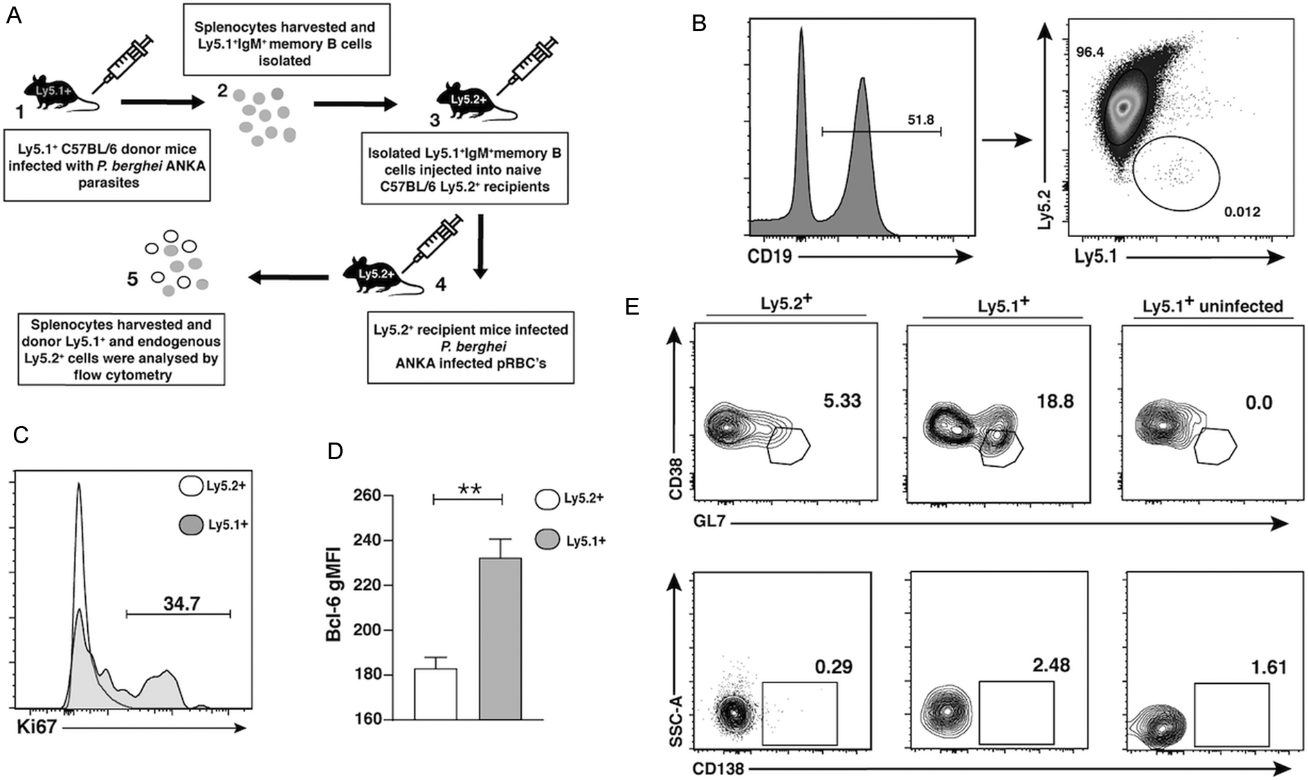
Having established that an IgM+ memory B cell compartment is detectable after infection with P. berghei, we next sought to understand the functional capacity of this cell subset upon re-infection. To that end, congenically marked Ly5.1+ C57BL/6 mice were infected with P. berghei, then the drug was cured at day 5 pi as described above. On day 21 p., IgM+ memory B cells were isolated by cell sorting following the gating strategy shown in Fig. 1A, and adoptively transferred into Ly5.2+ congenically marked recipient mice (Fig. 2A). Flow cytometry analysis indicated that the sorted population was ~93% pure (Supplementary Fig. 1). Recipient mice were then infected with P. berghei ANKA and on day 3 pi the phenotype of transferred CD19+ donor and recipient cells was examined by flow cytometry (Fig. 2B). Rapid proliferation upon re-stimulation is a key feature of antigen-experienced memory cells (Tangye et al., Reference Tangye, Avery, Deenick and Hodgkin2003; Good et al., Reference Good, Avery and Tangye2009). To determine if adoptively transferred IgM+ memory B cells from malaria exposed donors rapidly proliferated in response to secondary infection like conventional memory B cells do, the expression of Ki67 was assessed within Ly5.1+ and Ly5.2+ CD19+ cells. Consistent with a memory-like phenotype, Ly5.1+ donor cells readily proliferated only 3 days after re-infection with P. berghei ANKA (Fig. 2C) while Ly5.2+ cells did not.
IgM+ memory B cells adopt a GC B cell phenotype during a secondary P. berghei ANKA infection. (A) Schematic illustrating the adoptive transfer workflow used to assess the functional capacity of IgM+ memory B cells. IgM+ memory B cells isolated from infected C57BL/6 Ly5.1+ mice were adoptively transferred into Ly5.2+ recipient mice. Recipient mice were then challenged with P. berghei ANKA, followed by the analysis of both the Ly5.1+ donor and Ly5.2+ endogenous compartments 3 days pi by flow cytometry. (B) Congenically, labelled cells were identified among gated CD19+ B cells. (C) Percentage of proliferating cells was calculated by assessing Ki67 expression among gated CD19+ Ly5.1+ and Ly5.2+ cells. (D) Geometric mean fluorescence intensity (gMFI) for Bcl-6 expression in CD19+Ly5.1+ or Ly5.2+ cells. (E) Percentage of CD38lowGL7+ GC B cell-phenotype cells (top panels) and CD138+ plasma cells (bottom panels) was calculated among CD19+ Ly5.2+ cells from infected mice and CD19+Ly51+ cells from infected mice and uninfected controls. Data are representative of 2 experiments. Graphs depict means of 4 mice ± s.e.m., **P < 0.01. Representative histograms and dot plots are shown.

Blimp-1 and Bcl-6 are transcription factors required for the differentiation of B cells into plasma cells and GC B cells, respectively (Shapiro-Shelef et al., Reference Shapiro-Shelef, Lin, McHeyzer-Williams, Liao, McHeyzer-Williams and Calame2003; Crotty et al., Reference Crotty, Johnston and Schoenberger2010). Thus, the expression of these transcription factors was examined among Ly5.1+ B cells from malaria-exposed donors and Ly5.2+ recipient controls in order to determine the differentiating capacity of IgM+ memory B cells upon antigenic re-stimulation. No Blimp-1 upregulation could be detected among donor or recipient B cells on day 3 pi (not shown). In contrast, Bcl-6 expression could be readily detected among Ly5.1+ B cells from malaria-exposed donors and was significantly higher to that of recipient controls (Fig. 2D), suggesting that adoptively transferred IgM+ MBCs might adopt a GC B cell-like phenotype during re-stimulation. To investigate this further, frequencies of plasma cells and GC B cells were examined on day 3pi in Ly5.1+ donor and Ly5.2+ recipient B cells. Analysis of Ly5.1+ donor cells in recipient mice that were left uninfected was included as controls. No plasma cells (CD138+CD19+) could be detected emerging from the Ly5.1+ donor compartment or Ly5.2+ recipients upon malaria infection or from Ly5.1+ cells adoptively transferred into uninfected controls (Fig. 2E). Instead, nearly 20% of Ly5.1+ cells from exposed mice downregulated expression of CD38 and upregulated the GC B cell-specific marker GL7 when challenged with P. berghei but not when recipient mice were left uninfected (Fig. 2E). Together, these results indicate that IgM+ memory B cells induced in response to malaria adopt a GC B cell-like phenotype in response to a secondary infection.
Discussion
Recent evidence supports the concept that IgM+ memory B cells have the ability to remodel their B cell receptor specificity by accessing secondary GC reactions after encounter with cognate antigen during re-infection. This response has been postulated to be beneficial for responding to antigenic variants of the original pathogen; a desirable feature to efficiently recognize polymorphic malarial antigens. The present study provided evidence for the functional capacity of IgM+ memory B cells generated during malaria infection to acquire a GC B cell-like phenotype upon a secondary challenge.
Differentiation into antibody-secreting plasma cells is a widely accepted re-stimulatory role of isotype-switched memory B cells (Tangye and Tarlinton, Reference Tangye and Tarlinton2009) and a few studies have reported strong plasma cell responses also mediated by IgM+ memory B cells in response to viral infection (Zabel et al., Reference Zabel, Mohanan, Bessa, Link, Fettelschoss, Saudan, Kündig and Bachmann2014). In contrast, other work in parasitic and bacterial infections demonstrated the capacity of IgM+ memory B cells to re-enter GCs during recall responses (Yates et al., Reference Yates, Racine, McBride and Winslow2013; Kenderes et al., Reference Kenderes, Levack, Papillion, Cabrera-Martinez, Dishaw and Winslow2018). Thus, the available information supports the idea that different pathogens might direct IgM+ memory B cells towards various differentiation fates (McHeyzer-Williams et al., Reference McHeyzer-Williams, Dufaud and McHeyzer-Williams2018). In malaria, previous studies using the mild Plasmodium chabaudi murine infection model detected antigen-specific IgM+ memory B cells that appear to differentiate into plasma cells after re-stimulation (Krishnamurty et al., Reference Krishnamurty, Thouvenel, Portugal, Keitany, Kim, Holder, Crompton, Rawlings and Pepper2016). Unlike P. chabaudi, the P. berghei ANKA infection used in the current study results in the induction of a highly inflammatory response to infection, responsible for the development of severe disease symptoms (Hansen, Reference Hansen2012). In that context, IgM+ memory B cells induced by P. berghei ANKA infection failed to differentiate into antibody-secreting plasma cells upon re-encounter with antigen and adopted a GC B cell-like phenotype, raising the possibility that inflammatory responses and the cytokine environment-induced during infection might also play a role influencing the differentiation fate of IgM+ memory B cells upon re-encounter with a cognate antigen.
Our findings are consistent with a model in which the IgM+ memory B cell compartment plays an important role in populating secondary GCs after re-infection with Plasmodium. Similar to other infection settings (Pape et al., Reference Pape, Taylor, Maul, Gearhart and Jenkins2011), malaria-specific IgM+ memory B cells might mediate the rapid re-seeding of secondary GCs for earlier recall responses, thereby playing a critical role in the immune response by rapidly initiating the induction of B cell clones with enhanced affinity for antigen, at faster rates than naive B cells (Good et al., Reference Good, Avery and Tangye2009). Further work will be required to confirm whether IgM+ memory B cell-derived GC B cells localize in splenic GCs during re-exposure to malaria and if this process is required for the gradual acquisition of protective immunity. Understanding the role of IgM+ B cells in mediating immunological memory responses to Plasmodium parasites and the impact that infection has on the functional capacity of this compartment may be highly informative for the development of novel therapeutics and vaccines to target malaria.
Supplementary material
The supplementary material for this article can be found at https://doi.org/10.1017/S003118202000061X.
Financial support
This work was made possible through Victorian State Government Operational Infrastructure Support and Australian Government National Health and Medical Research Council IRIISS, Project Grant 1058665 and the Australian Academy of Science Regional Collaboration Grant. The funders had no role in study design, data collection and analysis, decision to publish, or preparation of the manuscript.
Conflict of interest
None.
Ethical standards
All experiments conducted with approval from the Walter & Eliza Hall Institute Animal Ethics Committee.