Case report
A 40-year-old woman presented with a right-sided cervical mass which had been growing slowly over 18 months. She was otherwise asymptomatic, and denied any pain, dysphagia, dyspnoea, cough or recent weight change.
On physical examination, the patient was noted to have a right neck mass spanning levels I to III. The mass was non-tender, non-compressible and laterally mobile. The patient's airway was patent, with normal landmarks identified on flexible laryngoscopy. The remainder of the head and neck examination was unremarkable.
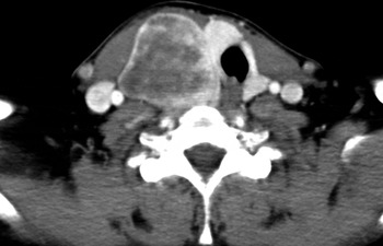
A contrast-enhanced computed tomography scan revealed a well circumscribed, right-sided neck mass measuring 9 × 4 × 5 cm, with heterogeneous enhancement, breaching the anterior and posterior cervical triangles and lying in close proximity to the carotid sheath (Figure 1).
Axial, contrast-enhanced computed tomography image of the neck showing a well circumscribed, heterogeneously enhancing mass lying in close proximity to the carotid sheath.

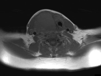
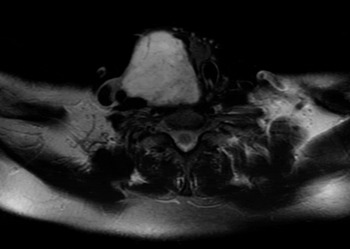
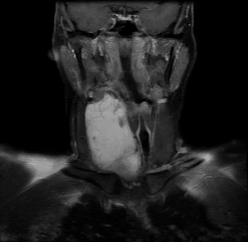
Further evaluation of the lesion by magnetic resonance imaging showed the mass to be heterogeneous with a ‘salt and pepper’ pattern. It was hyperintense on T1-weighted imaging with respect to the adjacent neck musculature (Figure 2), and markedly hyperintense on T2-weighted imaging (Figure 3). Following administration of gadolinium contrast, there was intense enhancement of the mass (Figure 4). Sharp demarcation was noted between the mass and the adjacent musculature, as well as between the mass and the thyroid gland.
Axial, T1-weighted magnetic resonance imaging scan, corresponding to the Figure 1 scan, showing a heterogeneous mass with hyperintense regions.

Axial, T2-weighted magnetic resonance imaging scan with fat saturation, showing markedly increased signal intensity.

Coronal, gadolinium contrast enhanced, T1-weighted magnetic resonance imaging scan, showing intense, heterogeneous enhancement of the mass.

Fine needle aspiration cytology was performed, but yielded non-diagnostic results.
Serum thyroid function tests and urinary vanillylmandelic acid and normetanephrine levels were all within the normal range.
The patient was scheduled for pre-operative angioembolisation of her neck mass. The superior thyroid artery and thyrocervical trunk branches supplying the cervical mass were successfully embolised.
The mass was successfully removed. Histopathological findings were suggestive of benign follicular adenoma.
Discussion
True benign thyroid masses very rarely present as a solitary lateral neck mass. Indeed, the aetiology of such masses has generated much controversy. In 1906, Schrager first coined the term ‘lateral aberrant thyroid’, which he defined as a ‘… normal pathology thyroid, … situated at some definite distance from the normal thyroid, with which it has no connection whatsoever’.Reference Schrager1 In 1988, Rubenfeld et al. reported the first case of lateral aberrant thyroid tissue in the absence of any other thyroid tissue.Reference Rubenfeld, Joseph, Schwartz, Weber and Jhingran2 These authors attributed their findings to a combination of ‘… agenesis of the medial primordial and dysembryoplasia of the lateral primordial’, thus highlighting the importance of thyroid gland evaluation and thyroid function testing in patients presenting with lateral neck masses.
In 1946, Lahey and Ficarra substantiated the concept by reporting a series of 47 cases of ‘lateral aberrant thyroid’ tissue.Reference Lahey and Ficarra3 They further defined these masses as ‘…thyroid tissue that persists outside of the thyroid gland proper, and [which] may be of lateral, median, or ectopic origin, arising from the ultimobranchial bodies as a result of departure from normal embryologic development’.Reference Lahey and Ficarra3 They reported cases originally thought to be carotid body tumours but subsequently discovered to be lateral aberrant thyroid tissue. These authors attributed the origin of such ‘lateral aberrant thyroid tissue’ to ultimobranchial bodies which had undergone conversion to thyroid tissue.
Also in the mid-1940s, Clay and Blackman proposed a differing theory of the origin of lateral aberrant thyroid tissue. They thought that these masses represented metastases to lymph nodes from a primary carcinoma of the thyroid.Reference Clay and Blackman4 This view has been further popularised by several other authors.Reference Maceri, Babyak and Ossakow5–Reference King and Perberton8 Maceri et al. reported a series of 268 cases of thyroid cancer managed between 1974 and 1984, of which 36 patients had, as the sole presenting sign, a lateral neck mass that proved to be metastatic thyroid cancer.Reference Maceri, Babyak and Ossakow5 Rabinov et al. presented a patient with a right neck mass who underwent excision for presumed branchial cleft cyst.Reference Rabinov, Ward and Pusheck9 However, post-operative histopathological examination revealed papillary thyroid carcinoma, necessitating further surgery for total thyroidectomy and right modified radical neck dissection.
The debate surrounding the origin of lateral aberrant thyroid tissue is probably based on a misunderstanding of the term as initially defined by Schrager. The term ‘lateral aberrant thyroid tissue’ has been commonly used to indicate thyroid tissue in the lateral cervical lymph nodes, and, more frequently, to indicate metastases to lymph nodes from a primary carcinoma of the thyroid gland.Reference Kozol, Geelhoed, Flynn, Kinder, Feind and Mack10
However, there are other situations in which benign thyroid tissue can be found in ectopic locations, in the presence of a normal, orthotopic thyroid gland. Akcam et al reported a case of ectopic thyroid tissue (with follicular adenoma) arising in the lateral neck.Reference Akcam, Talas, Gerek, Deveci and Ozkaptan11 A functional thyroid of normal size was noted to be separate from the neck mass. The authors concluded that the mass was the result either of cell rests derived from the ultimobranchial bodies, or of a detachment from the lateral lobe of the thyroid primordium during embryogenesis.
• Benign thyroid masses very rarely present as a lateral neck mass
• Aberrant thyroid embryogenesis may result in ectopic thyroid tissue
• Orthotopic thyroid tissue may be absent in patients presenting with lateral aberrant thyroid tissue
• Lateral aberrant thyroid tissue may be the presenting sign of metastatic thyroid carcinoma
In our patient, lateral aberrant thyroid tissue was located anteromedial to the carotid sheath, centred at the level of the carotid bifurcation. Intra-operatively, the mass was noted to be separate from the right thyroid lobe. The location of the mass in the lateral neck, its benign histopathology (follicular adenoma), and its distinct separation from the thyroid gland all support the diagnosis of ‘lateral aberrant thyroid’.
Conclusion
Lateral aberrant thyroid represents a rare yet important condition in the differential diagnosis of a lateral neck mass. Clinicians should be aware of the presentation and radiographic manifestations of lateral aberrant thyroid, in order to prompt suspicion of ectopic thyroid tissue when imaging reveals non-specific findings for a carotid body tumour. Further investigation should always include complete blood tests to characterise the orthotopic thyroid gland. In cases in which the definitive diagnosis is uncertain pre-operatively, patient consent should be obtained for the relevant definitive surgical therapy, should intra-operative findings and/or frozen section analysis indicate malignancy.