Introduction
The concept of intrauterine experience to disease in later life forms the basis of ‘foetal origins of disease’ phenomenon.Reference Barker 1 It is suggested that physiological, metabolic and structural adaptations (programming) are made by the foetus with manifestation of impaired or disproportionate foetal growth, when exposed to adverse early environment to promote survival. However, early programming conflict occurs when the postnatal conditions are improved.Reference Cottrell and Ozanne 2 In long term, these programming changes are subsequently translated into pathology and an increased likelihood of developing chronic diseases.Reference Langley-Evans 3
The concept of ‘intrauterine origins of obesity’ comes from the follow-up of offspring born to women during the Dutch famine. Female offspring who were exposed to prenatal undernutrition in early gestation are at a high risk for adult obesity.Reference Ravelli, van Der Meulen, Osmond, Barker and Bleker 4 However, the mechanisms governing foetal programming of obesity are complex and remain unresolved. Biologically plausible, oxidative stress programming could be the underlying factor in the developing foetus that contributes to higher risk of metabolic dysfunction and later obesity.
Over the course of development, maternal oxidative stress insults impose an adverse programming effect on foetal growth.Reference Luo, Fraser and Julien 5 Disruption of intrauterine redox status results in adaptive response to metabolic dysregulation, leading to pathophysiological alterations at critical developmental windows. In this respect, a growing body of evidence now exists in support of oxidative stress programming hypothesis. Oxidative stress has always been observed in preterm and low birth weight infants because these infants are very often exposed to high oxygen concentration, have immature antioxidant defence system and vulnerable to infection.Reference Gitto, D’Angelo, Cusumano and Reiter 6 Not limited to preterm infants,Reference Negi, Pande and Kumar 7 inverse relationship of maternal DNA damage, lipid peroxidation or protein oxidation with foetal growth was also noted in term infants.Reference Kim, Hong and Lee 8 – Reference Saker, Soulimane Mokhtari and Merzouk 11 Such foetal oxidative stress may be triggered by prenatal oxidative state or labour-induced oxidative stress.Reference Gitto, D’Angelo, Cusumano and Reiter 6 On the other hand, birth weight was found to be significantly increased with a high level of maternal antioxidant status.Reference Jain, Wise, Yanamandra, Dhanireddy and Bocchini 12 , Reference Osorio, Cruz and Milanés 13
Although there is considerable literature concerning the effects of prenatal oxidative stress on foetal growth and birth outcomes, much less is known about the long-term impact on postnatal growth and adiposity development. It has been shown that small for gestational age (SGA) infants born in a high oxidative stress intrauterine environment demonstrated altered lipoprotein metabolism at birth.Reference Saker, Soulimane Mokhtari and Merzouk 11 Another study indicated that increased oxidative stress and insulin resistance were found in SGA obese children.Reference Chiavaroli, Giannini and D’Adamo 14 These findings suggest that there is a link between oxidative stress, reduced birth weight and obesity. Therefore, by understanding the contributory role of oxidative stress on postnatal adiposity, it is hoped that the underlying mechanism of obesity development can be identified and suggests a preventive measure to fight against the obesity epidemic since early life. This study was therefore aimed to investigate the effect of maternal oxidative stress levels in the second trimester on infant adiposity development during the first year of life.
Methods
The Universiti Sains Malaysia (USM) Birth Cohort Study comprised two phases, which were prenatal phase and postnatal phase. This prospective cohort study was conducted from April 2010 to December 2012 in Kelantan state, Malaysia. Prenatal phase took place in the Obstetrics and Gynaecology (O&G) Clinic of Hospital Universiti Sains Malaysia (HUSM) and Kubang Kerian Health Clinic, Kota Bharu, whereas postnatal phase involved ward visiting in the HUSM and home visiting in the districts of Kota Bharu, Bachok, Pasir Mas, Tumpat, Pasir Puteh and Machang. The study protocol was approved by the Human Research Ethics Committee of USM and Medical Research Ethics Committee of Ministry of Health, Malaysia.
Pregnant women were recruited in the second trimester (⩽24 weeks of gestation) and followed up in the third trimester of pregnancy (⩾32 weeks of gestation), whereas infant growth was assessed at birth, 2, 6 and 12 months of age. Healthy pregnant women were recruited on the basis of convenience sampling technique. The selection criteria were defined as: (i) Malaysian and Malays ethnicity; (ii) aged 19–40 years; (iii) singleton pregnancy; (iv) gestational age 24 weeks and less, based on the last menstrual period or early ultrasound examination; (v) plan to give birth in HUSM; and (vi) live within a distance of 50 km from HUSM. The exclusion criteria included: (i) diagnosis with pre-existing chronic diseases or pregnancy complications and (ii) preterm delivery before 37 weeks of gestation. Sample size for the study was calculated using a priori Sample Size Calculator for multiple regression, Version 3.0,Reference Soper 15 based on the formula of Cohen et al. Reference Cohen, Cohen, West and Aiken 16 A total of 141 participants were required to achieve an anticipated effect size of 0.15 with the power of 90% and probability of 5%. Written informed consent was acquired from each pregnant woman at recruitment. The flow chart and selection procedure illustrating an overall study progress are shown in Fig 1.
Flow chart and sampling procedure of the study.

Maternal blood and hair samples measurements
Pregnant women fasted for 10–12 h before the blood collection in the second trimester (14–24 weeks of gestation). A total of 5 ml venous blood samples were drawn into the oxalate, EDTA and lithium-heparinized tubes for the analyses of glucose concentration and oxidative stress markers. Plasma was separated by centrifugation at condition of 3500 rpm for 10 min at 4ºC and stored in −80ºC freezer until further analyses. Fresh whole blood sample from the lithium-heparinized tube was used to analyse DNA damage on the same day. Plasma glucose concentration was determined by using commercial enzymatic kit (RX Series GL 3981, Randox, UK) and measured by RX Daytona Chemistry Analyzer. Protein oxidation marker as indicated by protein carbonyl and enzymatic antioxidant as indicated by glutathione peroxidase (GPx) activity were measured using the commercial kits (Cayman Chemical, Ann Arbor, Michigan, USA), according to the manufacturer’s instructions. The inter-assay coefficients of variability (CV) for protein carbonyl was 6.9%. The intra- and inter-assay CV for GPx were 5.1% and 8.7%, respectively. Plasma total antioxidant capacity (TAC) was measured by ferric reducing ability of plasma assay according to Benzie and Strain,Reference Benzie and Strain 17 which modified slightly by using 96-well plate. The absorbance readings were taken after 4 min at 593 nm. The intra- and inter-assay CV for TAC were 4.0% and 4.8%, respectively. DNA damage was measured by performing Comet Assay according to Singh et al.,Reference Singh, McCoy, Tice and Schneider 18 with slight optimization as reported in the study by Loy et al. Reference Loy, Sirajudeen and Hamid Jan 19 CometScoreTM software was used to determine the comet parameter: tail moment (percentage of DNA in comet tail×length). The intra-assay CV for tail moment was 3.0%.
Between 1 and 5 days after delivery, 15–20 strands of maternal hair were cut from the posterior vertex of the scalp to assess personal exposure to second-hand smoke exposure during pregnancy. The hair sample was tapped to a piece of white paper and kept in an envelope at room temperature until analysis.Reference Almeida, Koren, Platt and Kramer 20 Level of hair nicotine was analysed by gas chromatography-mass spectrometry with the use of internal standard (diphenylamine), according to Man et al. Reference Man, Ismail, Harn, Lajis and Awang 21
Maternal anthropometric measurement
Height was measured using a microtoise tape (Seca 206, Hamburg, Germany). Prepregnancy weight was based on the maternal recall. Prepregnancy body mass index (BMI) was computed according to the formula: weight (kg)/height (m2). BMI was categorized into underweight (<18.5 kg/m2), normal (18.5–24.9 kg/m2), overweight (25.0–29.9 kg/m2) and obese (⩾30.0 kg/m2) based on the World Health Organization (WHO) classification of BMI cut-offs. 22 Total gestational weight gain (GWG) was computed from the difference between prepregnancy weight and weight at last antenatal visit.
Maternal dietary assessment
A validated semi-quantitative food frequency questionnaire (FFQ) was used to assess maternal dietary patterns in pregnancy via interview basis. Process of the FFQ development and validation was described in the study by Loy et al. Reference Loy, Marhazlina, Nor Azwany and Hamid Jan 23 The FFQ was conducted in the third trimester of pregnancy, which included 82 food categories. Briefly, women were asked to recall their food intake during the past 6 months in pregnancy. Frequency of intake for each food category was then reported as either daily, weekly, monthly or never, followed by estimating the amount of intake based on standard portion sizes. Daily food intake (g/day) was computed according to the formula: frequency of intake per day×total number of servings×weight of food in one serving.Reference Fahmida and Dillon 24 , Reference Norimah, Safiah and Zuhaida 25 The 82 food categories were further classified into 23 foods or food groups based on the similarity of nutrient profiles.Reference Loy and Hamid Jan 26 Principle component analysis (PCA) with varimax rotation was performed to identify the dietary patterns based on the 23 food groups that were derived from the FFQ. Two major dietary patterns were finally determined that were labelled as Healthy pattern and Less-Healthy pattern. Dietary pattern score for each woman was computed by summing the intake of each food group weighted by the factor loading.Reference Kim and Mueller 27 Detailed procedures to perform PCA and compute dietary pattern score were reported by Loy and Hamid Jan.Reference Loy and Hamid Jan 26
Postnatal growth measurement
The growth of infants was assessed at birth, 2, 6 and 12 months of age. Infant weight and recumbent crown–heel length were measured using a baby weighing scale with attached measuring rod and head positioner (Seca 334, Hamburg, Germany). Infant skinfold thickness was measured at triceps and subscapular sites, using a Harpenden Skinfold Calliper (British Indicator, UK). Skinfold measurement was taken at 6 and 12 months of age. On the basis of the chronological age and anthropometric measurements, infant growth was assessed by using the WHO Child Growth Standards 2006 to derive weight-for-length Z-scores (WLZ), BMI-for-age Z-scores (BAZ) and triceps skinfold-for-age Z-scores (TSFZ). These growth indices were computed using the WHO Anthro Software (Version 3.2.2). Infant nutritional status was defined according to the WHOZ-scores. 28
Infant breastfeeding patterns
Infant feeding patterns were assessed using structured questionnaires at birth, 2, 6 and 12 months of age. Women were asked about the types of feeding given to their babies (breast milk, formula milk, plain water or other beverages), duration of breastfeeding (months) and introduction of complementary foods (months of age). Using these information, breastfeeding patterns comprised exclusive breastfeeding, partial breastfeeding and no breastfeeding. Exclusive breastfeeding was defined as the infant receiving only breast milk without any additional food or drink, not even water, except drops/syrups of vitamins, minerals or medicines. Partial breastfeeding was defined as the infant receiving breast milk and water-based drink, food-based fluid, semi-solid or solid food, or non-human milk. 29
Statistical analyses
Data analyses were performed using IBM SPSS Statistics, Version 20.0 (USA). Categorical data were presented as number and frequency. Continuous data with normal distribution were presented as mean and standard deviation, whereas skewed distributed data were presented as median and interquartile range. Log-transformation was performed on skewed data to reduce the leverage of outliers for statistical analyses.
Outcome variables
Infant weight and BAZ at birth, 2, 6 and 12 months of age, as well as infant TSFZ at 6 and 12 months of age, were used as the indicators of infant adiposity during the first year of life.
Exposure variables
Exposure variables for infant adiposity were the oxidative stress markers in the second trimester of pregnancy, including tail moment, protein carbonyl, TAC and GPx.
Associations of maternal oxidative stress markers in the second trimester of pregnancy with infant adiposity were examined by multiple linear regression models with stepwise selection procedure, adjusted for maternal age, prepregnancy BMI, total GWG, prenatal Less-Healthy pattern score, hair nicotine level, gestational age, infant sex, birth weight and breastfeeding patterns. These potential confounders were selected based on literature review.Reference Kim, Hong and Lee 8 , Reference Min, Park and Kim 10 , Reference Hong, Park and Kim 30 Finding at P<0.05 for a two-sided test was considered statistically significant. Multicollinearity, model assumptions of linearity, normality and equal variance were checked.
Results
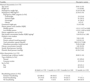
Table 1 shows the characteristics of the Malays pregnant women and infants. The mean age of the women was 29.01 years. On average, maternal nutritional status before pregnancy was within normal BMI category (18.5–24.9 kg/m2). Total GWG was at the mean value of 11.8 kg. None of the women reported smoking and alcohol intake before and during pregnancy. Dietary pattern scores were computed from the factor analysis. Healthy pattern was characterized by high intake of fish and other seafood, dairy products, fruit, vegetables, nuts and legumes. The Less-Healthy pattern reflected high intake of condiments, tea and coffee, meat and offal, confectioneries, oils and fats, and cereals.Reference Loy and Hamid Jan 26 Median hair nicotine content of 0.40 ng/mg indicated maternal nicotine exposure throughout the gestational period. Mean value of maternal DNA damage index in the second trimester, as defined by tail moment was 5.04 and the mean value of protein oxidation, as indicated by protein carbonyl content was 0.39 nmol/mg protein. Mean values of TAC and GPx were 1063.33 µmol/l and 76.96 nmol/min/ml, respectively. All women experienced pregnancies without maternal diabetes and they were deemed normotensive.
Characteristics of the pregnant women and infants

BMI, body mass index; IQR, interquartile range.
Note. Data were presented as mean (standard deviation), unless otherwise indicated.
a Sample size, n=143.
The infants were delivered on an average of 38.82 gestational weeks. The majority of infants were exclusively breastfed at birth (80.4%). However, rate of exclusive breastfeeding fell to 11.6% (n=17) by 6 months. A total of 53.1% (n=77) of the infants were still breastfed at 12 months of age (Table 1). Among infants, 14.4% (n=21) were introduced with non-milk foods in the first 4 months of age, whereas 49.3% (n=72) were given complementary foods started from 6 months of age.
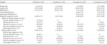
A total of 11 (7.2%) infants and 3 (2.0%) infants were shown to have low birth weight (<2.50 kg) and high birth weight (>4.0 kg), respectively. Birth weight (r=0.25, P=0.002) and birth length (r=0.19, P=0.019) increased with gestational age. All measurements, except skinfolds, were greater in boys than in girls during the first year of life (P<0.05). On average, WLZ and BAZ during infants’ first year of life were within the normal range. Infant BMI and skinfold thickness were reduced from 6 months to 12 months of age. Infants’ mean anthropometric measurements at birth, 2, 6 and 12 months of age are presented in Table 2.
Anthropometric measurements of the infantsFootnote a

BMI, body mass index.
a Data were presented as mean (standard deviation), unless otherwise indicated.
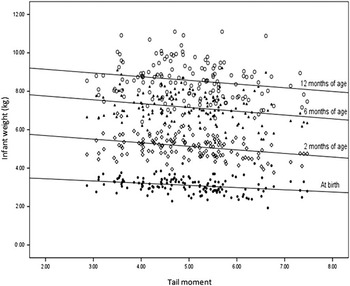
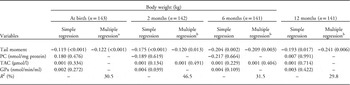
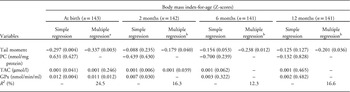
Table 3 shows the regression coefficients for infant weights during the first year of life by prenatal oxidative stress markers in the second trimester of pregnancy. Among the oxidative stress markers, infant weights at birth and after birth were better predicted by marker of DNA damage. As maternal tail moment increased, infant weights at birth (β=−0.122, p<0.001), 2 months (β=−0.120, P=0.013), 6 months (β=−0.209, P=0.003) and 12 months of age (β=−0.241, P=0.006) decreased, with confounders’ adjustment. The associations between maternal DNA damage and infant weights are illustrated in Fig. 2. Similar results were noted when infant BAZ (Table 4) was used as the adiposity indicator. Of the antioxidant markers, GPx activity in the second trimester was positively associated with infant BAZ (β=0.011, P=0.012) (Table 4) at birth.
Associations of maternal DNA damage in the second trimester of pregnancy and infant weights at birth, 2, 6 and 12 months of age.

Regression coefficients (P-values) for infant body weights by prenatal oxidative stress markers in the second trimester of pregnancy

PC, protein carbonyl; TAC, total antioxidant capacity; GPx, glutathione peroxidase.
a Adjusted for maternal age, prepregnancy body mass index, gestational weight gain, prenatal Less-Healthy pattern score, nicotine level, gestational age and infant sex.
b Adjusted for maternal age, prepregnancy body mass index, gestational weight gain, prenatal Less-Healthy pattern score, nicotine level, gestational age, infant sex, birth weight and breastfeeding patterns.
Regression coefficients (P-values) for infant body mass index-for-age by prenatal oxidative stress markers in the second trimester of pregnancy

PC, protein carbonyl; TAC, total antioxidant capacity; GPx, glutathione peroxidase.
a Adjusted for maternal age, prepregnancy body mass index, gestational weight gain, prenatal Less-Healthy pattern score, nicotine level and gestational age.
b Adjusted for maternal age, prepregnancy body mass index, gestational weight gain, prenatal Less-Healthy pattern score, nicotine level, gestational age, birth weight and breastfeeding patterns.
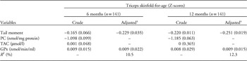
Associations of maternal oxidative stress markers in the second trimester with infant TSFZ are shown in Table 5. With confounders’ adjustment, increasing concentration of maternal tail moment was consistently associated with smaller infant TSFZ at 6 months (β=−0.229, P=0.035) and 12 months of age (β=−0.251, P=0.019). As GPx increased, TSFZ became significantly larger.
Regression coefficients (P-values) for infant triceps skinfold-for-age by prenatal oxidative stress markers in the second trimester of pregnancy

PC, protein carbonyl; TAC, total antioxidant capacity; GPx, glutathione peroxidase.
a Adjusted for maternal age, prepregnancy body mass index, gestational weight gain, prenatal Less-Healthy pattern score, nicotine level, gestational age, birth weight and breastfeeding patterns.
Discussion
The present study shows a consistent inverse association between maternal DNA damage in the second trimester and infant adiposity, as assessed by infant weight, BAZ and TSFZ during the first year of life. Greater prenatal DNA damage was associated with lower infant adiposity at birth, 2, 6 and 12 months of age. Of the antioxidant markers, increased GPx activity was associated with higher infant BAZ at birth and higher TSFZ at 6 and 12 months of age. The results indicate the phenomenon of reduced early postnatal growth and adiposity in term infants born to women with high oxidative stress during normal pregnancies.
Birth weight represents the summary measures of the interaction between maternal environment and genetic endowment in infants and remains an important clinical and global indicator of health.Reference Christian 31 In the current study, the mean birth weight (3.11 kg) of the term infants was similar to that reported in other Malaysian population (3.16 kg),Reference Ramesh, Vidyadaran and Goh 32 but markedly smaller than that of Western infants (3.60 kg).Reference Olsen, Halldorsson and Willett 33 On the other hand, prevalence of low birth weight (7.2%) was slightly lower than the reported figure (8.7%) in a study that was conducted in KelantanReference Norkhafizah, Norsa'adah, Zainuddin, Sosroseno and N Hazlina 34 and the rates that estimated by Unicef (10%) in the general Malaysia population, 35 attributable to the exclusion of preterm birth infants from the analysis.
The trends in anthropometry changes during infancy are consistent with the data that were reported by Kuzawa.Reference Kuzawa 36 Higher rates in body size increment observed in the first 6 months of life were mainly accounted for by fat deposition. Between the ages of 6 and 12 months, decreases in BMI and skinfolds were found. Such fat faltering is contributed by fat mobilization to buffer nutritional stress of weaning and infectious disease stress with the introduction of complementary foods, which are poorer in quality than breast milk.Reference Kuzawa 36 Although there were no differences in skinfolds between boys and girls, the lower average weight in girls implied that girls had a greater proportion of subcutaneous fat. It has been demonstrated that the growing male infants were more rapid to gain in fat-free weight and laid down more musculoskeletal and lean tissue compared with the female infants.Reference Dauncey, Gandy and Gairdner 37
A contributory role of maternal oxidative stress in affecting birth size has been reported previously. Most studies found an inverse association of maternal DNA damage during the second trimester with birth size in term pregnancies,Reference Kim, Hong and Lee 8 – Reference Min, Park and Kim 10 which is in line with our results. However, contrary to Saker et al.,Reference Saker, Soulimane Mokhtari and Merzouk 11 maternal protein oxidation did not show an effect on infant growth in this study. Because oxidative DNA damage mainly arises from the rapid growing cells of the trophoblast columns, which relate to foetal growth,Reference Takagi, Nikaido and Toki 38 it is therefore more sensitive than other oxidative stress markers in affecting infant growth and adiposity.
Increased oxidative stress during pregnancy has been shown to be caused by various factors, such as maternal nutritional status, dietary intake and pollutant exposure. As evidenced in our previous study,Reference Loy, Sirajudeen and Hamid Jan 19 pregnant women with greater body fat were found to have higher DNA damage levels. In addition, adherence to unhealthy dietary pattern (β=0.20, P=0.014) and exposure to second-hand smoke (β=0.05, P=0.036) during pregnancy was associated with increased DNA damage and protein oxidation (results not shown). These oxidant and pro-oxidant stimuli might promote an oxidative attack in maternal–foetal units, further exaggerating placental oxidative stress which can lead to poor foetal growth.
Few biological mechanisms are proposed to explain the causal linkage between oxidative stress and reduced birth size. Genomic instability resulting from DNA damage in pregnancy may alter cellular phenotypes and reduce proliferative potential of cells, causing intrauterine growth restriction associated with defective placentation.Reference Furness, Dekker and Roberts 39 This defective placentation may trigger placental hypoxia-ischaemia and/or hyperoxia, exaggerating placental oxidative stress that can restrict normal foetal growth.Reference Takagi, Nikaido and Toki 38 , Reference Furness, Dekker and Roberts 39 On the other hand, inflammatory response induced by exogenous oxidants is suggested to be associated with increased plasma viscosity, resulting in circulatory compromise and diminished placental blood flow.Reference Hong, Lee and Kim 40 Subsequently, this leads to lower growth trajectory of the placenta and retardation in foetal development.Reference Burton and Jauniaux 41 These findings reveal the potential role of oxidative state in mother on influencing the intrauterine oxidative balance and foetal growth.
Concordant with our results, a recent study confirmed a direct association between maternal enzymatic antioxidant activity and birth weight.Reference Osorio, Cruz and Milanés 13 The positive association between GPx and birth weight could be explained by the significant effect of GPx on embryonic development, by functioning as a major peroxide removing enzyme.Reference Dennery 42 A reducing environment is required for foetal growth and development to guarantee ∼65% of the birth weight.Reference Osorio, Cruz and Milanés 13 Nevertheless, sometimes, the rise in individual antioxidant may not result in high TAC concentration owing to the lack of dynamic equilibrium and cooperative effect among the antioxidants.Reference Adiga and Adiga 43 This provides an explanation for the non-significant association between maternal TAC and birth weight. The lack of GPx effect on infant BAZ after birth may be attributed to the influences of early life environmental factors.
With respect to the early postnatal growth, the first published reportReference Hong, Park and Kim 30 for the study on association between maternal oxidative stress and first-year infant growth coincided with our results. The author observed that a high level of oxidative stress tended to be related to low infant weight during the first year of life, although this relationship did not reach statistical significance.Reference Hong, Park and Kim 30 In the same study, it was pointed out that vitamin C was a strong predictor for infant growth.Reference Hong, Park and Kim 30 As maternal serum vitamin C in the second trimester of pregnancy increased, infant weight at birth, 6 months and 12 months of age became significantly higher.Reference Hong, Park and Kim 30
The strongest feature of this study was the use of longitudinal approach in studying the effect of oxidative stress on infant adiposity. The relationship between prenatal oxidative stress levels on infant growth was better evaluated. The present results serve as an important baseline data for infant nutritional status and oxidative stress concentrations during pregnancy that are currently lacking in Malaysia. Moreover, second-hand smoke exposure was validated by biological measure rather than relied on self-reported data. At present, there is no longitudinal study that has validated the ‘oxidative stress programming’ hypothesis to explain the development of obesity starting from the womb. This study clearly provides useful knowledge and becomes important data to clarify the underlying factor of obesity development via intrauterine oxidative stress.
There were several limitations in this study. All the Malays pregnant women were recruited from a particular region, which would limit the generalizability of the findings. Data on placenta were lacking, and therefore this study is unable to demonstrate a complete mechanistic link between prenatal oxidative stress levels and infant adiposity development. Maternal physical activity, genetic polymorphisms of metabolic and oxidative stress-related enzymes, which may affect redox status, were not considered in this study. Moreover, DNA damage was just measured in the second trimester. Hence, the DNA damage could not be generalized across the whole of the pregnancy period. It is difficult to assess infant body composition as there is a lack of simple and valid methods.Reference Olhager and Forsum 45 Infant weight, BMI and skinfold thickness were used as surrogate markers of adiposity. These indicators did not produce accurate estimates of infant total body fat. More precise techniques, such as body water dilution method as carried out by Olhager and ForsumReference Olhager and Forsum 45 in 45 full-term infants, might elucidate stronger relationships of infant adiposity to the in utero environment; however, it is tedious, costly and impractical to carry out in a large sample size. The use of multiple parameters in this study was believed to reveal reliable results about infant adiposity development.
In conclusion, higher prenatal DNA damage was found to be associated with smaller infant size in early life. Yet, it should be noted that these data represent birth size in normal pregnancy. The association between maternal oxidative stress and the range of birth size measured is a reflection of the oxidative stress that occurs in normal pregnancy only. Whether this contributes to the reduced birth size or an index of foetal growth restriction has not been demonstrated. Furthermore, the effects of prenatal oxidative stress on infant growth and adiposity development were not been well defined yet in the present study. The contributory role of oxidative stress on inducing infant or childhood adiposity may appear at a later age. As reported previously, infants born lighter and thinner may tend to experience catch-up growth during their preschool period and, subsequently, obesity development.Reference Ong 44 It is therefore crucial to continue follow-up on this group of children to investigate the long-term oxidative stress effect on obesity development, particularly focusing on body composition.
Acknowledgements
The authors thank the research team, hospital staff and pregnant women for their cooperation and support.
Financial Support
This study was supported by the Universiti Sains Malaysia Research University Grant Scheme (1001/PPSK/811087) and Tanita Healthy Weight Community Trust (JAPAN) Grant (304/PPSK/6150119/T122). Loy S.L. was supported by the Universiti Sains Malaysia fellowship scheme.
Conflicts of Interest
None.