Introduction
Yellow fever and dengue are two arboviruses that affect public health worldwide. Although an effective vaccine is available, yellow fever is endemic in the Tropics and re-emerges regularly, even after long periods of silence (Monath, Reference Monath2001). WHO has estimated that 200,000 new cases appear each year with 30,000 deaths mainly in sub-Saharan Africa, where 34 countries are at risk (OMS, 2011). Similarly, 70–500 million new cases of dengue are reported annually with two million serious cases and over 20,000 deaths, mostly among children (Aubry & Gaüzère, Reference Aubry and Gaüzère2015). Over the past decade, reported cases of both diseases have increased significantly in many countries of West African sub-region. Yellow fever cases were reported in Liberia in 2009, Senegal and Guinea in 2010, Sierra Leone in 2011 and Ghana in 2012 (WHO, 2016). Dengue cases were reported in Cape Verde and Senegal in 2009 (Franco et al., Reference Franco, Di Caro, Carletti, Vapalahti, Renaudat, Zeller and Tenorio2010).
In Côte d'Ivoire, yellow fever cases have been reported, respectively, in 2008, 2009, 2010 and 2011 in Abidjan, the economic capital (Konan et al., Reference Konan, Koné, Ekra, Doannio and Odéhouri2009; WHO, 2016) and in the northern part of the country in 2009 (Attoh-Touré et al., Reference Attoh-Touré, Dagnan and Taglianté-Saracino2010). Three dengue fever cases were also reported for the first time in 2008 in the country (WHO, 2009). Aedes aegypti, the main vector of yellow fever and dengue viruses (Gubler, Reference Gubler2004), was found in Abidjan through entomological investigations following the yellow fever outbreak of 2008 (Konan et al., Reference Konan, Koné, Ekra, Doannio and Odéhouri2009).
Aedes aegypti is a polytypic mosquito species (Trips & Hausermann, Reference Trips and Hausermann1978) with morphological, physiological and eco-ethological variability (Christophers, Reference Christophers1960; Mouchet et al., Reference Mouchet, Barthe and Sannier1972; Leahy et al., Reference Leahy, Vandehey and Booth1978) leading to a great genetic heterogeneity (Powell, Reference Powell1985). Early 1980s, studies reported significant variations in the ability of Ae. aegypti to transmit yellow fever or dengue virus depending on the geographical origin of mosquito strains (Beaty et al., Reference Beaty, Tesh and Aitken1980; Powell, Reference Powell1985). Such a finding is of great epidemiological importance with regards of recomposition of the different populations due to the increase and performance of transportation between cities and continents. The genetic diversity of Ae. aegypti populations is then an important factor to take into account when assessing vector competence. Ravel et al., working in 2002 with microsatellite markers found two different populations of Ae. aegypti in Bouaké, a city located at 400 km from Abidjan, where two different coloured larvae (brown and white) had been notified (Coulibaly, Reference Coulibaly2001). Recently, these kinds of larvae were also identified in Abidjan by Guindo-Coulibaly et al. (Reference Guindo-Coulibaly, Adja, Koffi, Diakité, Ahoua Alou, Bassa and N'Goran2014). The later authors shown that Aedes mosquitoes from both larvae presented different levels of resistance to insecticides commonly used in public health.
The polymorphism in the colouration is known for several animals. Roulin (Reference Roulin2004) and Gray & McKinnon (Reference Gray and McKinnon2007) have shown that alternative colour morphs often differ in features other than colour. In view of these findings, we propose to evaluate some biological parameters that can influence the capacity of transmission of pathogens by adult mosquitoes emerging from these two morphological larval forms of Ae. aegypti. We hypothesized that morphological types could exhibit distinct biology.
Materials and methods
Mosquito collection and breeding
Mosquito eggs were collected in Cité Portuaire, an urban district belonging to Treichville health district in the Southern part of Abidjan, using the WHO layer-traps technique. A layer trap is composed by a black box, pierced in the upper third, and in which is immersed a hardboard where female mosquitoes can lay their eggs. Collected eggs were dried in the laboratory of National Institute of Public Health, during 10 days and then wetted as previously described (Cordellier and Bouchite, Reference Cordellier and Bouchite1973). After hatching, larvae were reared up to emergence (F0 generation). Identification of adult mosquitoes was done using Edwards (Reference Edwards1941) and Huang (Reference Huang2004) identification keys.
Aedes aegypti species (F0 generation) were maintained in 30 × 30 × 30 cm cages covered with mosquito net where they had the opportunity to mate and fed ad libitum on a 10% glucose solution. In addition, guinea pigs were used to blood feed adult female mosquitoes 3 days after their emergence. After blood feedings, oviposition cups (5.5 cm diameter, 8 cm height, with a circular-shaped Whatman paper placed on a wetted hydrophilic cotton pad) were introduced in each cage to collect mosquito eggs.
Mosquitoes were kept under standard insectary conditions (27 ± 2°C, 80 ± 10% RH and 12 : 12 light and dark photoperiod).
Selection of Ae. aegypti populations from brown and white larvae
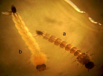
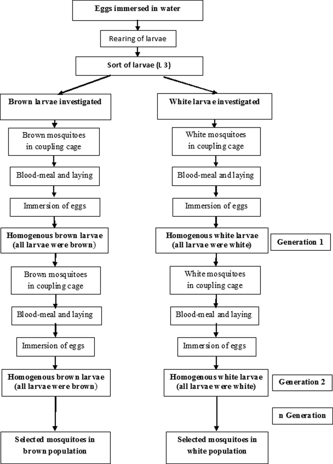
Aedes aegypti natural populations (F0 generation) were used to select brown (BL) and white larvae (WL) populations. Randomly selected eggs were taken and observed. The incubation occurred after immersion of the eggs in water, in white enamelled trays, as previously described (Silva et al., Reference Silva, Silva and Lira1998). After 24 h, trays were examined to check for hatching. Larvae were fed immediately with a finely ground cat food. The same amount of food was put inside each tray. Three days after hatching, larvae were sorted by colour using a plastic pipette and placed into two separate trays (fig. 1). Pupae from each tray were then gathered in a cylindrical plastic cup, which was placed into a corresponding cage where adult mosquitoes will have opportunity to mate after emergence. These adult mosquitoes (F1 generation) were fed ad libitum on a 10% glucose solution. Three days after emergence, females were allowed to feed on guinea pigs over 1 h for two consecutive days. After the blood meal, 100 females from each form were isolated each in a cylindrical plastic cup containing a circular-shaped Whatman paper placed on a wetted hydrophilic cotton pad where females can lay their eggs. After immersion of eggs from each female, trays were examined 3 days after hatching. Only females with a homogeneous descendant (i.e., a descendant composed only of WL or only BL) were used to continue the selection process. These homogeneous larvae in each population were reared until adult stage (F2 generation) and grouped in two cages according to their colour. Adult mosquitoes of F2 generation were subjected to the same process as those of F1 until their next generation. The selection process was carried on until a homogeneous descendant was obtained on two consecutive generations among both populations of Ae. aegypti (fig. 2).
Aedes aegypti brown and white larvae; (a) brown larva; (b) white larva.

Selection process of Aedes aegypti brown and white populations.

Gonotrophic cycle and fecundity
Selected mosquitoes were fed on guinea pigs over 1 h for two consecutive days. After blood feeding, 100 females of each population were isolated for oviposition. Females were followed up individually and regularly as described by Hervé et al. (Reference Hervé, Sà Filho, Travassos Da Rosa and Dégallier1985) with modifications. The gonotrophic cycle of Ae. aegypti was defined as the duration time between a blood meal and a complete egg laying. Fifty females from a natural population were used as control.
Three days after the last oviposition, eggs of each female were counted under a binocular microscope. These data were used to estimate the average number of eggs laid by each female in each population.
Fertility
The fertility was evaluated for 50 WF (‘white’ females: females from WL) and 50 BF (‘brown’ females: females from BL) of Ae. aegypti. Eggs laid by each female were immersed simultaneously in water in separate basins. Larvae were counted in order to determine the hatching rate (HR). Pupae were sorted and counted every day. They were then placed in plastic tubes containing distilled water covered with mosquito net. The number of pupae was used to determine the pupation rate (PR) and preimaginal yield (PIY) of each female's progeny. After emergence, adult mosquitoes (imagos) are removed from the plastic cups with an aspirator and isolated in haemolysis tubes. The number of imagos was used to evaluate the emergence rate (ER) and the overall yield (OY) in each population of Ae. aegypti. The imagos were separated by sex under a binocular microscope, following Rodhain & Perez (Reference Rodhain and Perez1985) criteria. They were then counted to evaluate the sex ratio in both populations of Ae. aegypti.
Life span
Three days after emergence of selected mosquitoes, 100 engorged females of each population were individually isolated in a cylindrical plastic cup containing a circular-shaped Whatman paper placed on a wetted hydrophilic cotton pad for oviposition. They were fed ad libitum on a 10% glucose solution. After oviposition, females were removed and gathered in two cages according to their larval colour origin. Females were again fed on guinea pigs. Twenty four hours after this blood feeding, they were again isolated for oviposition. This procedure was repeated until all females died. The number of dead mosquitoes was recorded daily during the experiment.
Data analysis
Data were double entered using Microsoft Office Excel 2007 spreadsheet, cross-checked and transferred into STATA version 8.04 (STATA Corporation, College Station, United Kingdom). T test was used to compare the means (i.e., number of eggs per female, life span). The Pearson χ2 was used to compare proportions (i.e., hatching, pupation and ERs, PIY and OY). The gonotrophic cycle and their 95% confidence intervals (CI95) were estimated with Win DL 2.0 software. All differences were considered significant either at P < 0.05 or when there were no-overlaps in confidence intervals.
Results
Population's selection, fecundity and gonotrophic cycle
Selected Ae. aegypti mosquitoes from WL and BL were obtained after five (F5) and seven (F7) generations, respectively.
Among 100 females of each population used for the experimentation, six WF and 11 BF died after their blood meal. The experimentation was thus based on 94 BF and 89 WF. Our results indicate that the mean number of eggs laid by BF (85.95 eggs (CI95 = 78.87–93.02)) was significantly higher than that laid by WF (64.40 eggs (CI95 = 55.27–73.54)).
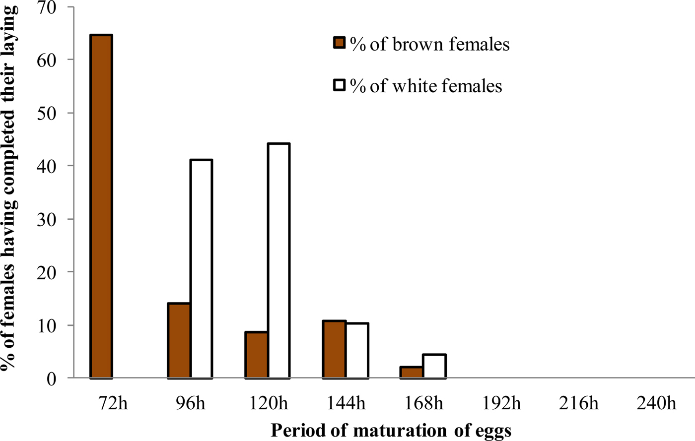
The gonotrophic cycle of both populations of Ae. aegypti established in laboratory was between 72 and 168 hours (fig. 3). One BF and 21 WF did not perform egg laying until the end of the study. Therefore, the gonotrophic cycle was estimated based on 93 BF and 68 WF. Gonotrophic cycle lasted 125.14 h (CI95 = 101.80–217.14) or 5.21 days in 95% of BF, while in 95% of WF, the cycle lasted 136.25 h (CI95 = 120.6–198.49) or 5.68 days. The mean duration of gonotrophic cycle was 74.77 h (CI95 = 54.99–89.90) or 3.11 days for BF, whereas it was 101.79 h (CI95 = 84.79–113.40) or 4.24 days for WF.
Gonotrophic cycle of brown and white females of Aedes aegypti populations.

Fertility
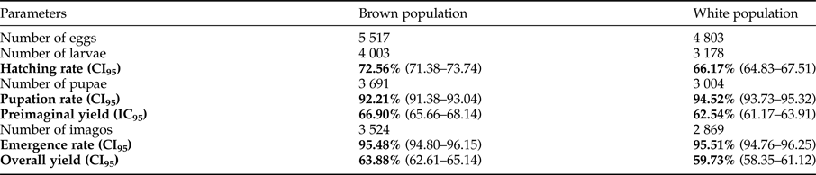
Overall, 5517 and 4803 eggs were laid, respectively, by 50 BF and 50 WF (table 1). The average HR in the BF group (72.56%) was significantly higher compared with the WF group (66.17%) (χ2 = 49.54; P < 0.001). The breeding of larvae provided a mean PR of 92.21% in the brown population and 94.52% in the white population with a significantly higher PR in white population (χ2 = 15.1; P < 0.001). The PIY was 66.90% in the brown population and 62.54% in the white one, and was significantly higher in the brown population compared with the white one (χ2 = 21.40; P < 0.001). However, no difference was observed in the average of ER between brown population (95.48%) and the white one (95.51%) (P > 0.05). The OY of mosquitoes breeding was 63.88% and 59.73%, respectively, in brown and white populations. The OY of mosquitoes breeding in brown population was significantly higher compared with the white one (χ2 = 18.69; P < 0.001).
Laboratory breeding parameters of 50 brown females and 50 white females of Aedes aegypti.

(CI95), confidence interval to 95%.
Sex ratio
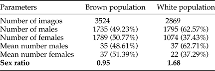
Assessment of the sex ratio showed that the progeny of a female in ‘brown’ population consisted an average of 35 males and 37 females while, in ‘white’ population an average of approximately 37 males and 22 females was observed (tables 2). Therefore, the sex ratio was 0.95 and 1.68, respectively, in the brown and white populations.
Sex ratio in brown and white populations of Aedes aegypti.

Life span
The life span among BF was ranging between 7 and 46 days with an average of 20.01 days, while among WF, life span was set between 7 and 31 days with an average of 17.58 days (fig. 4). The life span in both populations were statistically different (t = −2.332; P = 0.021).
Life span of brown and white females of Aedes aegypti populations.

The ratio between the average of life span and the average of gonotrophic cycle duration in each population was six and 4 blood feeding, respectively, among females from brown and white populations of Ae. aegypti.
Discussion
Aedes aegypti, the main vector of dengue and yellow fever, is a polytypical mosquito species (Trips & Hausermann, Reference Trips and Hausermann1978) and the various habitats it colonizes are at the origin of the individualization of ecotypes or geographical breeds. Thus, it presents a great morphological, physiological and eco-ethological variability (Mouchet et al., Reference Mouchet, Barthe and Sannier1972; Leahy et al., Reference Leahy, Vandehey and Booth1978), which explains its great genetic heterogeneity (Powell, Reference Powell1985). Several studies reported significant variations in its ability to transmit yellow fever or dengue virus depending on the geographical origin of the mosquito strain (Beaty et al., Reference Beaty, Tesh and Aitken1980; Powell, Reference Powell1985). Aedes aegypti genetic diversity is also an indispensable component of research about its vector competence. In prelude to genetic studies, we first looked for differences in biological parameters of two forms of Ae. aegypti larvae.
The WHO layer-traps technique was used to collect in the field the two different colours of Ae. aegypti larvae (brown and white). Different colours of Ae. aegypti larvae have already been reported in previous study elsewhere (Hopkins, Reference Hopkins1952; Christophers, Reference Christophers1960). This variability among populations could be due to a genetic divergence. Indeed, genetic studies on Ae. aegypti showed a probable existence of two twin species in Bouaké, central Côte d'Ivoire (Ravel et al., Reference Ravel, Hervé, Diarrassouba, Koné and Cuny2002). At the opposite, Hopkins in 1952 suggests that this variability is related to environmental and nutritional factors in the breeding sites. Thus, the white population was associated with breeding water containing few organic materials. However, in our study, BL and WL were reared in the same water and fed with the same amount of food. These conditions have not changed the colour of the larvae. In addition, in the current study, the same laboratory conditions were used for the selection of both Ae. aegypti populations.
It is usually accepted that most of the female that will complete a blood meal will lay eggs. However, in our study, one and 21 females did not lay eggs in brown and white populations, respectively. This may be due to the containment of the females in single containers, which may not be favourable to a normal behaviour (Goindin et al., Reference Goindin, Delannay, Ramdini, Gustave and Fouque2015). According to Morrison et al. (Reference Morrison, Costero, Edman, Clark and Scott1999), females of Ae. aegypti only fed with human blood laid more eggs than those fed with both human blood and glucose solution. Our work showed that BF laid more eggs than WF, although they were all fed guinea pig blood and glucose solution. BF could be less demanding on oviposition sites and food in terms of fecundity, whereas WF could have a preference for oviposition sites and meal only made with blood.
Gonotrophic cycle was 3 and 4 days, respectively, in brown and white populations. These results show that the duration of the gonotrophic cycle is variable in populations of the same species. These data are of great importance because they demonstrate that the risk of transmission could be variable within individuals of different populations of the same species. Similar results were observed in Martinique by Etienne (Reference Etienne2001). A study carried out in Bangkok showed that during hot season the gonotrophic cycle of Ae. aegypti females was 3 days, whereas there was a delay of 1 up to 2 days during the cool dry season (Pant & Yasuno, Reference Pant and Yasuno1973). The delay in taking a blood meal observed in females from WL could reduce the frequency of human–mosquito contact and consequently vector capacity of these mosquitoes. BF lives about 3 weeks, whereas the white ones live slightly more than 2 weeks. This important parameter indicates that both types of Ae. aegypti females are able to transmit yellow fever or dengue viruses because the extrinsic incubation time of these viruses in the mosquito vary from 10 to 14 days (Guillaumot, Reference Guillaumot2013).
Sex determination in the mosquito Ae. aegypti is governed by a dominant male-determining factor (M factor) located within the Y chromosome-like region called the M locus (Gilchrist & Haldane, Reference Gilchrist and Haldane1947; Newton et al., Reference Newton, Wood and Southern1978). Assessment of sex ratio revealed the same number of males and females in the brown population, while in the white one, females are only a third of the progeny. Similar results were reported in the United States where about five males for three females were reported in wild populations of Ae. aegypti (William & George, Reference William and George1966). Hall et al. (Reference Hall, Basu, Jiang, Qi, Timoshevskiy, Biedler, Sharakhova, Elahi, Anderson, Chen, Sharakhov, Adelman and Tu2015) also showed that the ectopic expression of an M locus (Nix gene) in Ae. aegypti female embryos caused the initial development of male genitals and testes in more than 69% (16/23) of genetic females. The M locus seems to be better expressed in the white population of Ae. aegypti, which could represent an interesting target as a new mosquito control strategy that converts female mosquitoes into harmless males.
Taken together, our results support the presence of subspecies among the Ae. aegypti populations that have been reported by several studies (Failloux et al., Reference Failloux, Vazeille and Rodhain2002). Further investigation is necessary to deepen our understanding including ecological, ethological, physiological and genetic aspects of both potential forms of Ae. aegypti. In addition, an evaluation of their vector competence for yellow fever and dengue viruses should be emphasized.
Conclusion
The present study revealed that Ae. aegypti populations from both coloured larvae differ in their fecundity, fertility, gonotrophic cycle, life span and sex ratio. In view of all biological differences between these two populations of Ae. aegypti, further investigations are needed in order to define their vector competence for dengue and yellow fever viruses in Côte d'Ivoire. In addition, molecular investigations are warranted to deepen our understanding of the genetic aspects of both populations.
Acknowledgements
The authors are thankful to the West Africa Economic and Monetary Union (WAEMU) for funding support. The authors specially thank the National Institute of Public Health, Abidjan, Côte d'Ivoire for the material and technical support. The authors are also thankful to Diallo Adama and Ziogba Jean-Claude for helping in mosquito collection.