Introduction
Doubly committed subarterial ventricular septal defect is relatively rare, accounting for approximately 5% of all ventricular septal defect cases in Western populations. Reference Lopez, Houyel and Colan1–Reference Schmidt, Cassidy, Silverman and Stanger2 However, doubly committed subarterial ventricular septal defect appears to be more common among Asian populations, with reports indicating that its proportion can be as high as 25% among patients undergoing ventricular septal defect repair. Reference Cao, Chen, Zhang, Chen, Qiu and Xu3–Reference Sim, Grignani and Wong4
Doubly committed subarterial ventricular septal defect locates adjacent to the aortic valve, thus aortic valve prolapse and aortic regurgitation are relatively common and tend to occur earlier in the disease course. Reference Lopez, Houyel and Colan1,Reference McCarthy, Ho and Anderson5 Due to its unique anatomical location, surgical repair remains the standard treatment for closing doubly committed subarterial ventricular septal defect, and current recommendations advocate for early closure to prevent aortic valve prolapse and aortic regurgitation. Reference Turner, Bouhout, Petit and Kalfa6–Reference Chiu, Wang and Lin8 To date, evidence on the aortic valve outcomes following doubly committed subarterial ventricular septal defect surgical repair largely consists of small-sample studies with relatively short follow-up periods, and the results are also inconsistent. Reference Devlin, Russell and Mongé7,Reference Chen, Chen, Chen, Liao, Chen and Chen9–Reference Shamsuddin, Chen, Wong, Le, Anderson and Corno10 Besides, for patients who received concurrent aortic valve surgeries during doubly committed subarterial ventricular septal defect surgical repair, aortic valve outcomes during follow-up are also poorly understood. Reference Chen, Chen, Chen, Liao, Chen and Chen9,Reference Trusler, Williams, Smallhorn and Freedom11–Reference Brauner, Birk, Sahar, Blieden and Vidne12 Furthermore, there is still an insufficient exploration of the risk factors associated with concurrent aortic valve surgery and the development of postoperative new-onset aortic regurgitation.
This study retrospectively analysed our patients’ data over the past decade, and based on a larger sample size, targeting to provide insights on the following two aims: 1. Clarify the postoperative aortic valve outcomes among doubly committed subarterial ventricular septal defect patients after surgical repair; 2. Explore potential risk factors of concurrent aortic valve surgery and postoperative new-onset aortic regurgitation.
Methods
Study population
This was a single-centre, retrospective study. The inclusion criterion was patients diagnosed with doubly committed subarterial ventricular septal defect and underwent surgical repair at our centre between January 2013 and October 2023. The diagnosis of doubly committed subarterial ventricular septal defect was initially made via transthoracic echocardiography, and it was further confirmed by intraoperative inspections.
Echocardiography
Transthoracic echocardiography was used for the preoperative diagnosis of doubly committed subarterial ventricular septal defect. The examinations were performed by 5 experienced cardiologists who had received specialised training in congenital heart disease. For uncooperative children, oral chloral hydrate at 3–5 mg/kg was used for sedation. Doubly committed subarterial ventricular septal defect was defined as being located at the 12:00 to 1:30 positions in the short-axis parasternal view and adjacent to the aortic valve on transthoracic echocardiography. When measuring the size of the defect, special attention was paid to avoid considering the prolapsed aortic valve as the upper margin of the defect, in order to ensure that the measured results reflected the true size of the defect as much as possible. Aortic regurgitation was assessed via two-dimensional and colour Doppler echocardiography on the parasternal long-axis view, and it was graded into none, mild, mild to moderate, moderate, and severe, with the relevant standards consistent with previous guidelines and studies. Reference Brauner, Birk, Sahar, Blieden and Vidne12–Reference Danial, Neily and Pontailler14
To provide more information on the exploration of risk factors of concurrent aortic valve surgery and postoperative new-onset aortic regurgitation, we reviewed the preoperative echocardiographic images of the included patients. Incorporated with the findings from previous studies, Reference Tomita, Arakaki, Ono, Yamada, Yagihara and Echigo15–Reference Leung, Chau, Chiu, Yung and Mok16 two assessments were established for evaluating the aortic valve prolapse: a. Systolic Prolapse: Defined as deformation of the aortic valve leaflets during systole, with partial obstruction of the defect (Figure E1). b. Diastolic Prolapse: Defined as deformation of the aortic valve leaflets and/or their sinuses during diastole, with partial obstruction of the defect or protrusion into the right ventricular outflow tract (Figure E1).
After general anaesthesia and before surgery, transesophageal echocardiography was performed to confirm the findings of TTE. Besides, immediate postoperative transesophageal echocardiography was also performed to evaluate the immediate residual shunt and valve regurgitation severity. Transesophageal echocardiography was performed by trained anaesthesiologists.
Surgical techniques
All patients were approached via median sternotomy, and cardiopulmonary bypass was established through ascending aortic and vena cava cannulation. Repair was typically performed via a pulmonary artery incision, with sutures placed through the base of the pulmonary valve. When the patient had any of the following conditions, aortotomy would be performed after ventricular septal defect repair to allow direct visualisation of the aortic valve, in order to assess whether aortic valve surgery was needed: 1. Preoperative echocardiography suggested moderate or greater aortic regurgitation; 2. Combined with obvious diastolic aortic valve prolapse, with herniation of the aortic valve leaflets and/or their sinuses into the right ventricular outflow tract (Figure E1); 3. Combined with ruptured sinus of Valsalva aneurysm. When the assessments revealed that the aortic valve had the following conditions, aortic valve surgery was performed: 1. The valve exhibited fibrosis, thickening, or calcification, with or without poor leaflet coaptation and aortic regurgitation; 2. The valve exhibited redundancy, torsion, or prolapse, with poor leaflet coaptation, with or without aortic regurgitation; 3. Significant annular dilation, with aortic regurgitation. The aortic valve repair techniques we employed included leaflet shaving, leaflet plication, leaflet extension, and single leaflet reconstruction. Details on aortic valve repair techniques have been well discussed by others, Reference Trusler, Williams, Smallhorn and Freedom11,Reference Ozaki, Kawase and Yamashita17–Reference Baird, Cooney, Chávez, Sleeper, Marx and Del Nido18 and a brief description is provided in the supplementary material.
Postoperative follow-up
Our centre routinely required all patients to undergo echocardiography and electrocardiography follow-up at 1, 3, 12, and 24 months postoperatively. For patients with satisfactory 24-month follow-up results, subsequent follow-up was recommended every 2–3 years.
Study outcomes
The primary outcome of this study was the incidence of new-onset aortic regurgitation during follow-up. New-onset aortic regurgitation was defined as the development of aortic regurgitation ≥ mild during follow-up compared to the immediate postoperative results. Besides, considering that new-onset aortic regurgitation > mild indicates a higher risk of reintervention, so we reported it separately. The secondary outcomes included: a. The incidence of postoperative adverse events, including residual shunts, immediate new aortic regurgitation, new-onset pulmonary regurgitation, and new-onset left ventricular outflow tract or right ventricular outflow tract obstruction; b. Risk factors for concurrent aortic valve surgery and postoperative new-onset aortic regurgitation.
Statistical analysis
For continuous variables, skewness was assessed using the Shapiro–Wilk test. Continuous variables were presented as mean ± standard deviation (SD) or median (interquartile range, IQR). Student’s t-test or the Wilcoxon rank-sum test was used for comparisons of continuous variables. Categorical variables were presented as frequencies (percentages) and were compared using the Chi-Square test or Fisher’s exact test. Kaplan–Meier analysis was utilised to evaluate the incidence of new-onset aortic regurgitation, and the log-rank test was applied to assess significance. Univariate logistic regression and Cox regression were performed to identify potential risk factors of concurrent aortic valve surgery and postoperative new-onset aortic regurgitation, respectively. Subsequently, multivariate logistic regression and Cox regression were conducted to evaluate the risk factors after adjusting for age. Multiple imputation with five iterations was employed to address missing values. The significance level (α) was set at 0.05, and all P-values were two-sided. Statistical analysis was performed using R (version 4.3.1, R Development Core Team). This study was approved by the ethics committee of our centre [2024(129)].
Results
Baseline characteristics
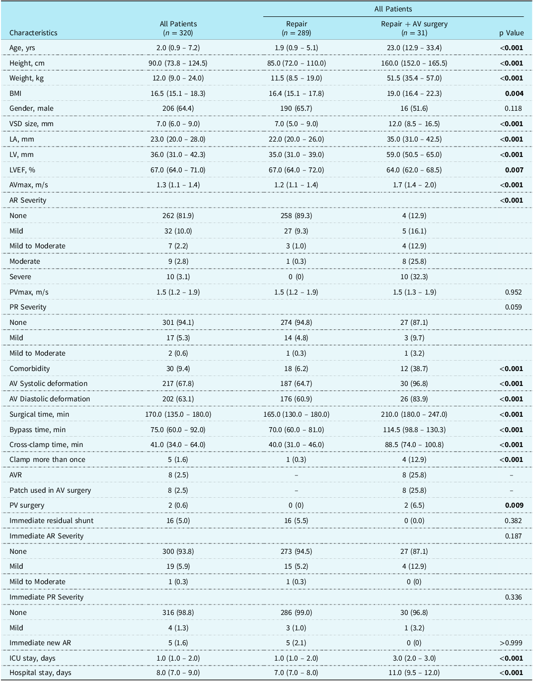
A total of 320 patients were included, with 289 in the repair group and 31 in the repair + aortic valve surgery group. The baseline characteristics of the included patients are presented in Table 1. The median age of the included patients was 2.0 (0.9–7.2) years, and 206 (64.4%) of them were male. Patients in the repair group were significantly younger (P < 0.001). The overall median ventricular septal defect size was 7.0 (6.0–9.0) mm, with patients in the repair group having significantly smaller defects (P < 0.001). A total of 58 (18.1%) patients had preoperative aortic regurgitation ≥ mild, and 26 (8.1%) had preoperative aortic regurgitation > mild, aortic regurgitation was more severe in the repair + aortic valve surgery group (P < 0.001). A total of 30 (9.4%) patients had preoperative comorbidities (Table E1), with patent ductus arteriosus being the most common (9/30, 30.0%), followed by ruptured sinus of Valsalva aneurysm (8/30, 26.7%). The proportion of patients with preoperative comorbidities was higher in the repair + aortic valve surgery group (6.2 vs 38.7%, P < 0.001). In terms of preoperative aortic valve prolapse, 217 (67.8%) patients had systolic prolapse and 202 (63.1%) had diastolic prolapse. Both types of AV prolapse were more common in the repair + AV surgery group (both P < 0.001).
Baseline characteristics and surgical outcomes

BMI = body mass index; VSD = ventricular septal defect; LA = left atrium; LV = left ventricle; LVEF = left ventricle ejection fraction; AVmax = Maximum aortic valve flow velocity; AV = aortic valve; AR = aortic regurgitation; PVmax = Maximum pulmonary valve flow velocity; PR = pulmonary regurgitation; AVR = aortic valve replacement; ICU = intensive care unit.
Surgical outcomes
The surgical outcomes of the included patients are shown in Table 1. The repair group had significantly shorter operation time, cardiopulmonary bypass time, and aortic cross-clamp time (all P < 0.001). A total of 5 (1.6%) patients required a second pump run, including 1 patient in the repair group (due to a large residual shunt) and 4 patients in the repair + aortic valve surgery group (all due to suboptimal initial repair outcomes) (P < 0.001). In the repair + aortic valve surgery group, 23 (74.2%) patients underwent aortic valve repair (Table E2 and Table E3), while the remaining 8 (25.8%) patients received mechanical aortic valve replacement (More surgical and postoperative information for these 8 patients are presented in Table E4). The reasons for aortic valve replacement were: severe aortic regurgitation with poor valvular conditions (n = 5) and poor initial repair outcomes (n = 3). There were 4 patients without preoperative aortic regurgitation who received aortic valve repair, all of them had ruptured sinus of Valsalva aneurysm and diastolic aortic valve prolapse, and after closing the defect, the leaflet distortion became more obvious, thus leaflet plication was performed.
Immediate postoperative aortic regurgitation improved compared to the preoperative status (Figure 1), with only 20 (6.2%) patients having aortic regurgitation ≥ mild, and the severity of aortic regurgitation was similar between the two groups (P = 0.187). A total of 5 (1.6%) patients had immediate new aortic regurgitation (Figure 1a), all of whom were in the repair group (P > 0.999), and aortic regurgitation was mild in all cases.
( a ) Changes of aortic regurgitation (AR) severity for the repair alone group. ( b ) Changes of AR severity for the repair + AV surgery group. Severity of AR before surgery (first row), immediately after surgery (second row), and at the last follow-up (third row) are exhibited. The number in blue indicates immediate new AR, while the numbers in red indicate new-onset AR.

Follow-up outcomes
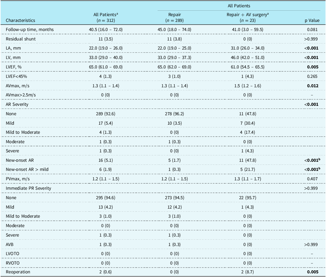
In the following analysis, 8 patients who received aortic valve replacement were excluded, and the follow-up outcomes are presented in Table 2. The overall median follow-up duration was 40.5 (16.0–72.0) months (P = 0.081).
Follow-up outcomes

LA = left atrium; LV = left ventricle; LVEF = left ventricle ejection fraction; AVmax = Maximum aortic valve flow velocity; AV = aortic valve; AR = aortic regurgitation; PVmax = Maximum pulmonary valve flow velocity; PR = pulmonary regurgitation; AVB = atrioventricular block; LVOTO = left ventricular outflow tract obstruction; RVOTO = right ventricular outflow tract obstruction; A = Patients with aortic valve replacement (n = 8) were excluded; b = The p values were from log-rank tests.
At the last follow-up, 23 (7.4%) patients had aortic regurgitation ≥ mild (3.8 vs 52.2%, P < 0.001), and 6 (1.9%) had aortic regurgitation > mild (0.3 vs 21.7%, P < 0.001). Five patients with immediate new aortic regurgitation naturally resolved during the follow-up period. 16 (5.1%) patients developed new-onset aortic regurgitation during follow-up (1.7 vs 47.8%, log-rank P < 0.001, Figure 2a), and 6 (1.9%) of them developed new-onset aortic regurgitation > mild (0.3 vs 21.7%, log-rank P < 0.001, Figure 2b). For the 10 patients who developed only mild new-onset aortic regurgitation, the median follow-up duration after the aortic regurgitation occurrence was 19.9 months (range: 0–93 months), and no progression in aortic regurgitation severity was observed. Besides, for the 6 patients who developed new-onset aortic regurgitation > mild, the aortic regurgitation in 4 patients was confirmed at the most recent follow-up, with total follow-up durations of 3 months, 3 months, 6 months, and 125 months respectively; in the other 2 patients, new-onset aortic regurgitation was detected at the 12th month (mild to moderate) and 36th month (severe) during the follow-up period, after which they were further followed for 30 months and 23 months, with no progression in aortic regurgitation for the former, and the latter received reoperation. For the 5 patients in the repair + aortic valve surgery group who developed new-onset aortic regurgitation > mild, the initial aortic valve repair techniques were: leaflet plication, leaflet shaving, leaflet shaving + leaflet extension, leaflet shaving + leaflet plication, and single leaflet reconstruction. Besides, among the 262 patients without preoperative aortic regurgitation, 3 (1.1%) developed new-onset aortic regurgitation during follow-up, all of which were mild.
Kaplan-Meier analysis and the log-rank test results of free from new-onset aortic regurgitation (AR) during follow-up. ( a) Free from new-onset AR among the included patients. ( b) Free from new-onset AR > mild among the included patients.

In the repair group, 1 (0.3%) patient developed severe pulmonary regurgitation at 72 months postoperatively. This patient had no pulmonary regurgitation preoperatively, and he did not receive any intraoperative pulmonary valve intervention. A total of 2 (0.6%) patients received reoperation, both of whom were in the repair + aortic valve surgery group (P = 0.005). The reasons for reoperation were residual shunt after ruptured sinus of Valsalva aneurysm repair (n = 1, underwent device closure of the residual shunt) and new-onset severe aortic regurgitation (n = 1, underwent redo aortic valve repair). No patient had new-onset left ventricular outflow tract or right ventricular outflow tract obstruction.
Risk factors for aortic valve surgery and new-onset AR during follow-up
The results of risk factors of concurrent aortic valve surgery are presented in Table E5. In the univariate logistic regression, age, ventricular septal defect size, preoperative aortic regurgitation > mild, systolic aortic valve prolapse, preoperative AVmax, and preoperative comorbidities were significant. After adjusting for age, the significance remained unchanged.
The results of risk factors of new-onset aortic regurgitation during follow-up are presented in Table E6. In the univariate Cox regression, age, ventricular septal defect size, preoperative aortic regurgitation > mild, preoperative AVmax, and concurrent aortic valve repair were significant. After adjusting for age, the significance also remained unchanged.
Discussion
Due to the unique anatomical location of doubly committed subarterial ventricular septal defect, its natural history and treatment strategies differ considerably from those of other types of VSD. Surgical repair remains the standard treatment for dscVSD, and concurrent aortic valve surgery is sometimes needed for those with significant aortic valve deformation or aortic regurgitation. Currently, the most used incision in doubly committed subarterial ventricular septal defect surgical repair is the pulmonary artery incision, with sutures placed through the base of the pulmonary valve. There have been concerns that this technique might affect the pulmonary valve’s function; however, current findings partly alleviate these concerns. Reference Devlin, Russell and Mongé7,Reference Miwa, Iwai, Kanaya and Kawai19 Because doubly committed subarterial ventricular septal defect is relatively uncommon, there is a lack of large-scale, high-quality clinical studies summarising the aortic valve outcomes after doubly committed subarterial ventricular septal defect surgical repair. Reference Devlin, Russell and Mongé7,Reference Chen, Chen, Chen, Liao, Chen and Chen9–Reference Shamsuddin, Chen, Wong, Le, Anderson and Corno10,Reference Brauner, Birk, Sahar, Blieden and Vidne12 Drawing on a relatively larger sample size, this study reports the mid-term aortic valve outcomes among doubly committed subarterial ventricular septal defect patients, and risk factors for concurrent aortic valve surgery and new-onset aortic regurgitation were also explored.
Preoperative aortic valve prolapse is common among doubly committed subarterial ventricular septal defect patients, with a reported prevalence of 63.1– 100.0%, whereas preoperative aortic regurgitation ≥ mild is less frequent, occurring in about 4.9– 41.0% of the patients. Reference Devlin, Russell and Mongé7,Reference Shamsuddin, Chen, Wong, Le, Anderson and Corno10,Reference Jung, Cho and Lee20 The high rate of preoperative aortic valve prolapse and relatively low rate of preoperative aortic regurgitation among patients in this study aligned with the previous findings, indicating that aortic regurgitation tends to occur in the prolapsed valves. Besides, we further assessed preoperative aortic valve prolapse in both systolic and diastolic phases, and we found that the incidence of both types of aortic valve prolapse was similar.
About 0.0 – 40.2% of patients required concurrent aortic valve surgery during doubly committed subarterial ventricular septal defect repair, with aortic regurgitation and ruptured sinus Valsalva aneurysm being the main reasons. Reference Devlin, Russell and Mongé7,Reference Chen, Chen, Chen, Liao, Chen and Chen9,Reference Brauner, Birk, Sahar, Blieden and Vidne12,Reference Jung, Cho and Lee20,Reference Krishnasamy, Sivalingam, Dillon, Mokhtar, Yakub and Singh21 Valve repair is the primary choice when aortic valve surgery is needed, and only about 0.0 – 18.0% of doubly committed subarterial ventricular septal defect patients received aortic valve replacement. Reference Chen, Chen, Chen, Liao, Chen and Chen9,Reference Brauner, Birk, Sahar, Blieden and Vidne12,Reference Jung, Cho and Lee20,Reference Krishnasamy, Sivalingam, Dillon, Mokhtar, Yakub and Singh21 Because leaflet elongation and prolapse are the main aortic valve pathologies in doubly committed subarterial ventricular septal defect patients, leaflet plication is the most commonly used repair technique. Reference Devlin, Russell and Mongé7,Reference Chen, Chen, Chen, Liao, Chen and Chen9,Reference Trusler, Williams, Smallhorn and Freedom11,Reference Brauner, Birk, Sahar, Blieden and Vidne12,Reference Krishnasamy, Sivalingam, Dillon, Mokhtar, Yakub and Singh21 Unlike previous studies, however, leaflet extension (n = 2) and single-leaflet reconstruction (n = 6) were also adopted among our patients. We chose the two techniques for two reasons: a. In some patients with long disease courses, the aortic valve leaflets might exhibit significant contraction, thickening, and calcification, which could not be repaired via leaflet plication; b. In cases where attempted repair produced suboptimal results, single leaflet reconstruction often served as our last resort. Currently, studies on single leaflet reconstruction are mostly single-centre small-sample studies, Reference Marathe, Chávez and Sleeper22–Reference Sivalingam, Haranal and Pathan24 including one specifically targeting doubly committed subarterial ventricular septal defect patients. Reference Sivalingam, Haranal and Pathan24 In these studies, the short-term outcomes are generally satisfactory, but the mid- to long-term outcomes are still needed.
Based on the limited existing reports, concurrent aortic valve surgery during doubly committed subarterial ventricular septal defect surgical repair appears to be related to preoperative aortic regurgitation, patients’ age, and comorbidities (such as ruptured sinus of Valsalva aneurysm). Reference Chen, Chen, Chen, Liao, Chen and Chen9,Reference Brauner, Birk, Sahar, Blieden and Vidne12,Reference Krishnasamy, Sivalingam, Dillon, Mokhtar, Yakub and Singh21 In our analysis, apart from the aforementioned factors, ventricular septal defect size, preoperative systolic aortic valve prolapse, and AVmax were also found to be linked to concurrent aortic valve surgery.
Regarding the aortic valve outcomes during follow-up, existing reports indicates that the incidence of new-onset aortic regurgitation after doubly committed subarterial ventricular septal defect surgical repair is about 0.0 – 13.5%, whereas the incidence of new-onset aortic regurgitation > mild is only around 0.0 – 1.0%. Reference Chen, Chen, Chen, Liao, Chen and Chen9–Reference Shamsuddin, Chen, Wong, Le, Anderson and Corno10,Reference Jung, Cho and Lee20 In our study, 5.1% of patients developed new-onset aortic regurgitation during follow-up, mostly in the early postoperative period and were mild in severity, additionally, the mild new-onset aortic regurgitation appeared stable during the subsequent follow-up, but further observation is still needed. Specifically, among doubly committed subarterial ventricular septal defect patients without preoperative aortic regurgitation, the incidence of new-onset aortic regurgitation was 0.0 and 1.3%, Reference Shamsuddin, Chen, Wong, Le, Anderson and Corno10,Reference Jung, Cho and Lee20 similar to our findings. These results indicate that patients without preoperative aortic regurgitation need not be concerned about the new-onset aortic regurgitation during mid- to long-term follow-up. Nineteen patients in this study who had preoperative mild aortic regurgitation and 1 patient who had mild-to-moderate aortic regurgitation experienced a spontaneous and persistent resolution without aortic valve intervention, which is also consistent with the previous reports. Reference Devlin, Russell and Mongé7,Reference Chen, Chen, Chen, Liao, Chen and Chen9,Reference Brauner, Birk, Sahar, Blieden and Vidne12,Reference Jung, Cho and Lee20,Reference Krishnasamy, Sivalingam, Dillon, Mokhtar, Yakub and Singh21 This indicates that, with the elimination of the shunt and the improvement of the subvalvular haemodynamics, aortic regurgitation can naturally resolve. Among the 5 patients with immediate new aortic regurgitation, natural resolution was also observed, suggesting that mild immediate new aortic regurgitation is relatively uncommon and appears acceptable.
Data on new-onset aortic regurgitation after doubly committed subarterial ventricular septal defect repair + aortic valve surgery is very limited. One study on ventricular septal defect patients (46% of whom had doubly committed subarterial ventricular septal defect) noted that 22.2% of those who had concurrent aortic valve repair developed new-set aortic regurgitation ≥ moderate during follow-up. Reference Krishnasamy, Sivalingam, Dillon, Mokhtar, Yakub and Singh21 In our study, 47.8% of patients who received aortic valve repair developed new-onset aortic regurgitation, but only 1 (4.3%) progressed to ≥ moderate aortic regurgitation. However, whether the mild new-onset aortic regurgitation will worsen in the remaining patients requires ongoing surveillance.
According to the existing studies, older age at operation, ventricular septal defect size, preoperative aortic regurgitation, and concurrent aortic valve surgery appear to be risk factors for new-onset aortic regurgitation. Reference Devlin, Russell and Mongé7,Reference Chen, Chen, Chen, Liao, Chen and Chen9–Reference Shamsuddin, Chen, Wong, Le, Anderson and Corno10,Reference Jung, Cho and Lee20 Additionally, our study found that a higher preoperative AVmax was also a risk factor. We speculate that a higher preoperative AVmax might indicate more pronounced leaflet thickening and contraction, thereby adversely affecting the valve’s durability after repair. Moreover, prior studies have also suggested that the use of pericardial patch during aortic valve repair might undermine the durability of the repair results. Reference Aicher, Kunihara, Abou Issa, Brittner, Gräber and Schäfers25–Reference Ram, Bouhout, Karliova, Schneider, El-Hamamsy and Schäfers26 In our study, there were 8 patients who received aortic valve repair with a pericardial patch, and 3 developed new-onset aortic regurgitation (1 mild aortic regurgitation and 2 mild-to-moderate aortic regurgitation).
This study has several limitations: a. Although the sample size here is relatively large, it is still a single-centre, retrospective study and only mid-term follow-up outcomes are provided. b. Due to the limited sample size, we did not undertake more extensive multivariate analysis to explore risk factors for concurrent aortic valve surgery and new-onset aortic regurgitation. c. For doubly committed subarterial ventricular septal defect patients, there are two strategies for treatment in our centre: device closure and surgical repair. Reference Gan, Peng and Liang27–Reference Wang, Xie, Ruan, Li, Gan and Lin28 Patients with relatively mild disease severity are more likely to undergo device closure; therefore, the results presented in this study more reflect the postoperative outcomes of doubly committed subarterial ventricular septal defect patients with more severe conditions (larger defect size and more significant aortic valve deformity).
Based on the findings of this study, we draw the following conclusions: a. According to our single-centre, retrospective data, the mid-term aortic valve outcomes after doubly committed subarterial ventricular septal defect surgical repair are relatively satisfactory, with a low incidence of new-onset aortic regurgitation during follow-up. b. For patients who require concurrent aortic valve surgery during doubly committed subarterial ventricular septal defect surgical repair, the aortic valve outcomes are less satisfactory. c. Age, ventricular septal defect size, preoperative aortic regurgitation severity, and AVmax appear to be common risk factors for both concurrent aortic valve surgery and new-onset aortic regurgitation.
Supplementary material
The supplementary material for this article can be found at https://doi.org/10.1017/S1047951125109608.
Acknowledgements
The authors declare that they have no known competing financial interests or personal relationships that could have appeared to influence the work reported in this paper.
Financial support
None.
Competing interests
None.