Percutaneous closure with self-centering nitinol devices is the standard of care in oval fossa defects with acceptable margins. Reference Du, Hijazi, Kleinman, Silverman and Larntz1 The self-expanding nature and shape memory of nitinol revolutionised this field of interventional cardiology. Nevertheless, deformations seen in some devices during deployment pose technical challenges. While abnormalities are variably described as cobra-head, tulip, conal, holy-grail, or hour-glass deformities, the former is the most commonly characterised deformation. Reference Mazic, Gavora and Masura2,Reference Yip and Chan3 Incidence of such deformities varies from 0 to 9.5% with different occluder designs. Reference Masura, Gavora and Podnar4–Reference Aaron, Mainzer and Lorber9 While some deformations are subtle and transient, rectifying spontaneously or with minimal manipulation, a few are extreme and persistent needing extensive strategies and occasionally force a change of the device. The clinical impact of these shape-memory abnormalities has not been systematically analysed in any large study so far.
The two peculiar properties of Nitinol (Nickel Titanium Naval Ordnance Laboratory) that enables its medical applications are shape memory that recovers its shape on heating and superelasticity that returns it to its original shape after unsheathing. Reference Buehler and Cross10 It exhibits two forms at different temperatures. As the temperature increases, its herring-bone shape of the Martensite form changes to the cubic shape of the Austenite form. Reference Otsuka and Ren11 An increased proportion of Nickel reduces this transition temperature. Reference Otsuka and Ren11 In addition to temperature, external force is another reason for a martensitic transformation that remains temporary as long as the strain is below 8%. Reference Otsuka and Ren11 As human body temperature is relatively constant at 37oC during the device deployment in most patients, these external forces often lead to the shape-memory abnormalities observed in the nitinol septal occluders. Surface treatment with oxides and nitrides reduces leaching of nickel, makes it less corrosive, and improves its resistance to cyclic fatigue. Reference Shabalovskaya, Anderegg, Rondelli, Vanderlinden and De Feyter12 Nickel ion hinders mitosis of human fibroblasts and is said to be carcinogenic. Reference Cameron, Buchner and Tchounwou13 The alloy form of nitinol overcomes these adverse features of nickel ion. Medical use of nitinol is promoted due to its biocompatibility, unique stress–strain behaviour, high recoverable strain (up to 8%), and high fatigue resistance. Reference Spenciner and Scutti14
Reasons attributed for loss of shape memory of devices include restraint of the left disc by the left atrial structures; use of delivery sheaths larger than the recommended size that causes in-folding of the disc edge or twist of the device while advancing within the sheath; angulation or kink in the delivery system; larger devices as they have a longer length of the wires between the edges compromising the memory; quality checks missing the manufacturing defects; repeated release and retraction within the sheath; and lower the ambient temperature of the saline in the catheterisation laboratory. Reference Cooke, Gelman and Harper15,Reference Hoole, McNab, Rana, Davies and Shapiro16 A simple retrieval of a deformed device sometimes proves challenging when the concave configuration of the left atrial disc in tulip deformity fails to get retracted back into the delivery sheath. Reference Kohli17,Reference Hayes and Rosenthal18
A study of the shape-memory abnormalities among occluders used for oval fossa defects will throw light on the incidence, characterisation of the different deformities, identification of predictors, correlation with the design and size of the device, measures taken to overcome the abnormal memory, and clinical outcomes after the occurrence of deformity.
Methods
This is a retrospective, single-institution study evaluating the deformation of nitinol atrial septal occluders in all consecutive device closures of atrial septal defects. Two independent observers carefully reviewed the procedural fluoroscopic records of all patients and included all cases that showed device deformations. The Institutional Review Board permitted this analysis and anonymized reporting of the results. The Institutional Ethical Committee approved the study.
Patient and device selection
Patients with oval fossa defects were selected for device closure after echocardiography based on their size and margins. Device closure was performed often from femoral veins and extremely rarely from jugular veins. The commonly chosen devices included Amplatzer Septal Occluder (ASO, Abbott Medical, Plymouth, MN), Cera Septal Occluder (CSO, Lifetech Scientific, Shenzhen, PRC), and Figulla Septal Occluder (FSO, Occlutech International, Helsingborg, Sweden). Reference Bhattacharjya, Pillai and Doraiswamy5 The choice of the device was based on the size, availability, and discretion of the operator. In comparison to ASO, CSO was available in additional sizes of 42–46 mm and customised FSO was available in additional sizes of 44–48 mm. Even though the size of the delivery sheath was largely guided by the manufacturer’s recommendations, it was sometimes influenced by the discretion of the operator.
Device deployment
In most patients, a conventional deployment of the distal disc in the left atrium was followed by the release of the proximal right atrial disc, guided by fluoroscopy and echocardiography. Special techniques like the pulmonary vein deployment technique were used if conventional methods failed or difficulty was anticipated due to large defect size and deficient margins. Reference Bhattacharjya, Pillai and Doraiswamy5
Device deformity
If a shape-memory abnormality was observed in fluoroscopy, a continued observation looked for spontaneous resolution with or without minimal manipulations. In refractory memory abnormalities that failed all in vivo manipulations within the cardiac chambers, the device was withdrawn out of the sheath and shaped again in warm saline. Being a retrospective analysis, whenever a shape-memory abnormality was noted, the appearances of the delivery sheath and the slenderized devices observed in the fluoroscopic loops were reviewed repeatedly to identify probable pre-disposing factors during the deployment steps.
Data collection
Procedures associated with shape-memory deformities during device closures were assessed for patient parameters including age, gender, weight, and defect size; technical parameters including type and size of the device, device size indexed for body surface area, the extent of device oversizing as measured by device:defect ratio, sheath size, and extent of sheath oversizing (French size of the sheath compared to the manufacturer recommendation); and procedural parameters including deployment technique, sheath kinking or angulation, and number of attempts needed to complete the procedure. As the nitinol wire thickness increased beyond 26 mm ASO, larger devices (>26 mm) were compared with smaller devices (<24 mm). Additional information included the shape of the deformity, disc involved (right, left, or both), whether the deformity was transient or persistent and methods deployed to rectify the deformity.
Statistical analysis
Data were entered in Microsoft Excel and analysed using SPSS version 23. The categorical data were expressed as frequency and percentage. Comparison between the groups was done using Chi-square or Fisher’s exact test. The continuous variables were expressed in mean and standard deviation or median with range whichever was appropriate. Comparison of the continuous variables was done by independent Student’s t-test or Mann–Whitney U-test based on the distribution. The factors associated with deformation were analysed by univariate analysis initially followed by multivariate analysis for those factors that were found significant in univariate analysis.
Results
A total of 936 consecutive device closures of oval fossa defects performed over a 6-year period commencing from May 2013 were included in this analysis. As 922 patients received 1 device and 14 patients received 2 devices, the total number of devices analysed in the study was 950. A retrospective observation of the fluoroscopic records showed 112 procedures (11.8%) associated with shape-memory abnormalities and they formed the study group. Among the 14 patients who received 2 devices, deformation was observed in 1 device. The demographic, device-related, and procedural parameters of devices that showed deformations were compared to those that did not show deformations (Table 1). Anatomical peculiarities in the 112 cases included 1 patient with a thick vertical band partitioning the defect, 7 patients had multi-fenestrated defects (of whom one needed transjugular closure), and 2 patients with a septal aneurysm.
Comparison between procedures with and without deformations

* Pearson Chi-square test
** Mann–Whitney U-test
*** Logistic regression analysis
Device-related parameters
Device deformation was noted in 13.9% of 374 ASO, 15.6% of 262 FSO, and 6.6% of 286 CSO devices. Twenty-eight devices of other designs, namely Cribriform septal occluder (Abbott, Plymouth, MN), Coccoon septal occluder (Vascular Innovations, Nonthapuri, Thailand), and Memopart septal occluder (Lepu Medical, Beijing, PRC) did not show any deformations, but the numbers were too small. Shape-memory abnormalities were seen in 9.8% of devices smaller than 30 mm compared to 18.9% of larger devices. The mean device size indexed for body surface area for those with deformation was significantly higher than those without deformation. The extent of oversizing of a device for a given defect size analysed as device:defect ratio was not significantly different between the groups.
Delivery sheath details
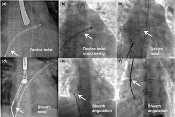
Information about the size of the delivery sheath was available in 928 out of 950 devices. Among the procedures associated with deformations, the delivery system was significantly oversized more than the manufacturer recommendation in 57% of instances and they exceeded by 1 F in 23 patients, 2 F in 31 patients, 3 F in 6, and 4 F in 1 patient. Among those without deformations, the sheath oversizing was noted in 43.1% of instances. Angulation and kinking of the delivery sheath were observed in 28 (25%) and 2 (1.8%) procedures, respectively. A sharp angle between the delivery sheath and the delivery cable was responsible for right atrial deformities in 15 patients. While observing the slenderized device during its advance through the sheath, an eccentric location of the device within the oversized sheath was observed in one and a twist of the device within the long sheath in either the right atrial end (Fig 1a), left atrial end (Fig 1b) or waist (Fig 1c) of the device, kink in the long sheath (Fig 1d), sharp angulation between the tip of the sheath and the delivery cable (Fig 1e, f) was observed in 15 procedures.
Twist of a long device either at the right atrial end (a), left atrial end (b) or the waist (c), a kink in the long sheath (d) or a sharp angulation between the tip of the sheath, and the delivery cable (e, f) also led to the loss of shape memory.

Deployment methods
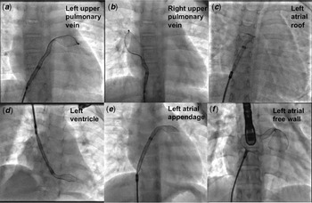
Special deployment techniques especially left pulmonary vein deployment was observed in 64% of procedures associated with deformation compared to 41% of the procedures without deformations. The possibility of trapping the distal disc against a left atrial structure such as left upper pulmonary vein (Fig 2a), right upper pulmonary vein (Fig 2b), left atrial roof (Fig 2c), left ventricle (Fig 2d), left atrial appendage (Fig 2e) or left atrial free wall (Fig 2f) was observed by replaying the fluoroscopic record loops. The distal disc was deployed against the left atrial free wall or the roof in 53, left atrial appendage in 6, and against the left ventricular free wall (transjugular deployment) in 1 patient. Among these different possible predictors, two or more factors were observed together in 35 out of 112 procedures.
Restraint of the left atrial disc by a left heart structure prevented recovery of shape memory of the device. The restraint originated from the left upper pulmonary vein (a), right upper pulmonary vein (b), left atrial roof (c), left ventricle (d), left atrial appendage (e), or left atrial free wall (f). Deformities occurred commonly in Amplatzer septal occluders (a, b) and Figulla septal occluders (c–f). The vascular access was from the femoral vein in all cases unless inferior caval vein interruption warranted jugular venous access (d).

Details of the deformities
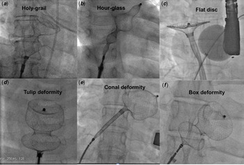
Forty-five deformations (40%) were transient with spontaneous resolution or needed minimal manipulations. Persistent deformities occurred in 67 patients (60%). The deformations involved left disc alone in 54 cases (48.2%), both right and left discs in 43 cases (38.3%), and right disc alone in 15 cases (13.4%). Cobra malformations were the commonest (90.2%) accounting for 101 deformities. Tulip deformity of the left disc in two patients (1.8%), hour-glass deformity in one (0.9%) and unclassifiable deformity in eight patients were the other abnormalities. Figure 3 shows the various shapes such as holy-grail (Fig 3a), hour-glass (Fig 3b), disc (Fig 3c), tulip (Fig 3d), conal deformity (Fig 3e) and box shape (Fig 3f). Since each of the non-cobra shapes was confined to 1–2 patients, we could not identify a specific provocative factor leading to these specific shapes. It was noteworthy that all these very rare shapes such as Tulip, hour-glass, conal, disc, box, and holy-grail deformities were confined to non-ASO devices (Fig 3).
While cobra-head was the most common deformity, other shapes were holy-grail (a), hour glass (b), exaggerated flat disc (c), tulip deformity (d), conal deformity (e), and box deformity (f). Figulla septal occluder was involved in the holy-grail deformity (a) and while Cera septal occluder was involved in the other shapes (b–f).

Measures to recover the original shape
The procedural manipulations that recovered the original memory included repeated re-sheathing and redeployment in 26 patients (23.2%), vigorous wiggle within the atrial cavity in 15 patients (13.5%), adopting a rapid deployment technique in 10 patients (9%), gentle forward push in 9 patients (8.1%), gentle backward traction in 6 patients (5.4%), changing to balloon-assisted technique (when the left upper pulmonary vein deployment resulted in deformations) in 6 patients (5.4%), and spontaneous correction without any manipulation in 14 patients (12.6%), but a complete exteriorisation of the device to allow in vitro reformation followed by re-deployment of the same device was required in 19 patients (17.1%). Four patients (3.6%) needed a complete change of the device. In one patient, the device was deployed completely within the left atrium allowing it to reform within LA followed by re-sheathing of its RA disc and redeployment. Two patients showed mild persistent loss of shape memory after the release of the device without any clinical consequence.
A number of attempts needed for achieving a recovery of the device memory was single in 28; two attempts in 32, three attempts in 26, and four or more attempts in 26 procedures. The composite mean of attempts for a final successful deployment in procedures that showed deformities was 2.76 ± 1.7 (with a range from 1 to 9 attempts) compared to 1.12 ± 0.4 (with a range from 1 to 4 attempts) in patients without any deformations.
Complications
Inadvertent unscrewing of the delivery cable within the delivery sheath in three procedures during the repeated manipulations was successfully managed with snare without any clinical consequence or embolisations. Two patients developed transient supraventricular tachycardia during the device manipulations within the atrium, needing antiarrhythmic amiodarone, and metoprolol. None of the patients showed any major sustained clinically relevant adverse effects.
Impact on procedural duration
The mean procedure and fluoroscopic time in these 112 procedures were significantly higher than those without deformations. The mean cumulative dose area product was also significantly higher in this group. However, the long procedural time in those with deformations was not associated with any increase of complications like thrombus formation or air embolism.
Analysis of pre-disposing factors
While univariate analysis showed a higher incidence of deformities with FSO and ASO compared to CSO (p = 0.006), it was not significant on multivariate analysis. A similar higher incidence with larger devices compared to smaller devices on univariate analysis (p = 0.021) was also not substantiated on multivariate analysis (Table 1). However, many factors related to delivery sheath characteristics continued to retain significance on univariate and multivariate analysis. An oversized large sheath (p = 0.004), kinked/angulated sheath (p =< 0.001), and a twist of the device waist within the sheath (p = 0.011) were strong associations on multivariate analysis. Special deployment techniques that restrained the discs of the occluders within the cardiac structures also showed significance (p < 0.001) both on univariate and multivariate analysis. None of the patient characteristics such as age, weight, defect size, and the extent of oversizing shown by device:defect ratio showed any significance.
Discussion
Incidence of shape-memory abnormality
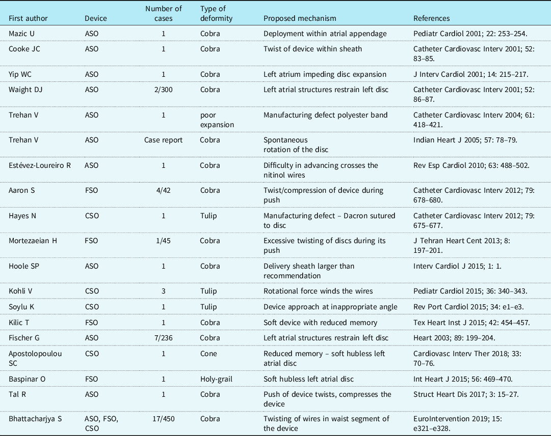
Shape-memory abnormality in nitinol septal occluders used for closure of oval fossa defects was not an unusual observation. Reference Aaron, Mainzer and Lorber9 The first two documentations of “cobra-head deformity” after the initial 200 implants of ASO were attributed to the left atrial disc constrained within the left atrial appendage in the first instance and against the left atrial posterior wall in the second instance. Reference Mazic, Gavora and Masura2,Reference Yip and Chan3 Despite their frequent occurrence, the literature on these deformities were confined to isolated reports and listed in Table 2. Our meticulous retrospective targeted search of all the fluoroscopic loops identified even the subtlest deformations in almost 11.8% of the procedures, which were higher than the previous reports quoting upto 9.5% while using FSO. Reference Aaron, Mainzer and Lorber9 However, 40% of the deformations observed in this focused search were transient getting corrected spontaneously or with minimal manipulations and were unlikely to be reported otherwise.
Overview of previous reports of device deformations

Impact on procedure
The procedural and fluoroscopic times were significantly longer in procedures associated with deformations. The longer procedural time was known to lead to more complications including thrombus formation and air embolism, though no additional complications other than transient supraventricular tachycardia in two instances were observed in our group. Reference Chessa, Carminati and Butera19 Four devices had persistent deformities necessitating a change of device. Multiple manipulations including re-sheathing and redeployments led to the inadvertent unscrewing of the device in three patients but were snared out from the delivery sheath before embolisation. Supraventricular tachycardia secondary to repeated manipulations of the device and delivery system in two patients were also medically controlled with antiarrhythmic drugs. Adopting standard operative procedures prevented thrombus formation, air embolisms, and cardiac injury.
Difference between the devices
Among the different devices, deformations were noted in 13.9, 15.6, and 6.6% with ASO, FSO, and CSO, respectively. Shape-memory abnormalities of atrial septal occluders were previously reported in 2–9.5%, with a relatively higher incidence for FSO compared to other devices. Reference Aaron, Mainzer and Lorber9 Reduced memory of FSO was attributed to the reduced metal on the left atrial hub. Reference Bhattacharjya, Pillai and Doraiswamy5,Reference Aaron, Mainzer and Lorber9 Multivariate analysis in our cohort failed to observe any significance between the different occluders.
Deformations common in large devices
Larger devices have longer nitinol wires that pre-dispose to their twist and compromises the device memory. Reference Cooke, Gelman and Harper15 The device size indexed for body surface area and usage of larger sized devices (>26 mm) had a strong correlation with device deformation only on univariate analysis but failed to show any significance on multivariate analysis. However, the device:defect ratio was not different between those with and without deformity, indicating device oversizing was not a factor.
Role of a large delivery sheath
A large delivery sheath allows excess space within its lumen that allows the twist and kink of the nitinol wireframe pattern. Reference Hoole, McNab, Rana, Davies and Shapiro16 Larger sheaths are selected to facilitate easy deployment and recapture when modified deployment strategies are attempted to facilitate device alignment and increase the success of transcatheter closure. Reference Kammache, Mancini, Ovaert, Habib and Fraisse20 Large sheaths are also commonly used when snares assist to safeguard against early embolisation. Reference Pavithran and Sivakumar21 Sheath oversizing was observed in 57% (oversizing by 1 F in 23, 2 F in 31, 3 F in 6. and 4 F in1) of our patients. The frequency of sheath oversizing was significantly higher in those with deformation (57%) compared to those without deformation (43.1%) on both univariate and multivariate analysis. Sheath oversizing may be an avoidable provocation.
Modified deployment techniques
Modified techniques like pulmonary vein deployment are followed when difficulty is anticipated in aligning the left disc to the atrial septal plane leading to its prolapse through the defect. Reference Kammache, Mancini, Ovaert, Habib and Fraisse20 Pulmonary vein deployment was more frequently used and was commonly associated with deformations in our group. Restraint of the left atrial disc either by the walls of the pulmonary veins, atrial appendage, or the atrial wall distorts the device leading to such malformations. Reference Mazic, Gavora and Masura2,Reference Yip and Chan3 An alteration in the relationship of the tip of the sheath and the screw end of the right atrial disc similarly leads to deformation of the right atrial disc. Reference Kilic, Ural and Sahin22 Isolated right disc deformities were noted in 15 patients when there was a sharp angulation between the delivery sheath and cable. Kinks and angulations in a delivery system, a commonly observed pre-disposing factor was observed in 28 patients who developed deformations. These deployment techniques, kinks/angulations of the sheath, and sharp angulation between the sheath tip and the screw end of the cable, identified as strong associations on multivariate analysis in this study are potentially avoidable factors.
Limitations
This study has inherent bias related to any retrospective study. We could not get details of the temperature of saline used while loading the device or temperature of catheterisation laboratory. Difficulties encountered by an operator during loading and advancing of the device were not traced in this retrospective analysis. The relationship between left atrial size and device size might have played a role, but details of left atrial size were not available from the records. The minimum delivery sheath size recommendations were obtained from the instructions for use, but operators had identified difficulties to advance devices while using small sheaths. Even though a multi-institutional study would validate the observations, this single-centre study had the advantage of uniform standardised procedural protocols.
Conclusion
Deformation of nitinol septal occluders during percutaneous device closure of oval fossa defects was seen in 11.8% of procedures. Persistent deformities in 60% prolonged the procedure, but they did not alter the procedural success. A change of device was very rarely needed, as most deformities could be reformed in vivo or in vitro with manipulations. Oversized and kinked/angulated delivery sheaths and restraint of the discs by left atrial structures during special deployment techniques were potential provoking factors. Awareness about deformations and their provoking factors should prepare interventional cardiologists for manipulations to reform the deformities.
Acknowledgements
None.
Financial support
This research received no specific grant from any funding agency, commercial, or not-for-profit sectors.
Conflicts of interest
None.
Ethical standards
The authors assert that all procedures contributing to this work comply with the ethical standards of the Indian council of medical research and with the Helsinki Declaration of 1975, as revised in 2008, and has been approved by the institutional committee of Madras Medical Mission, Chennai, India.