Introduction
The narrow, steep-walled Camarones Valley in Chile's Arica Province is known locally for its small-scale sulfate outcrops, which have been exploited in the past as small quarries and diggings (Salas, Reference Salas1964, Reference Salas1965). In late 2011, while searching the valley floor for rocks that had tumbled down from outcrops on the steep valley walls, one of the authors (AAMD) encountered a small boulder with unusual sulfate mineralisation ~9 km NE of the village of Cuya. This boulder yielded a variety of rare sulfates, including massive intergrowths of crystals of the new mineral camaronesite, [Fe3+(H2O)2(PO3OH)]2(SO4)⋅1–2H2O, (Kampf et al., Reference Kampf, Mills, Nash, Housley, Rossman and Dini2013a). In the same vicinity, also ~9 km NE of the village of Cuya, AAMD encountered a deposit on the valley wall rich in arsenite mineralisation. This yielded the 3R polytype of gajardoite, KCa0.5As3+4O6Cl2⋅5H2O, (Kampf et al., Reference Kampf, Nash, Dini and Molina Donoso2016) and, in association with the gajardoite-3R, another new species, cuyaite, which is described herein.
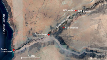
On a second visit to the Camarones Valley in early 2012, AAMD found a second small boulder containing camaronesite ~100 m west of the first discovery. The two boulders containing camaronesite evidently originated from a leached outcrop, high on the NW side of the steep valley. We have designated the site of the sulfate deposit as the Cuya NE9 sulfate occurrence. The arsenite-rich deposit is designated as the Cuya NE9 arsenite deposit. On the same trip in early 2012, AAMD also located a small deposit rich in copper chloride mineralisation ~5 km NE of the village of Cuya. This deposit, which we have designated the Cuya NE5 chloride deposit, produced the new mineral paratacamite-(Mg) (Kampf et al., Reference Kampf, Sciberras, Leverett, Williams, Malcherek, Schlüter, Welch, Dini and Molina Donoso2013b). The locations of the three deposits noted above are shown in Fig. 1.
Map showing locations of NE5 chloride, NE9 sulfate and NE9 arsenite deposits in the Camarones Valley (satellite base map from Google Earth).

The new arsenite mineral described in the present study is named cuyaite (/ˈkoɪ ɑ: aɪt/) for the village of Cuya, Camarones Valley, Arica Province, Chile, the nearest populated place to the site of the deposit. The new mineral and name have been approved by the Commission on New Minerals, Nomenclature and Classification of the International Mineralogical Association (IMA2019-126, Kampf et al., Reference Kampf, Nash, Dini and Molina Donoso2020). One holotype specimen is deposited in the Natural History Museum of Los Angeles County under catalogue number 74462.
Occurrence
As noted above, cuyaite was found at the Cuya NE9 arsenite occurrence (Fig. 1). Cuyaite is associated with anhydrite, native arsenic, arsenolite, calcite, claudetite, ferrinatrite, leiteite, magnesiocopiapite, phosphosiderite, pyrite, realgar, talmessite and the 3R polytype of gajardoite (Kampf et al., Reference Kampf, Nash, Dini and Molina Donoso2016).
Information on the geology and mineralogy of the Camarones Valley area is very limited. The most pertinent studies are those by Salas (Reference Salas1964, Reference Salas1965) and Thomas (Reference Thomas1971), which indicate that the rocks in this area consist principally of andesite and metamorphosed lavas, lithic tuffs and marine sediments. Sulfide accumulations (mostly pyrite, but also locally chalcopyrite and/or arsenopyrite) are present in the marine shales; deposits of secondary sulfates, arsenates, arsenites and chlorides appear to be directly related to the near-surface, low-temperature oxidisation of those deposits under increasingly arid conditions.
The secondary assemblage containing cuyaite and gajardoite-3R is interpreted as having formed from the oxidation of native arsenic and possibly other As-bearing primary phases and alteration by saline fluids derived from evaporating meteoric water under hyperarid conditions (cf. Cameron et al., Reference Cameron, Leybourne and Palacios2007). It is also worth noting that the Cuya 9NE arsenite deposit is only 19 km from the Pacific coast and the dense coastal camanchaca fogs frequently extend into the Camarones Valley. It seems likely that the camanchaca fogs have also played a role in the alteration of the veins and the formation of the secondary minerals, particularly in the recent past (see Kampf et al., Reference Kampf, Nash, Celestian, Dini and Molina Donoso2019). The Mn in cuyaite is presumably sourced from the marine shales and/or other country rocks. Although, we have not identified any other Mn-bearing secondary minerals at this deposit, we did detect a small amount of Mn in the paratacamite-(Mg) from the Cuya NE5 chloride deposit (Kampf et al., Reference Kampf, Sciberras, Leverett, Williams, Malcherek, Schlüter, Welch, Dini and Molina Donoso2013b). Finally, it should be noted that the coexistence of Mn3+ and As3+ in the same mineral is particularly perplexing, considering that Mn3+ generally requires much more oxidising conditions than does As3+. Indeed, cuyaite is the only mineral known containing both Mn3+ and As3+ as species determining constituents. We have no good explanation for this; however, the crystal structure determination leaves no doubt regarding the presence of Mn3+ and As3+ in the mineral.
Physical and optical properties
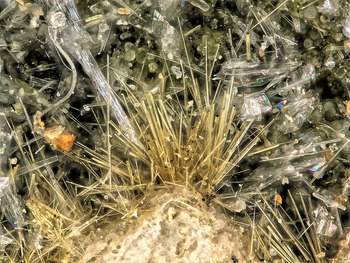
Cuyaite occurs as pale brown thin needles to ~300 μm long and 15 μm wide, typically in divergent sprays and subparallel intergrowths (Fig. 2). Mn3+ is generally a strong chromophore, commonly imparting deep red–brown or green colours, depending on the crystal field. The relatively pale colour in cuyaite is attributable to the thinness of the needles. A distinctive feature of the needles is that their tips appear very dark brown when viewed down the length of the needle.
Cuyaite needles with colourless lamellar claudetite on the holotype specimen (NMHLAC#74462). Field of view 0.84 mm across.

Needles are elongated on [010], more or less flattened on {101} and exhibit the prism forms {101}, { $\bar{1}$0
 $\bar{1}$}, {10
 $\bar{1}$} and {
 $\bar{1}$01}; terminal forms could not be measured, but needles appear to be terminated by either the {010} basal pinacoid or a combination of the pyramid forms {111}, {
 $\bar{1}$1
 $\bar{1}$}, {21
 $\bar{2}$} and {
 $\bar{2}$12}. Merohedral twinning is indicated by the structure refinement.
The streak is white or very pale brown. Crystals are transparent with adamantine lustre; subparallel intergrowths have silky lustre. Cuyaite does not fluoresce under long- or short-wave ultraviolet light. Scratch tests indicated a Mohs hardness of 2½. The mineral is brittle with no cleavage and irregular fracture. The density could not be measured because of the difficulty in seeing the thin fibres in Clerici solution. The calculated density is 4.140 g cm–3 for the empirical formula and 4.018 g cm–3 for the ideal formula. Cuyaite crystals dissolve slowly (within several minutes) in dilute HCl at room temperature.
Cuyaite is optically biaxial (–), with the indices of refraction α = 1.87(1), β = 1.956(calc) and γ = 1.98(1), determined in white light. The measured 2V is 60(1)° based on extinction data analysed with EXCALIBRW (Gunter et al., Reference Gunter, Bandli, Bloss, Evans, Su and Weaver2004). The thinness of the needles prevented the measurement of β; consequently, it was calculated from α, γ and 2V. Dispersion could not be observed. The optical orientation is X = b and Y ^ a = 53° in the obtuse angle β. No pleochroism was observed. The Gladstone–Dale compatibility index, 1 – (K p/K c), is 0.017 for the empirical formula, in the range of superior compatibility (Mandarino, Reference Mandarino2007).
Raman spectroscopy
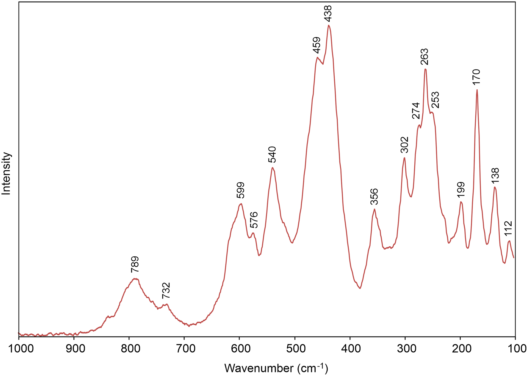
Raman spectroscopy was done on a Horiba XploRa+ micro-Raman spectrometer using an incident wavelength of 532 nm, 50 μm slit, 2400 gr/mm diffraction grating and a 100× (0.9 NA) objective. The mineral was stable under the laser. The spectrum was obtained on a flat-lying needle (⊥ [010]) that was otherwise randomly oriented. The spectrum from 4000 to 900 cm–1 was essentially featureless, confirming cuyaite to be anhydrous. The range from 1000 to 100 cm–1 is shown in Fig. 3.
Raman spectrum of cuyaite recorded using a 532 nm laser.

Arsenite As3+–O stretching and bending vibrations undoubtedly produce many of the bands observed in the Raman spectrum of cuyaite, particularly between 900 and 300 cm–1. The complexity of the spectrum in this region is attributable, in a large part, to the number of independent AsO3 groups (14) and the significant variation in the As3+–O bond lengths. Past studies have shown that Raman modes associated with arsenite groups vary considerably depending upon their As–O–As linkages (cf. Bahfenne and Frost, Reference Bahfenne and Frost2010; Szymanski et al., Reference Szymanski, Marabella, Hoke and Harter1968). Consequently, we have not endeavoured to make specific band assignments. Bands between 300 and 100 cm–1 are probably related to Mn3+–O and Ca–O bonds and to lattice modes.
Chemical composition
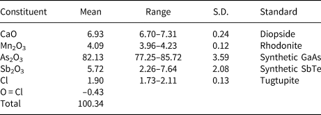
Quantitative analyses (six points on three crystals) were performed at the University of Utah on a Cameca SX-50 electron microprobe with four wavelength-dispersive spectrometers utilising Probe for EPMA software. Analytical conditions were 15 kV accelerating voltage, 10 nA beam current and a beam diameter of 3 μm. Because of the thinness and fragility of the thin needles, polishing was not possible; analyses were performed on unpolished, but smooth and flat, crystal faces. No elements other than Ca, Mn, As, Sb, Cl and O were detected by EDS. Other likely elements were sought, but none was above the detection limits. Raw X-ray intensities were corrected for matrix effects with a ϕ(ρz) algorithm (Pouchou and Pichoir, Reference Pouchou, Pichoir, Heinrich and Newbury1991). Analytical data are given in Table 1. The empirical formula based on 24 O atoms per formula unit (apfu) is Ca2.03Mn3+0.95 (As3+13.66Sb3+0.65)Σ14.31O24Cl0.88. The simplified formula is Ca2Mn3+(As3+,Sb3+)14O24Cl. The ideal formula is Ca2Mn3+As3+14O24Cl, which requires CaO 6.99, Mn2O3 4.92, As2O5 86.37, Cl 2.21, O = Cl –0.50, total 100 wt.%.
Electron microprobe analytical data (in wt.%) for cuyaite.

S.D. – standard deviation
X-ray crystallography and structure determination
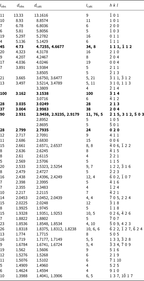
Powder X-ray diffraction data were obtained on a Rigaku R-Axis Rapid II curved imaging plate microdiffractometer utilising monochromatised MoKα radiation. Observed d values and intensities were derived by profile fitting using JADE 2010 software (Materials Data, Inc.). The observed powder data fit well with those calculated from the structure, also using JADE 2010 (Table 2). The unit-cell parameters refined from the powder data using JADE 2010 with whole–pattern fitting are: a = 14.7231(6), b = 5.58709(19), c = 17.4185(12) Å, β = 112.451(8)° and V = 1324.23(14) Å3.
Powder X-ray data for cuyaite. Only calculated lines with I > 3 are shown.

The strongest lines are given in bold. Data for I > 1 are provided in the supplementary material.
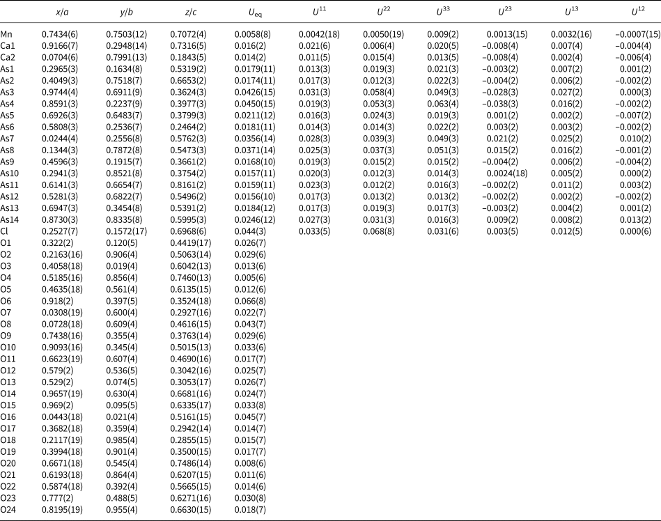
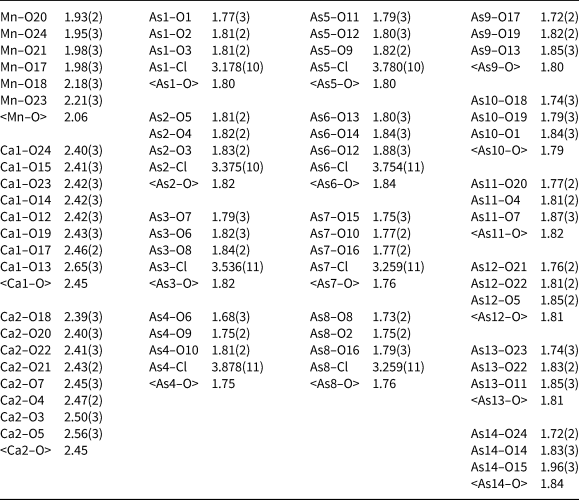
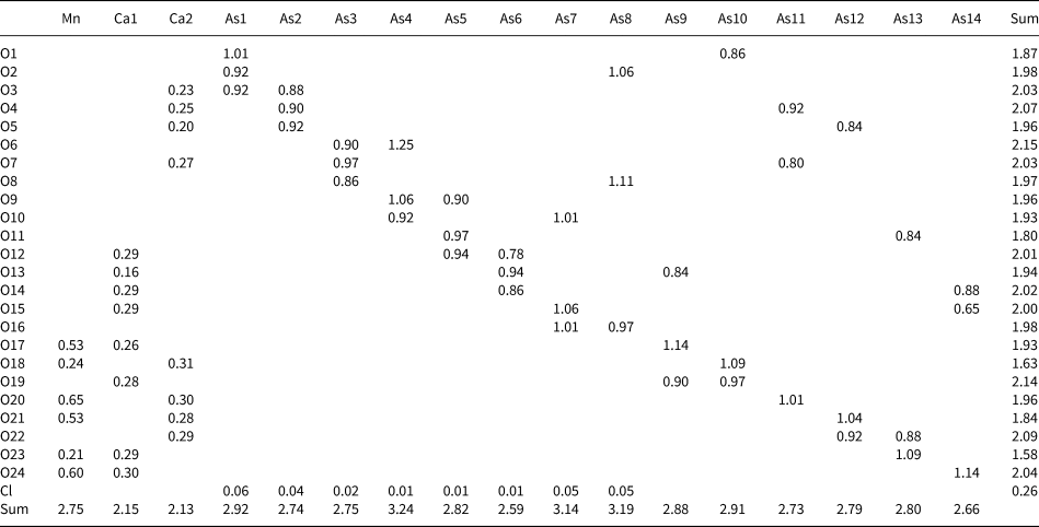
Single-crystal X-ray studies were carried out using the same diffractometer and radiation noted above. The small size and poor quality of crystals limited the usable data to 2θ < 40°, which is probably responsible for our inability to refine O atom sites with anisotropic displacement parameters (see below). The Rigaku CrystalClear software package was used for processing the structure data, including the application of an empirical multi-scan absorption correction using ABSCOR (Higashi, Reference Higashi2001). The structure was solved by the charge-flipping method using SHELXT (Sheldrick, Reference Sheldrick2015a). Refinement proceeded by full-matrix least-squares on F 2 using SHELXL-2016 (Sheldrick, Reference Sheldrick2015b). Initial attempts to solve the structure in space group P21/n indicated two split As sites and the refinement provided a high R 1 of ~0.11. A much better refinement (R 1 = 0.0369) was obtained in space group Pn. Merohedral twinning was indicated with nearly equal twin components, 0.48/0.52(3). All non-oxygen sites were successfully refined with anisotropic displacement parameters; most O sites became non-positive-definite (NPD) when refined anisotropically, so they were refined with isotropic displacement parameters. Efforts to refine the occupancies of the As sites did not show significant variance from full As occupancies, and all sites were assigned full occupancies by As in the final refinement. The occupancy of the Cl site refined to slightly greater than full occupancy; it was assigned full occupancy in the final refinement. The data collection and refinement details are given in Table 3, atom coordinates and displacement parameters in Table 4, selected bond distances in Table 5 and bond-valence sums (BVS) in Table 6. The crystallographic information files have been deposited with the Principal Editor of Mineralogical Magazine and are available as Supplementary material (see below).
Data collection and structure refinement details for cuyaite.

*R int = Σ|F o2–F o2(mean)|/Σ[F o2]. GoF = S = {Σ[w(F o2–F c2)2]/(n–p)}½. R 1 = Σ||F o|–|F c||/Σ|F o|. wR 2 = {Σ[w(F o2–F c2)2]/Σ[w(F o2)2]}½; w = 1/[σ2(F o2) + (aP)2 + bP] where a is 0.0243, b is 25.1977 and P is [2F c2 + Max(F o2,0)]/3.
Atom coordinates and displacement parameters (Å2) for cuyaite.

Selected bond distances (Å) and angles (°) for cuyaite.

Bond-valence analysis for cuyaite. Values are expressed in valence units (vu).

Bond-valence parameters for cation–O bonds are from Gagné and Hawthorne (Reference Gagné and Hawthorne2015). As3+–Cl bond strengths are from Brese and O'Keeffe (Reference Brese and O'Keeffe1991).
Although attempts to refine the occupancies of the As sites suggested full occupancies by As alone, the As–O bond lengths for some sites are longer than is typical for As3+–O bonds, as evidenced by their low BVS values (see Table 6). The As6 and As14 sites, in particular, have very low BVS values of 2.59 and 2.66 valence units (vu), respectively. While the EPMA indicates that Sb accounts for <5% of the content of the As sites, it is reasonable to assume that the As sites with the lowest BVS values accommodate more Sb than do the other As sites because Sb3+–O bonds are generally longer than As3+–O bonds. At the same time, it is worth noting that the unusually wide range in BVS values for the As sites may, in part, be due to the small size (low data to parameter ratio) and relatively low-quality of the dataset, as well as by refinement issues introduced by the merohedral twinning. In this regard, it should be noted that the average BVS for the As sites (2.87 vu) is not unreasonable for As3+.
Description of the structure
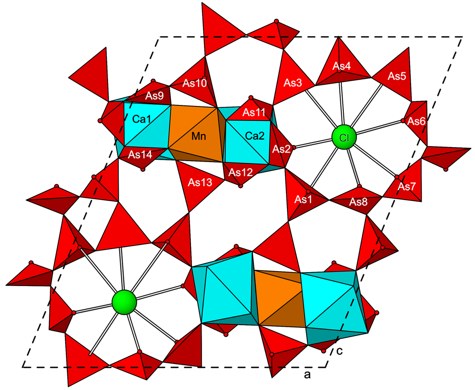
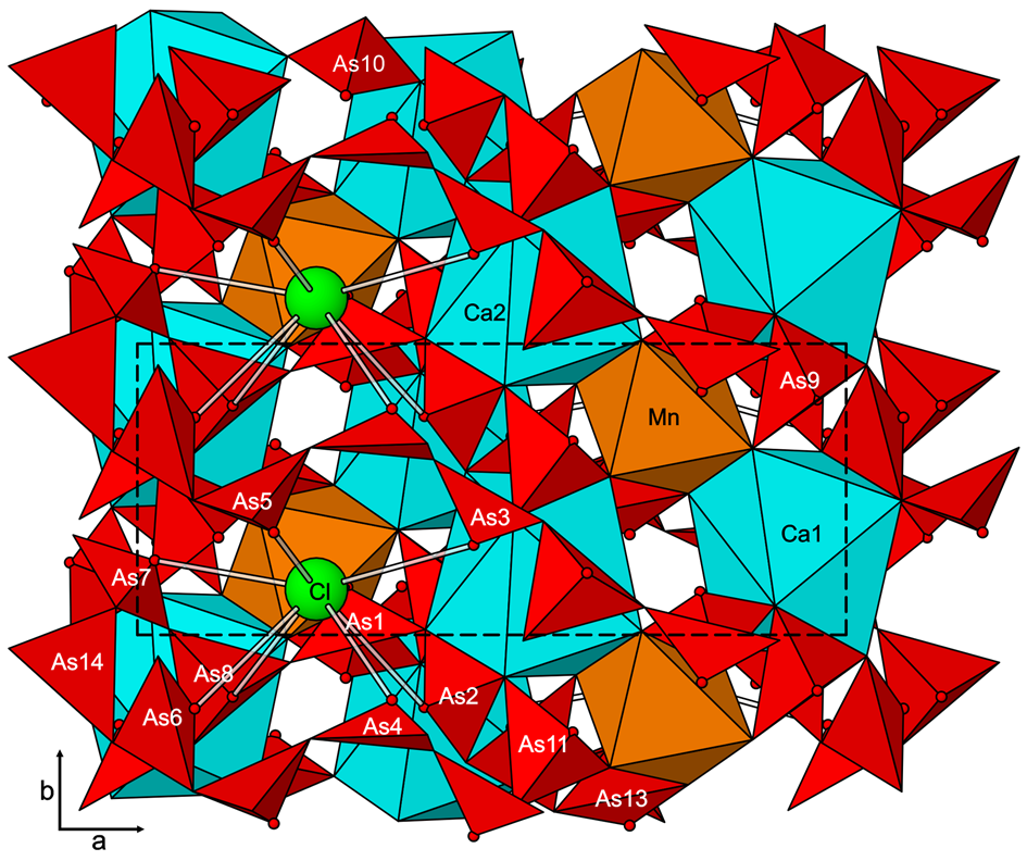
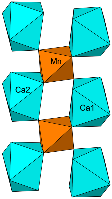
The structure of cuyaite (Figs 4 and 5) contains 14 independent sites that accommodate As3+ cations. Each of these sites forms short (<2 Å) links to three O sites resulting in AsO3 pyramids, the typical coordination geometry for As3+ and O due to the lone-pair electrons of the As3+ cation. The AsO3 pyramids share O corners to form a ‘loose’ 3D framework. The structure also contains one Jahn–Teller distorted Mn3+O6 octahedron and two different CaO8 polyhedra, which link by edges and corners to form columns along [010] (Fig. 6). The polyhedra in these columns also link by edge- and corner-sharing to most of the AsO3 pyramids (all except As4 and As8) in the framework. A channel in the framework along [010] contains a Cl site, which forms long bonds (3.175–3.878 Å) to eight different As3+ sites (As1–As8) in the surrounding framework.
The crystal structure of cuyaite viewed down [010]. The As3+ site at the apex of each AsO3 pyramid is shown with a small sphere. The unit cell is shown by the dashed outline.

The crystal structure of cuyaite viewed down [001]. The unit cell is shown by the dashed outline.

Chain of Mn3+O6 octahedra and CaO8 polyhedra along [010] (vertical) in the structure of cuyaite.

The BVS values are mostly consistent with the site assignments; however, there are significant deviations from ideal values. Much of the variability in BVS values is probably attributable to the inability of basic bond-valence theory to account for the effects of lone-pair electrons. This is especially demonstrated by the Cl site (BVS = 0.26 vu), which is located at the centre of the channel that also accommodates lone-pair electrons from the As1–As8 sites to which the Cl forms eight long bonds.
The structure of cuyaite is unique and we are not aware of any structure to which it is closely related. There are other structures that are based on infinite frameworks of AsO3 pyramids in combination with other Mϕ4–6 polyhedra (cf. Majzlan et al., Reference Majzlan, Drahota, Filippi, Bowell, Alpers, Jamieson, Nordstrom and Majzlan2014); however, none of these structures contains a framework of AsO3 pyramids when other polyhedra are excluded.
The distinctive squat pyramidal AsO3 group can be thought of as a tetrahedral coordination in which the lone-pair electrons of the As3+ comprise the fourth corner. The lone-pair corner of this tetrahedron, rather than bonding to nearby cations, instead effectively repels nearby anions. This type of coordination severely discourages the formation of 3D linkages involving only the AsO3 group. The same situation is seen for the 3+ oxidation states of two other period-7 semi-metal elements, Sb and Bi, which also commonly form squat pyramidal MO3 coordinations with lone-pair electrons forming the fourth tetrahedral corner. We have been unable to find reports of any other structures that contain 3D frameworks assembled of As3+O3, Sb3+O3 or Bi3+O3 pyramids.
The strongest bonds in the structure of cuyaite are the short As–O bonds of the AsO3 pyramids, as evidenced by the bond strengths provided in Table 6. The framework of AsO3 pyramids is, therefore, the most strongly bonded portion of the cuyaite structure; however, it is clear that this framework would not be stable, nor would it have formed, without the presence of the other structural components, which have effectively served as templating agents. Most important in this respect is probably the column comprised of Mn3+O6 octahedra and CaO8 polyhedra (Fig. 6), which effectively defines the b cell dimension. Topologically identical structural components in other phases appear to be relatively rare, although they may simply be difficult to identify, ‘buried’ within more complex structures. An example is found in another Chilean mineral, joteite (Kampf et al., Reference Kampf, Mills, Housley, Rossman, Nash, Dini and Jenkins2013c), Ca2CuAl[AsO4][AsO3(OH)]2(OH)2(H2O)5, in which the column consists of AlO6 octahedra and CaO8 polyhedra. In the joteite structure, adjacent columns are joined by shared edges of CaO8 polyhedra, as well as by corner linkages with AsO4 tetrahedra and Cu2+O5 square pyramids.
Supplementary material
To view supplementary material for this article, please visit https://doi.org/10.1180/mgm.2020.31
Acknowledgements
The paper benefited from constructive reviews by Uwe Kolitsch and an anonymous reviewer. A portion of this study was funded by the John Jago Trelawney Endowment to the Mineral Sciences Department of the Natural History Museum of Los Angeles County.