Awareness about normal cardiac dimensions and volumes is very important for understanding the cardiac function in childhood. Several two-dimensional echocardiographic examinations have been performed to obtain information on the longitudinal development of the left ventricular end-diastolic dimension and left ventricular end-systolic dimension using body height as an index in children and newborns, including premature neonates.Reference Nagasawa, Arakaki, Yamada, Nakajima and Kamiya 1 – Reference Nagasawa and Arakaki 4 Linear relationships between height and other dimensions of the heart have been reported in these studies. However, in neonates <1 week of age, the normal dimensions of the left ventricle remain undefined, as physiological pulmonary hypertension exists during the 1st week of life, making the left ventricle appear elliptical on short axis views. The left ventricular end-diastolic volume is altered according to the left ventricular position, as confirmed on echocardiography. The Pombo method is used to calculate left ventricular volumes and ejection fractions because it is a simple method for determining both measurements and calculations. However, this method is not applicable to early neonates because it assumes the presence of a round shape on short axis views. Few studies have estimated left ventricular end-diastolic and end-systolic volumes in normal neonates,Reference Foran, Fitzpatrick and Allsop 5 as performing invasive examinations is not feasible.
Three-dimensional echocardiography is used to evaluate cardiac volumes in children.Reference Acar, Marx, Salibe, Sidi and Kachaner 6 This method is performed to assess left ventricular volumes in the early neonatal period.
The objectives of this study were: (1) to assess the normal ranges of left ventricular end-diastolic and end-systolic volumes using body weight and body surface area as indexes, (2) to evaluate volume changes over time after birth and to ascertain the causes, if any, of these changes, (3) to detect any significant gender differences, (4) to compare volume data using the velocity–time integral method and to calculate the results using the Pombo method, and (5) to compare the results with previously reported left ventricular volumes.
Methods
Study participants
Neonates who met all of the following requirements to be “normal” were recruited in this study: (1) a gestational age between 37 weeks 0 days and 41 weeks 6 days, (2) an adequate weight for the gestational age, (3) no foetal distress and an Apgar score of more than 8 points at 5 minutes after birth, (4) no requirements for oxygen supplementation or respiratory or circulatory support, (5) good sucking and urination, (6) no congenital heart disease, normal arterial and mitral valve functions, and no evidence of coronary artery lesions, and (7) blood flow through the oval foramen from left to right on echocardiography.
Only neonates who underwent echocardiographic examinations at Gifu Prefectural General Medical Center were included in this study. The Ethics Committee of Gifu Prefectural General Medical Center approved this study in advance. The aims of the study were explained to the parents or guardians of the neonates and signed permissions were obtained.
The necessary number of neonates was determined according to the report by CohenReference Cohen 7 showing that more than 88 participants each are needed to compare groups with a type one error of 0.05 in two-sided tests and a type two error of 0.75 when the absolute difference divided by the standard deviation is presumed to be 0.4.
Study group
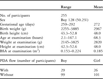
The study group consisted of 255 consecutive Japanese neonates – 128 boys and 127 girls. The participant characteristics are shown in Table 1. No significant weight loss was observed between the boys (5.2% and 2.3% (mean and standard deviation)) and the girls (5.6% and 2.4%). The adequate body weight criterion was determined based on a statistical analysis of children performed in Japan in 2008.
Characteristics of the 255 neonatal participants undergoing three-dimensional echocardiographic examinations.

BSA = body surface area; PDA = persistent ductus arteriosus
Data acquisition
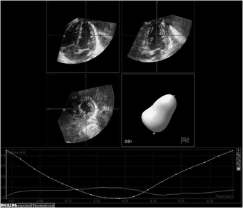
The three-dimensional echocardiographic studies were performed in an apical four-chamber view with a 7–2 MHz iE33 transducer (Philips Electronics, Bothell, Washington, United States of America) over four cycles during natural sleep. The datasets were post-processed offline using a software program supplied by Philips Medical – the Cardiac 3D Quantification Advanced software program, which is a Q-LAB application (Philips Ultrasound). The 3D Quantification Advanced has the diagnostic power of 3D full-volume imaging based on its ability to provide semiautomated analyses of left ventricular volumes using all voxels in order to generate a full three-dimensional endocardial border. Endocardial markers were placed manually on either side of the mitral annulus in two orthogonal views and at the apex; the semiautomated endocardial border detection software program then traced the endocardium and calculated the volume automatically. The endocardial detection accuracy was assessed and optimised manually (Fig 1).
Representative images of a participant, including an initialised boundary in three planes and a reconstructed three-dimensional cast.

The maximum and minimum values of the sequenced data were classified as the end-diastolic and end-systolic volumes, respectively. The final values were obtained from the average of more than three examinations in every neonate.
Conventional two-dimensional echocardiographic examinations with a 12–4 MHz iE33 transducer were performed immediately after three-dimensional echocardiography. The left ventricular end-diastolic dimension, the left ventricular end-systolic dimension, that is, the short dimensions, and the distance between the apex and the mitral orifice at end-diastole and end-systole, that is, the long dimensions, were measured. The Pombo method involves the following steps: the left ventricular volumes are calculated from the ventricular dimensions using the volume formula of a prolate ellipse, expressed as V = π/6 × D2L, where D is the shorter diameter and L is the longer diameter. If the longer diameter is assumed to be twice the length of the shorter diameter, the formula is further simplified to V = π/3 × D3. This formula assumes the algebraic cancellation of the factor π/3. The cube of the end-diastolic dimension is used as an estimation of the end-diastolic volume and the end-systolic dimension is used as an estimation of the end-systolic volume.Reference Pombo, Troy and Russell 8 In this study, the aortic flow velocity was measured from the suprasternal notch view using the pulsed Doppler technique and a region of interest was located just above the Valsalva sinus of the aorta, where the aortic diameter was measured. The measurement convention of the aortic diameter from one leading edge to another was used. The diameter was measured at least four times and the mean value was used as the aortic diameter. Care was taken to minimise the angle of insonation. No angle correction was performed. Velocity time–integrals were obtained manually by tracing the area under the Doppler recordings using the built-in electrical program of iE33. The aortic diameter was measured in the parasternal view with electronic callipers. The stroke volume was calculated as the product of the aortic cross-section area and the velocity–time integral using the two-dimensional M-mode technique. The cardiac output was calculated as the product of the stroke volume and the actual heart rate measurement.
All measurements were obtained by two observers H.N. and J.M. The inter-observer error was 3.7%. The intra-observer errors were 2.8% and 3.2%, respectively, calculated based on data obtained from 30 participants undergoing three measurements each. The examination time after birth was determined randomly. A three-dimensional echocardiographic examination was performed once in each neonate during the early neonatal period.
Clinical information on these participants was collected from inpatient records. Body height in the prone position and weight were measured at the time of echocardiography. The body surface area was calculated according to the formulas given by Haycock et alReference Haycock, Schwartz and Wisotsky 9 and DuBois and DuBois.Reference DuBois and DuBois 10
Statistical analyses
The F-test and Student's t-test were used to determine whether the sample variance and mean values of two categories were statistically equivalent. The Sheffé method was used to compare the mean values of three or more categories. A level of p < 0.05 was considered statistically significant.
Results
Body surface area data calculated by the formulas reported by Haycock et al and by DuBois and DuBois are indicated by adding H and D at the end of the results, respectively.
Normal left ventricular end-diastolic and end-systolic volumes in all participants
The average end-diastolic volumes were 30.33 (2.32; standard deviation) ml/m2 (H) and 32.26 (2.52) ml/m2 (D). The average end-systolic volumes were 12.85 (1.71) ml/m2 (H) and 13.67 (1.83) ml/m2 (D). The stroke volumes and ejection fraction were estimated to be 17.48 (1.28) ml/m2 (H) and 18.59 (1.40) ml/m2 (D) and 0.577 (0.034), respectively. The cardiac indexes were calculated to be 2.04 (0.30) L/minute/m2 (H) and 2.17 (0.32) L/minute/m2 (D). However, as the blood flow changes dramatically in the early neonatal period, more concise evaluations are needed, especially with respect to the effects of persistent ductus arteriosus, physiological pulmonary hypertension, and so forth.
Volume changes over time after birth: the influence of persistent ductus arteriosus
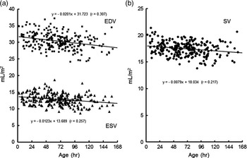
Changes in the end-diastolic and end-systolic volumes of the left ventricle over time are shown in Figure 2a, whereas those in the stroke volume are shown in Figure 2b. The volumes were seen to gradually decrease over time.
Relationships between the left ventricular end-diastolic volume, end-systolic volume and stroke volume divided by body weight and time after birth in 255 participants. EDV, left ventricular end-diastolic volume (square); ESV, left ventricular end-systolic volume (triangle); SV, stroke volume (circle).

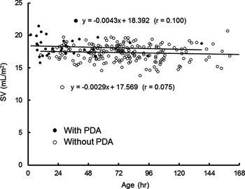
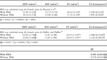
To determine the cause of this phenomenon, the participants were divided into two groups: the ductus group, which had a ductus arteriosus flow with persistent ductus arteriosus (n = 55, 29 boys and 26 girls), and the non-ductus group, which had no ductus arteriosus flow and no persistent ductus arteriosus (n = 200, 99 boys and 101 girls), based on the findings of conventional colour Doppler echocardiography. The results are shown in Figure 3 and Table 2. Very minor volume changes over time were observed in the non-ductus group (slope: −0.0029). There were no significant differences in the end-diastolic volumes, end-systolic volumes, stroke volumes, or ejection fractions according to the F-test. There were significant differences between the ductus group and the non-ductus group, respectively, in terms of the end-diastolic volume, (31.05 (2.51) ml/m2 (H)) and (30.19 (2.27) (H)) (p < 0.042); stroke volume, (18.32 (1.32) ml/m2 (H)) and (17.35 (1.24) (H)) (p < 0.0001); and ejection fraction (0.585 (0.034)) and (0.576 (0.033)) (p = 0.040). However, there were no significant differences in the end-systolic volume (p = 0.243). It is presumable that the ductus arteriosus flow is attributable to increases in the stroke volume, the primary cause of the aforementioned gradual volume decreases.
Stroke volumes of the participants with and without a persistent ductus arteriosus flow. Each volume is represented by closed and open circles, respectively. There were no stroke volume changes in the participants without a ductus arteriosus flow. PDA, persistent ductus arteriosus.

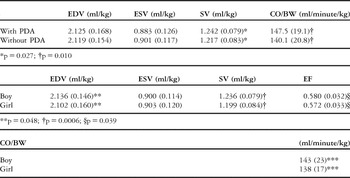
There was a significant difference in the end-diastolic volume between the ductus group and the non-ductus group (p = 0.042(H), 0.017(D)) when indexed to body surface area, although there were no significant differences when it was indexed to body weight (p = 0.401; Tables 2 and 4).
Left ventricular volumes measured using three-dimensional echocardiography in normal neonates with and without a ductus arteriosus flow.

CI = cardiac index; EDV = end-diastolic volume; EF = ejection fraction; ESV = end-systolic volume; PDA = persistent ductus arteriosus; SV = stroke volume
Values within parentheses represent SD values
Significant differences were found in EDV, SV, CI, and EF
§§§p = 0.040
The average end-diastolic and end-systolic volumes of the participants without a ductus arteriosus flow were 30.19 (2.27) ml/minute/m2 (H) and 12.85 (1.68) L/minute/m2 (H), respectively. The stroke volume and ejection fraction were estimated to be 17.35 (1.24) ml/minute/m2 (H) and 0.576 (0.033), respectively.
Comparison of stroke volume between genders
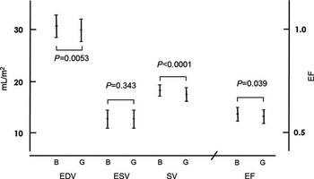
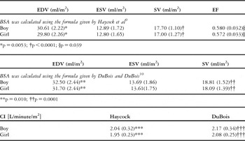
A comparison of the stroke volume of the participants without persistent ductus arteriosus was made between the 99 boys and the 101 girls included in this study. The results are shown in Figure 4 and Table 3. No significant differences in variance were observed in any item according to the F-test. There were significant gender differences in terms of the end-diastolic volume (30.61 (2.22) ml/m2 (H), boys) (29.80 (2.26) ml/m2 (H), girls) (p = 0.0053); stroke volume (17.70 (1.10) ml/m2 (H), boys) (17.00 (1.27) ml/m2 (H), girls) (p <0.0001); and ejection fraction (0.580 (0.032), boys) (0.572 (0.033), girls) (p = 0.039). However, the end-systolic volume did not differ significantly between the genders (12.89 (1.72) ml/m2 (H), in boys and 12.80 (1.65) ml/m2 (H) in girls) (p = 0.343). These results indicate the gender differences to be primarily attributable to the end-diastolic volume.
Left ventricular end-diastolic, end-systolic, and stroke volumes divided by body surface area in the participants without persistent ductus arteriosus according to gender. Each bar shows the mean and 95% confidential interval. There was a significant gender difference in the stroke volume. B, boy; EDV, left ventricular end-diastolic volume; ESV, left ventricular end-systolic volume; G, girl; SV, stroke volume.

Left ventricular volumes measured using three-dimensional echocardiography in normal neonates without persistent ductus arteriosus according to gender.

CI = cardiac index; EDV = end-diastolic volume; EF = ejection fraction; ESV = end-systolic volume; PDA = persistent ductus arteriosus; SV = stroke volume
Values within parentheses represent SD values
Significant gender differences were found in EDV, SV, EF, and CI
***p = 0.013; †††p = 0.017
Left ventricular volumes measured using three-dimensional echocardiography in normal neonates without persistent ductus arteriosus when indexed to body weight (kg)

BW = body weight; CO = cardiac output; EDV = end-diastolic volume; EF = ejection fraction; ESV = end-systolic volume; PDA = persistent ductus arteriosus; SV = stroke volume
Values within parentheses represent SD values
***p = 0.045
The data indexed to body weight are shown for reference in Figure 4.
Comparison of the stroke volume of the left ventricle measured using three-dimensional echocardiography and the velocity–time integral and Pombo methods
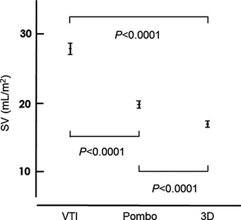
The results are shown in Figure 5. The stroke volume obtained with the velocity–time integral method was highest at 27.69 (95% confidence interval, 27.10–28.29) ml/m2 (H), followed by 19.91 (19.42–20.39) ml/m2 (H), with the Pombo method and at 17.48 (17.32–17.64) ml/m2 (H) on three-dimensional echocardiography. There were significant differences in the values obtained with these methods (p < 0.0001).
Comparisons among left ventricular stroke volumes measured using 3D echocardiography and the velocity–time integral and Pombo methods. Each bar shows the mean and 95% confidential interval. There were significant differences in the values obtained with these three methods. SV, stroke volume; VTI, velocity–time integral; 3D, three-dimensional echocardiography.

No gender differences in stroke volume were found with either the velocity–time integral method (p = 0.225) or the Pombo method (p = 0.070).
Discussion
Approaches to evaluating the left ventricular volume include biplane angiocardiography, the Fick method, dye dilution techniques, the velocity–time integral method, and the Pombo method. However, it is not possible to evaluate normal neonatal left ventricular volumes using invasive methods. The three-dimensional echocardiography modality has been used in children to evaluate morphological structures and estimate the volumes of heart chambers.Reference Acar, Marx, Salibe, Sidi and Kachaner 6 However, the use of three-dimensional echocardiography is limited in neonates, partially because the probes are too large and heavy for application in neonates and because the small size of the neonatal heart makes it impractical to obtain clear and accurate images. Advances in three-dimensional echocardiography apparatuses have allowed the application of three-dimensional echocardiographic examinations in neonates. However, there are no reports of neonatal left ventricular volume assessments using this modality. The present report is therefore the first to evaluate left ventricular volume assessments in the early neonatal period. A comparison was made among the results obtained with three-dimensional echocardiography and those obtained using the velocity–time integral and Pombo methods.
Influence of persistent ductus arteriosus on the stroke volume
The end-diastolic and end-systolic volumes changed over time after birth, whereas there were no apparent volume changes in the neonates examined more than 72 hours after birth. This can be attributed to the presence of a ductus arteriosus flow. The stroke volumes of the participants with and without a ductus arteriosus flow were compared. The stroke volumes of those with a ductus arteriosus flow were 2.9% higher than those of the participants without a ductus arteriosus flow. In those without a ductus arteriosus flow, the stroke volume demonstrated only a minor change in slope through the early neonatal period. These observations prompted the speculation that a ductus arteriosus flow contributes to a higher stroke volume in neonates with a ductus arteriosus flow.
It is not unexpected for the stroke volume to be higher in participants with a ductus arteriosus flow than in those without it. However, it was quite interesting to observe that the increased stroke volume was attributable not to an increased end-diastolic volume, but rather to a decreased end-systolic volume. This may have been because of lower end-systolic pressures in the participants with a continuous persistent ductus arteriosus flow than in those without it, although no direct evidence explaining this phenomenon was found.
Gender differences
There was also a significant difference in stroke volume between boys and girls in the early neonatal period. This indicates a significant gender difference in the internal dimension of the left ventricle starting from the beginning of human life.
It has previously been reported that there is a significant gender difference in the left ventricular end-diastolic volume measured using conventional two-dimensional echocardiography in children and infants with body heights of more than 75 cmReference Nagasawa, Arakaki, Yamada, Nakajima and Kamiya 1 and that there are no significant differences in the left ventricular end-diastolic volume between boy and girl neonates and in infants <75 cm in height.Reference Nagasawa 3 The results of this study do not support the former conclusion that there are no gender differences. The gender difference in end-diastolic volumes observed in this study was 0.034 ml, which is 1.60% of the mean end-diastolic volume in boys. This was converted to a 0.54% difference in the left ventricular end-diastolic dimension when using two-dimensional echocardiography. This result was acquired using the following calculations: the end-diastolic volumes in boys and girls and the gender difference in the left ventricular end-diastolic volume were expressed as Vm (ml), Vf (ml), and ΔDd (%), respectively. (Vm − Vf)/Vm = 0.0160 and Vf/Vm = 1 – 0.0160 if the end-diastolic volume is proportionate to left ventricular end-diastolic volume3. The latter formula can be changed to (1 − ΔDd/100)3 = 1 – 0.0160, that is, ΔDd = 0.54%. For example, if the left ventricular end-diastolic volume is 18.33 mm – a mean left ventricular end-diastolic dimension of 48.0 cm height = the average height of the participants in this study – the gender difference is only 0.10 mm.Reference Nagasawa 3 It was expected that three-dimensional echocardiography alone would have the capacity to identify the gender difference in left ventricular end-diastolic volumes at birth because ascertaining extremely small differences is quite difficult and even impossible using two-dimensional echocardiography. The gender difference at a height of 75 cm was estimated to be 0.15 mm according to the regression equations shown in earlier reports.Reference Nagasawa, Arakaki, Yamada, Nakajima and Kamiya 1 , Reference Nagasawa 3 These observations suggest that the gender difference in the left ventricular end-diastolic volume continues to increase throughout the infantile period. It can readily be imagined that the gender difference persists throughout human life, that is, from the stage of human cardiac development through adulthood.Reference Nagasawa, Arakaki, Yamada, Nakajima and Kamiya 1 – Reference Nagasawa and Arakaki 4 It was speculated that the gender disparity in exercise capacity could be attributable to this dimensional difference, at least in terms of the circulatory capacity. The cardiac indexes were 2.04 (0.32) L/minute/m2 (H) and 2.17 (0.34) L/minute/m2 (D) in boys and 1.95 (0.23) L/minute/m2 (H) and 2.08 (0.25) L/minute/m2 (D) in girls (Table 3).
Evaluation of the differences in left ventricular volumes measured using three-dimensional echocardiography and the velocity time integral and Pombo methods
The region of intensity is usually placed at the centre of the vessels when measuring the velocity–time integrals. The flow velocity in the peripheral area of the vessel is always lower than that observed in the centre. According to the Hagen–Poiseuille law, the average velocity within the vessel is two-thirds of the maximum velocity. In addition, the direction of the pulsed wave and the flow of blood are not always parallel. If the angle is estimated to be ∼20°, the true velocity time integral is 2/3/cos (20°) = 0.70 of the time of the original velocity–time integral. The stroke volume was calculated to be 19.10 mL/m2 on using the velocity–time integral method.
When applying the Pombo method, there are two points to consider. First, the calculation in this method is a rough approximation. For example, the major dimension is usually less than twice the left ventricular end-diastolic volume – it was estimated to be ∼1.72 times the actual left ventricular end-diastolic volume. Second, this method assumes that the anteroposterior and lateral lengths of the minor dimension are equal. These lengths are usually not equal in the early neonatal period at end-diastole. In general, a tendency to overestimate the end-diastolic volume and underestimate the end-systolic volume is observed, as the fraction shortening of the major dimension (=0.161 [0.153–0.169, 95% confidence interval]) is significantly smaller than that of the minor dimension (=0.332 [0.326–0.338]). The true stroke volume is seen to be lower than that reported by Pombo et al.
It is assumed that the value of the ejection fraction measured on three-dimensional echocardiography (0.58) corresponds better to the clinical impression compared with that obtained with the Pombo method (0.70).
The stroke volume estimated on three-dimensional echocardiography is considered to be more accurate and sensitive than that obtained with the other two methods.
Comparison of the volumes estimated in this study and those of former studies
Only a few studies have examined the neonatal stroke volume. Mandelbaum–Isken et alReference Mandelbaum-Isken and Linderkamp 11 reported the cardiac index to be 167 ml/minute/kg (modal) using the pulsed Doppler method with a sampling point in the ascending aorta. Patel et alReference Patel, Dodsworth and Mills 12 obtained a cardiac index of 216 ml/minute/kg (95% confidence interval, 179–253 ml/minute) in healthy neonates using conventional two-dimensional echocardiography of the suprasternal notch. Foran et alReference Foran, Fitzpatrick and Allsop 5 estimated the stroke volume of premature infants to be 1.3 ml/kg or 1.6 ml/kg per stroke based on two methods using magnetic resonance imaging. The estimated stroke volume and cardiac index in this study were 1.22 ml/kg and 141 ml/minute/kg or 2.16 L/minute/m2, respectively, using three-dimensional echocardiography. These values are 6–33% lower than those estimated by the above-mentioned reports. This discrepancy is primarily attributable to the differences in the methods of measuring and calculating the stroke volume and cardiac index. Friedberg et alReference Friedberg, Su, Tworetzky, Soriano, Powell and Marx 13 reported that there are no significant differences in the end-diastolic volume between the values obtained on three-dimensional echocardiography and magnetic resonance imaging, whereas the three-dimensional echocardiographic ejection fraction is significantly smaller (9.3%) than the magnetic resonance imaging ejection fraction in neonates and infants with congenital heart disease. These results indicate that the stroke volume obtained on three-dimensional echocardiography is smaller than that obtained on magnetic resonance imaging. Lytrivi et alReference Lytrivi, Bhatla and Ko 14 reported that the mean left ventricular end-diastolic volume indexed to (body surface area)1.38 is 70.4 ml/m2.6, which corresponds to 30.79 ml/m2 in neonates. These data are similar to those reported in this study. The true cardiac index could possibly be lower than that reported using the three-dimensional echocardiographic results obtained in this study. This low cardiac index may contribute to the circulatory system vulnerability that often appears in the early neonatal period.
The significance levels indexed to body surface area were lower than those indexed to body weight in all comparisons. This indicates that body height is much more informative than body weight when indexing.
Conclusion
In this study, the left ventricular volumes in the early neonatal period were measured using three-dimensional echocardiography. The cardiac index for this age is smaller than that previously recognised. Significant gender differences in the end-diastolic volume, stroke volume, and ejection fraction, but not in end-systolic volume, can be seen.
Further observations are warranted to confirm the points shown in this study.
Acknowledgements
The author thanks Junko Miyamoto and Masaki Sawa for their devoted coordination towards this manuscript.
Disclosures
None.