Obesity is a global health problem caused by urbanisation, sedentary habits and increased consumption of high-energy processed foods. The prevalence of obesity continues to increase in the mid-2020s(Reference Kanter and Caballero1,Reference Alegria Ezquerra, Castellano Vazquez and Alegria Barrero2) , indicating an imminent increase in the incidence of obesity-related noncommunicable diseases such as type 2 diabetes, cardiovascular disease and metabolic syndrome(Reference Ahmed and Mohammed3).
Research has indicated that obesity and metabolic disturbances are exacerbated in a sex-specific manner across the lifespan(Reference Kanter and Caballero1,Reference Alegria Ezquerra, Castellano Vazquez and Alegria Barrero2) . National Academy of Medicine guidelines stipulate that a balanced daily diet should consist of a minimum of 10 % of calories from fat and approximately 45–65 % from carbohydrates, with added sugars, including sucrose and fructose, not exceeding 10 % of the total caloric intake(4,Reference Romero-Martínez, Shamah-Levy and Franco-Núñez5) . Currently, the increasing consumption of sweetened beverages, such as soft drinks, fruit juices and energy drinks, also contributes to the development of obesity(Reference Alwahsh, Xu and Itmezeh6).
This effect is largely attributed to fructose, the principal carbohydrate source in these drinks, due to its organoleptic properties(Reference Lisbona Catalan, Palma Milla and Parra Ramirez7). Moreover, the excessive intake of both fat and fructose, particularly when combined, can negatively impact health, leading to obesity, insulin resistance and other metabolic disorders(Reference Vargas-Vargas, Gonzalez-Montoya and Torres-Isidro8).
The evidence indicates that the excessive consumption of trans-fats and ultra-processed meals causes an imbalance between free radical production and the antioxidant capacity in multiple organs, including the brain, adipose tissue, pancreas and liver. This imbalance results from the accumulation of oxidative products(Reference Longhi, Almeida and Pettenuzzo9,Reference Yan, Li and Meng10) . Pancreatic β-cells have a weak antioxidant defense system, which makes them highly susceptible to oxidative stress(Reference Wongmanee, Rojanaverawong and Boonsong11). Consequently, the glycolipotoxicity associated with obesity has several negative effects on pancreatic function. These negative effects include increased oxidative stress and endoplasmic reticulum stress, inflammation and an impairment of the endocannabinoid system(Reference Huang, Quinn and Frampton12), all of which contribute to impaired insulin synthesis, in addition to apoptosis(Reference Keane, Cruzat and Carlessi13).
Numerous studies conducted in rats have demonstrated that diets rich in fat and fructose lead to glucose intolerance and insulin resistance. These diets increase the expression of Srebp1c (sterol regulatory element binding transcription factor 1) while reducing the pancreatic expression of Pax6 (paired box 6), Pdx1 (pancreatic and duodenal homeobox 1), Mafa (MAF bZIP transcription factor A) and Glut2 (glucose transporter 2) in the pancreatic tissue of pregnant rats(Reference Wu, Liu and Wang14) and in pancreatic β-cells in male Wistar rats(Reference Balakumar, Raji and Prabhu15). Studies indicate that both short-term (14 d) and long-term (120 d) consumption of a Western diet contributes to the development of hepatic steatosis and changes in glucose homeostasis(Reference Balakumar, Raji and Prabhu15,Reference Crescenzo, Bianco and Coppola16) . Similarly, fructose consumption has been associated with accelerated islet dysfunction through increased oxidative stress and inflammation(Reference Cummings, Stanhope and Graham17). Research has also indicated that the consumption of water sweetened with 25 % dextrose or 25 % fructose by rats over a 20-week period leads to significant weight gain, increased abdominal circumference and fat deposits, elevated oxidative stress in the liver, glucose intolerance and dyslipidaemia. These factors are associated with the development of metabolic syndrome(Reference Moreno-Fernandez, Garces-Rimon and Vera18). Human studies have shown that the intake of fructose (75–168 g.kg–1 body weight) by overweight adults over a period of 10–12 weeks led to increased TAG levels, de novo lipogenesis and hepatic insulin resistance(Reference Stanhope, Schwarz and Keim19,Reference Taskinen, Soderlund and Bogl20) . In addition, fructose supplementation constituting 25 % of energy requirements for a period of 2–3 weeks is sufficient to increase the concentrations of free fatty acids and LDL-cholesterol and to induce insulin resistance in healthy individuals aged 21–25 years(Reference Aeberli, Hochuli and Gerber21,Reference Hieronimus, Medici and Lee22) .
Although studies have reported the effects of high-energy diets of fat and sugar on pancreatic dysfunction, the cellular and molecular mechanisms underlying glucolipotoxicity have received little attention. The present study aimed to determine whether high-fat and high-fructose diet (HFFrD) consumption induces sex-specific alterations in the expression of key genes related to insulin synthesis, inflammation and apoptosis in pancreatic β-cells and to elucidate the molecular mechanisms regulating glucose homeostasis.
Materials and methods
All procedures were approved by the Animal Experimentation Ethics Committee of the Instituto Nacional de Ciencias Médicas y Nutrición Salvador Zubirán (INCMNSZ), Mexico City, Mexico (ethical approval code, BRE-2019-20-22-1) and adhered to the ARRIVE reporting criteria for animal studies(Reference Grundy23,Reference Kilkenny, Browne and Cuthill24) . Wistar rats were born and maintained at the INCMNSZ animal facility, accredited by the Association for Assessment and Accreditation of Laboratory Animal Care International (AAALAC).
Animals
Twenty-six male and twenty-six female Wistar rats (Rattus norvegicus), aged 21 d and weighing 92 ± 4 g, were maintained in a room with 22 ± 2°C, 40–70 % humidity and a 12 h light/12 h dark cycle. The rats were paired and housed in acrylic cages with virgin wood sawdust bedding, which was regularly cleaned and replaced.
Experimental design
Male and female rats were divided into two groups (n 13) and fed two different diets that have been previously used(Reference Martínez-Orozco, Reyes-Castro and Lomas-Soria25) a control diet (11 % kcal from fat and 0 % kcal from fructose), while the other group was fed an HFFrD (48 % kcal from fat and 33 % kcal from fructose) for a period of 22 weeks. Food and water were provided ad libitum. The animals were provided a pellet diet daily.
Tissue collection
Because the technique for extracting and isolating pancreatic islets requires the entire pancreas to be inflated, the experiment was performed on two sets of animals. The first set was used for islet isolation (gene expression analysis) and biochemical analyses (n 7 rats × sex and group), and the second set was used for the immunohistochemical analysis (n 6 rats × sex and group).
At the end of the 22-week feeding period, all the animals fasted for 6 h. Body weight was recorded, and the animals were euthanised by an experienced individual under standardised conditions via aortic puncture under general anaesthesia with isoflurane (SOFLORANVET) and by inhalation of 3 % isoflurane until the animal showed no response to painful stimuli. Fat depots located inside the thorax (mediastinal depots) and abdomen (omental, perirenal, retroperitoneal and epididymal depots) were excised and weighed to determine the total fat mass.
Blood sampling and biochemical analysis
Seven animals per sex and group were used for the biochemical analysis. Blood samples for biochemical analyses were collected in gel collection tubes and centrifuged at 3500 × g for 15 min at 4°C to remove red blood cells. Serum samples were aliquoted into Eppendorf tubes, labelled and stored at −70°C until further analysis. Fasting serum glucose concentrations were determined enzymatically using an automated SYNCHRON CX 5 Delta system (Beckman Coulter Co.). Serum insulin concentrations were determined by ELISA using commercial rat kits from Merck-Millipore (Cat. No. EZRMI–13K) according to the manufacturer’s instructions. The inter- and intra-assay coefficients of variation were < 4 and < 6 %, respectively. The homeostatic model assessment for insulin resistance was calculated as follows: [glucose (mmol/l) × insulin (µU/ml)]/22·5(Reference Cacho, Sevillano and de Castro26).
Cytokine quantification
Serum cytokine concentrations were determined by ELISA using the following commercial kits for rat proteins from Invitrogen™: TNFα (Cat. No. KRC3011-TNFα), IL-6 (Cat. No. ERA31RB-IL-6) and IL-10 (Cat. No. ERA23RB-IL-10). The intra- and inter-assay coefficients were < 7·9 and < 8·7 %, < 8·4 and < 9·6 % and < 8·8 and < 10·2 %, respectively, for TNFα, IL-6 and IL-10.
Advanced glycation end-products measurement
The levels of two types of advanced glycation end-products (AGE) were measured in serum using two radioimmunoassays developed and validated in the INCMNSZ Reproductive Biology Department. The concentrations of glucose-derived AGE (nontoxic AGE), which are indicators of long-term glycaemic control, were quantified using a previously described immunoassay(Reference Jiménez, Díaz-Díaz and Castro27). The radioimmunoassays of glyoxal-derived AGE (toxic AGE) and indicators of oxidative stress were normalised and validated using the same methods employed for the development of glucose-derived AGE radioimmunoassay. The developed immunoassay showed a linear dynamic range of 469–30 000 µU/ml, which corresponds to a mass concentration of 1·9–120·0 ng/ml of the glycated BSA standard. This dynamic range allowed the quantification of serum glyoxal-derived AGE concentrations in both control rats and those fed an HFFrD. The method exhibited a detection limit of 70 µU/ml, with intra- and inter-assay precision coefficients of 8·0 and 7·4 %, respectively. The joint measurement of these two types of AGE can provide us with complementary information on the metabolic changes induced by the diets studied and the possible differences between sexes.
Pancreatic islet isolation
Pancreatic islets were isolated using a modified collagenase digestion procedure, as described by Lacy and Kostianovsky(Reference Lacy and Kostianovsky28). Following the previously described fasting and anaesthesia protocol, the abdomen was opened, and the pancreatic duct was cannulated. The pancreas was then distended with cold Hanks’ balanced salt solution containing 10 mg/pancreas of collagenase V (C9263, Merck, Mexico). The excised pancreas was cut into approximately 1 mm fragments and incubated at 37 ± 1·0°C and 120 rpm for 15 min. Digestion was terminated by the addition of cold Hanks’ balanced salt solution (24020117, Gibco BRL). Pancreatic islets were manually selected under a stereoscopic microscope and collected in tubes containing 1 ml of TRIzol® reagent (Invitrogen™), which were subsequently stored at –70°C until further analysis.
Gene expression and quantitative PCR
Total RNA was extracted from 300 to 500 pancreatic islets (n 7 animals x sex and group). The quality and integrity of the RNA were analysed by spectrophotometry with a BioDrop instrument (BioDrop Inc.) and agarose gel electrophoresis, respectively. Only samples whose A260/280 ratios ranged from 1·8 to 2·1 were used for reverse transcription. Complementary DNA (cDNA) was synthesised using the Transcriptor First-Strand cDNA Synthesis Kit (04379012001, Roche Life Science) according to the manufacturer’s specifications. cDNA samples were analysed for differential gene expression using qPCR with TaqMan probes on a Roche LightCycler 480. The qPCR cycling conditions were 95°C for 10 s, 60°C for 30 s and 72°C for 40 s (40 cycles). The primers used in the present study are shown in online Supplementary Table 1. The gene expression levels were normalised to the expression of Rpl32, a constitutively expressed control gene, and the relative gene expression was determined using the 2-ΔΔCT method(Reference Livak and Schmittgen29).
Immunohistochemistry
A portion of the pancreatic tail (n 6 animals x sex and group) was fixed with 4 % w/v formaldehyde in PBS for 24 h. Paraffin-embedded tissue sections (5 μm thickness) were mounted on glass slides (DAKO Corp.), dewaxed in xylene (Sigma-Aldrich) and rehydrated in graded ethanol solutions. The activity of endogenous peroxidase was blocked with 0·3 % H2O2/methanol at room temperature for 30 min. All the sections were incubated with protein-blocking buffer diluted in 1 % BSA (Sigma-Aldrich) to reduce the background signals. Diva Decloaker (901-DV2004X-071017) [BioCare Medical, Pacheco] was used for antigen retrieval. The sections were incubated overnight at 4°C with primary antibodies for the detection of insulin (INS) (2D11-H5) [sc-8033, diluted 1:100], glucagon (GCG) (4j80) [sc-71152, diluted 1:100] and TNFα (52B83) [sc52746; diluted 1:250] obtained from Santa Cruz Biotechnology, Inc. Additionally, the antibodies against CASP3 (31A1067) [GTX13585, diluted 1:150], CNR1 [GTX100219, diluted 1:100] and CNR2 [GTX23561, diluted 1:100] were acquired from GeneTex; all the antibodies were diluted with Da Vinci Green antibody diluent (901-PD900-071322) [BioCare Medical, Pacheco]. The sections were subsequently washed with 1× TBS for 5 min each, after which the signals were detected with the appropriate secondary antibody (mouse/rabbit-immunoDetector (HRP)) (cat. BSB0003 Bio SB, Inc., Santa Barbara, CA, USA) according to the manufacturer’s instructions. The sections were incubated with 3,3′-diaminobenzidine tetrahydrochloride (DAB) as a chromogen (Zymed®/Invitrogen Inc.). Finally, the sections were rinsed with distilled water, and nuclear counterstaining was performed with Mayer’s haematoxylin solution (MHS-1) [Sigma–Aldrich Co., St. Louis, MO, USA]. The sections were mounted with synthetic mounting medium (Entellan, Merck; OB046327) and coverslipped. In each case, negative controls incubated without the primary antibody were included (data not shown). All the sections were examined using a Leica DM1000 light microscope (Leica Microsystems), and images were obtained at 40× magnification. The number of cells/islet area (mm2) was calculated using ImageJ software (Image-Pro Plus Version 3.1; Media Cybernetics, Inc., Rockville MD, USA). The results are presented as percentages (%) of the stained areas.
TUNEL assay to apoptosis detection
TUNEL assay was performed to assess the apoptosis index using a Click-iT® TUNEL Alexa Fluor® 488 Imaging Assay kit (Thermo Fisher) according to the manufacturer’s instructions (n 5 animals per sex and group). Briefly, 5 μm pancreatic tissue sections were deparaffinised, permeabilised, and subjected to TUNEL staining. DNA breaks were detected through positive nuclear staining using a fluorescence microscope (Leica Q 500W) according to the manufacturer’s instructions. The number of apoptotic nuclei was counted in 6–10 fields for each rat at a magnification of 40×. The nuclei were counterstained with 4′,6-diamidino-2-phenylindole (DAPI). The results are reported as the percentage of TUNEL-positive cells per field.
Statistical analyses
The data are presented as the means (sems) (n 7 animals x sex and group). All the statistical analyses were performed using SigmaStat 3·5 software (Systat) for Windows. Body weight, total fat content, lipoperoxidation (MDA levels), the levels of glucose and lipid metabolism markers, cytokine levels and immunoassays were analysed using two-way ANOVA (sex × diet), followed by Tukey’s post hoc test when appropriate. Gene expression data were log-transformed before the statistical analysis to stabilise variance, improve normality and allow valid parametric comparisons(Reference Bustin, Benes and Garson30). The effects of the diet were significant for most variables, with P < 0·001. Diet affected a smaller number of variables at lower significance levels, including serum glyoxal-derived AGE concentrations, insulin concentrations, Glut2 gene expression and insulin immunostaining at P < 0·01 and Khk, Ins2, Tnfα and Anxa5 gene expression at P < 0·05.
Results
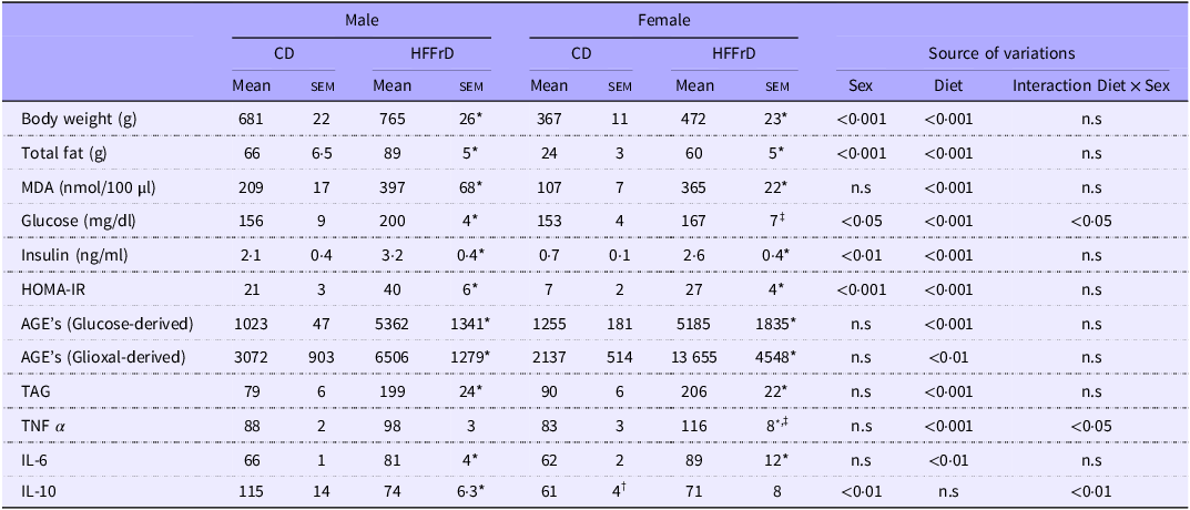
Body weight, total fat content, MDA levels, serum insulin concentrations and the homeostatic model assessment for insulin resistance increased in rats of both sexes of the HFFrD group compared with those in the CD group. Although males weighed more than females, they had higher total fat content and higher serum insulin levels and homeostatic model assessment for insulin resistance. However, these effects were global (sex effect P < 0·05), as no interaction between sex and the diet was observed.
Serum glucose concentrations increased in males but not in females in the HFFrD group compared with those in the CD group. A significant increase (P < 0·05) was observed in males fed the HFFrD compared with females, and an interaction between sex and diet was observed (P < 0·05). Compared with those in the CD group, the MDA levels in both males and females in the HFFrD group increased (P < 0·001). No sex differences or sex × diet interactions were detected.
Compared with those in the CD group, the serum glucose-derived AGE, glyoxal-derived AGE, TAG and IL-6 levels in male and female rats in the HFFrD group were increased (P < 0·001). No differences were observed between the sexes, and no sex × diet interactions were detected.
Serum TNFα concentrations increased in females but not in males in the HFFrD group compared with those in the CD group. A significant sex × diet interaction was observed (P < 0·05), although no overall sex effect was detected (P < 0·05).
Serum IL-10 concentrations decreased in males but did not change in females in the HFFrD group compared with those in the CD group. A sex effect (P < 0·01) and a sex × diet interaction (P < 0·01) were observed (Table 1).
Body weight, total fat, lipoperoxidation (MDA), glucose and lipid metabolism and cytokines
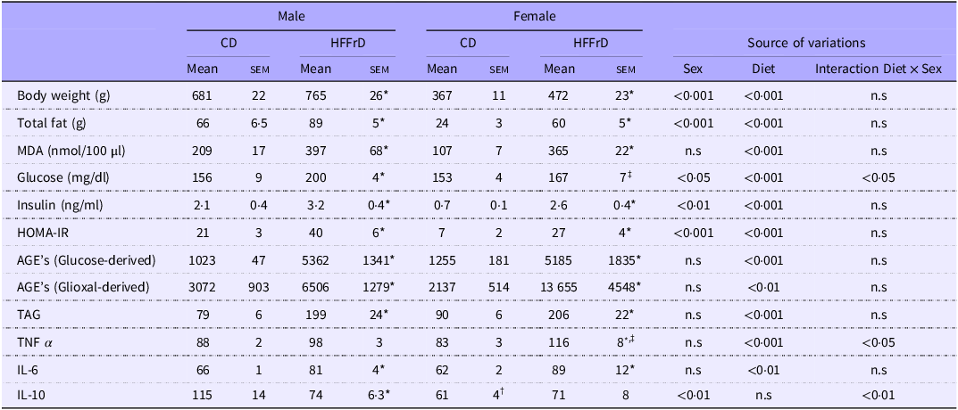
Table 1 Long description
A table comparing various health metrics between male and female subjects on different diets. The table has 12 rows and 13 columns. The columns are labeled as Male CD Mean, Male CD SEM, Male HFFrD Mean, Male HFFrD SEM, Female CD Mean, Female CD SEM, Female HFFrD Mean, Female HFFrD SEM, Source of variations Sex, Source of variations Diet, Source of variations Interaction Diet x Sex. The rows are labeled as Body weight (g), Total fat (g), MDA (nmol/100 µl), Glucose (mg/dl), Insulin (ng/ml), HOMA-IR, AGE's (Glucose-derived), AGE's (Glioxal-derived), TAG, TNF α, IL-6, IL-10. Each row provides mean and standard error of the mean (SEM) values for male and female subjects on control diet (CD) and high-fat high-fructose diet (HFFrD), along with the sources of variations due to sex, diet, and their interaction.
HOMA-IR, homeostatic model assessment for insulin resistance; AGE, advanced glycation end-products; n.s, non-significant.
Values are mean (sem), n 7 rats x sex and group. Control group (CD: 11 % kcal from fat and 0 % kcal from fructose) or a high-fat and high-fructose diet (HFFrD: 48 % kcal from fat and 33 % kcal from fructose). P < 0·05: *v. CD within male or female; † v. male CD; ‡ v. male HFFrD.
Higher basal levels of some variables were observed in males than in females within the CD group, which contributed to the magnitude of the percentage changes observed in animals fed the HFFrD. In the HFFrD group, the percentage of accumulated total fat was higher in females than in males, with an average increase of 150 % in females and 35 % in males compared with that in the CD group. Serum MDA concentrations increased by 241 % in females and 90 % in males in the HFFrD group compared with those in the CD group. Similarly, serum glyoxal-derived AGE levels increased by 539 % in females and 112 % in males compared with their respective CD groups. When females and males in the HFFrD group were compared, the glyoxal-derived AGE concentration was 110 % higher in females. In contrast, glucose-derived AGE level increased by 313 % in females and 424 % in males in the HFFrD group compared with those in the CD group, with serum glucose-derived AGE concentrations being slightly higher (3·4 %) in males than in females (Table 1).
Expression of genes involved in insulin synthesis in pancreatic islets
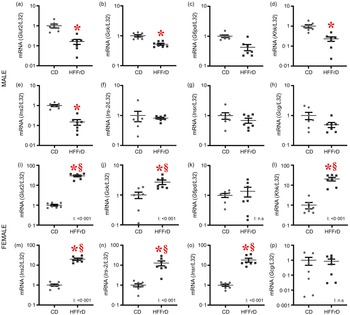
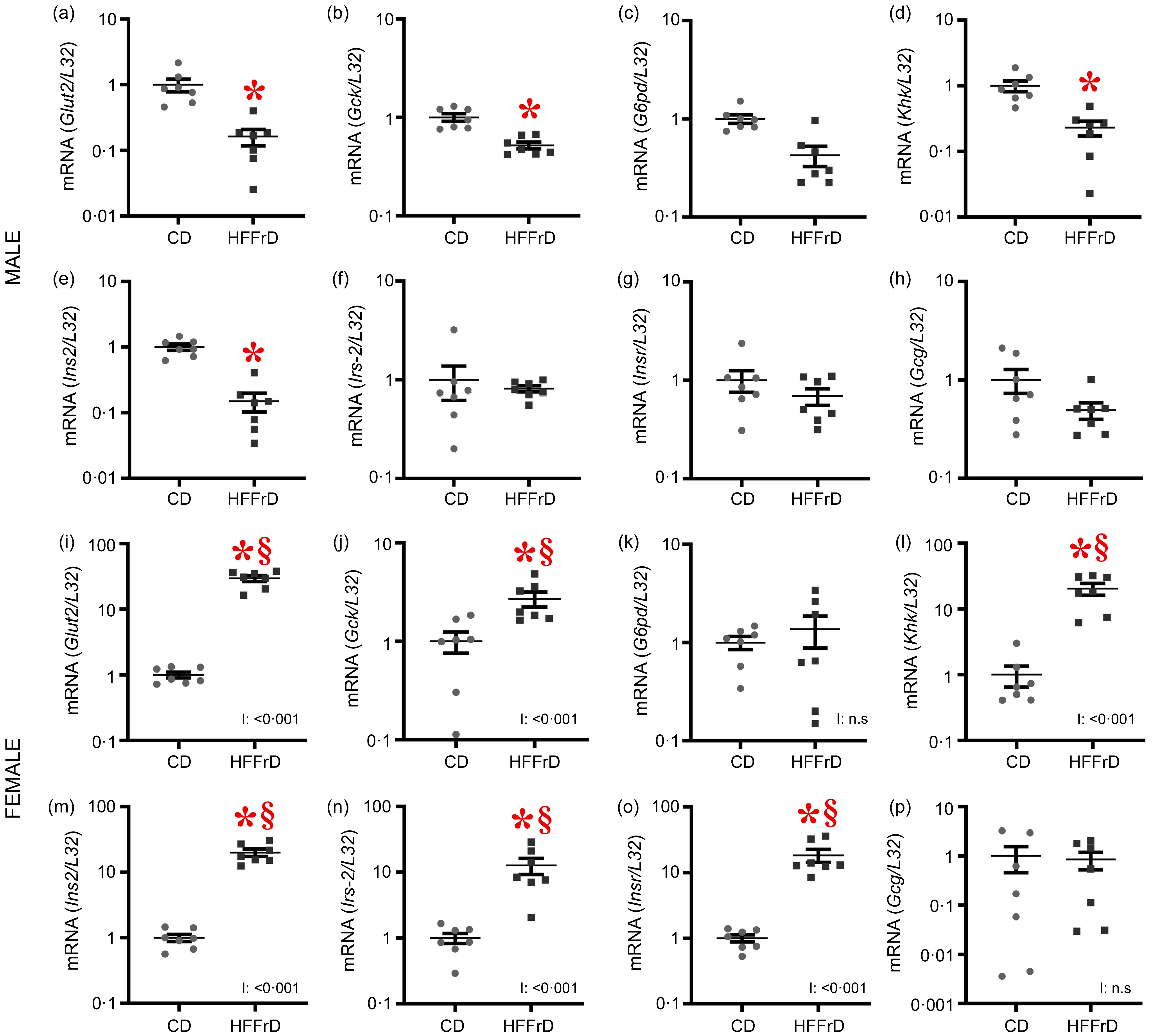
The mRNA expression of Glut2 (glucose transporter-2), Gck (glucokinase), Khk (ketohexokinase) and Ins2 (insulin 2) was lower in males (Figure 1(a, (b), (d), (e)) and higher in females (Figure 1(i), (j), (l), (m)) in the HFFrD group than in the CD group, and a sex × diet interaction was observed (P < 0·001). A significant sex effect was detected for the genes analysed (P < 0·001), including Gck, although with a smaller magnitude (P < 0·01), and a robust sex × diet interaction was also observed (P < 0·001). No differences were observed in G6pd (glucose-6-phosphate dehydrogenase) expression between the groups (Figure 1(c), (k)), although the comparison between males in the HFFrD and CD groups revealed a borderline statistical trend (P = 0·06) (Figure 1(c)). No sex differences or sex × diet interactions were detected. The mRNA expression of Irs-2 (insulin receptor substrate-2) and Insr (insulin receptor) increased in females but remained unchanged in males from the HFFrD group compared with those from the CD group (Figure 1(n), (o)). An overall significant sex effect (P < 0·001) and a sex × diet interaction were observed (P < 0·001). No differences in the effects of diet, sex, or the sex × diet interaction on Gcg (glucagon) expression were observed (Figure 1(h), (p)).
mRNA expression of genes involved in insulin secretion in pancreatic islets. Male: (a) Glut2, (b) Gck, (c) G6pd, (d) Khk, (e) Ins2, (f) Irs-2, (g) Insr and (h) Gcg; Female: (i) Glut2, (j) Gck, (k) G6pd, (l) Khk, (m) Ins2, (n) Irs-2, (o) Insr and (p) Gcg. Mean (sem), n 7 rats x sex and group. Control group (CD: 11 % kcal from fat and 0 % kcal from fructose) or a high-fat and high-fructose diet (HFFrD: 48 % kcal from fat and 33 % kcal from fructose). P < 0·05; * v. CD within male or female. § v. male HFFrD. I: interaction; n.s, non- significant.
Expression of genes encoding transcription factors associated with insulin synthesis in pancreatic islets
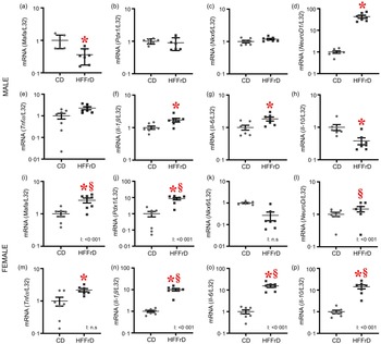
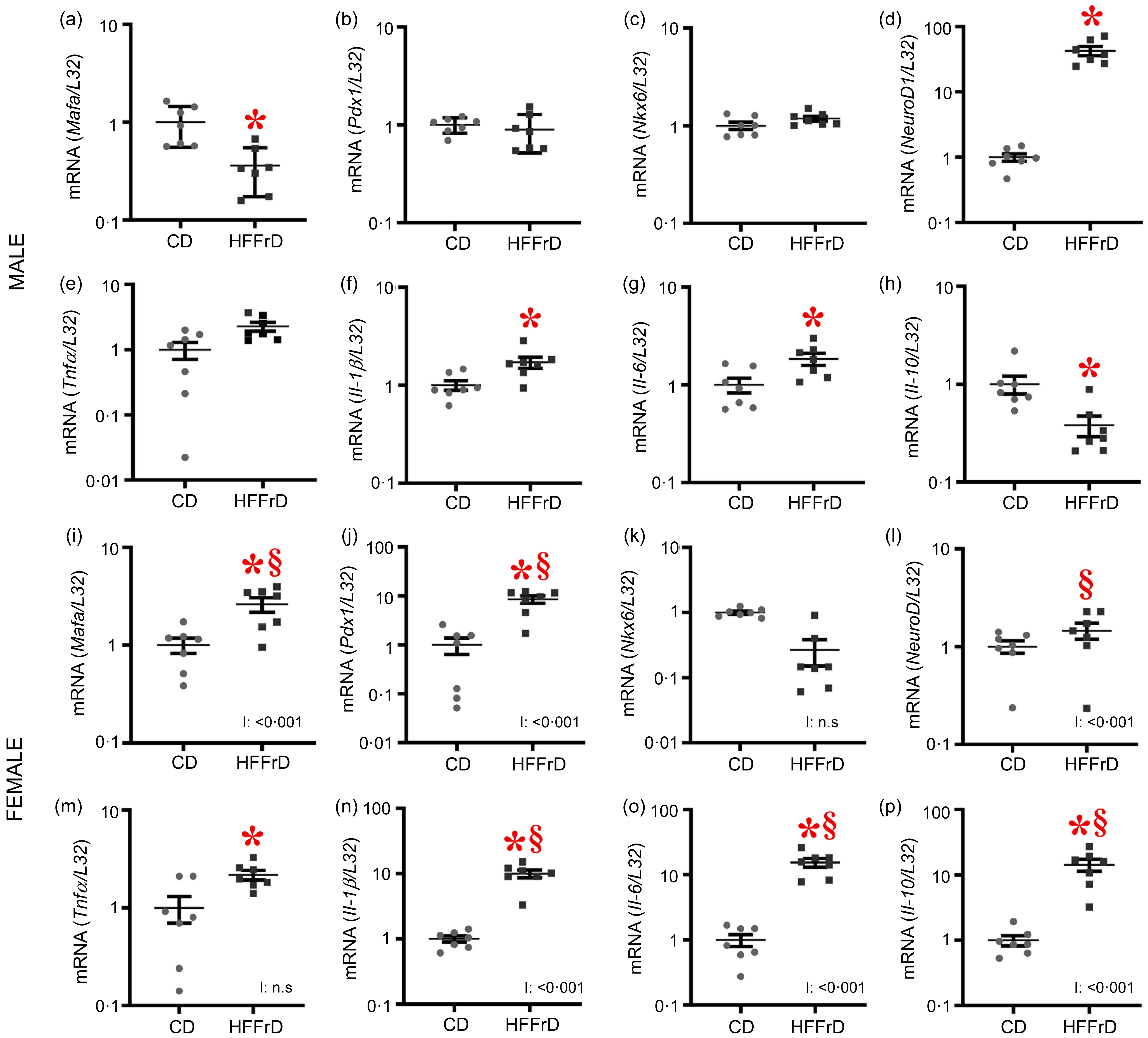
Mafa (MAF bZIP transcription factor A) mRNA expression was lower in males (Figure 2(a)) and higher in females (Figure 2(i)) in the HFFrD group than in the CD group. Pdx1 (pancreatic and duodenal homeobox 1) expression increased in females and remained similar in males from the HFFrD group compared with those from the CD group (Figure 2(b), (j)). NeuroD1 (neuronal differentiation 1) expression was increased in males and unchanged in females from the HFFrD group compared with the CD group (Figure 2(d), (l)). An overall significant sex effect was detected for the genes (P < 0·001), except for Pdx1, which showed a borderline statistical trend (P = 0·06), and a sex × diet interaction was also observed (P < 0·001). No differences in the effects of the diet, sex or sex × diet interaction on Nkx6-1 (NK6 homeobox 1) expression were observed (Figure 2(c), (k)).
mRNA gene expression of transcriptional factors involved in insulin secretion and inflammation in pancreatic islets. Male: (a) Mafa, (b) Pdx1, (c) Nkx6, (d) NeuroD1, (e) Tnfα , (f) Il-1β , (g) Il-6 and (h) Il-10; female: (i) Mafa, (j) Pdx1, (k) Nkx6, (l) NeuroD1, (m) Tnfα , (n) Il-1β , (o) Il-6 and (p) Il-10. Mean (sem), n 7 rats x sex and group. Control group (CD: 11 % kcal from fat and 0 % kcal from fructose) or a high-fat and high-fructose diet (HFFrD: 48 % kcal from fat and 33 % kcal from fructose). P < 0·05; * v. CD within male or female. § v. male HFFrD. I: interaction; n.s, non-significant.
Expression of genes involved in inflammation in pancreatic islets
In terms of proinflammatory genes, Tnfα mRNA expression was increased in females (Figure 2(m)) but remained unchanged in males (Figure 2(e)) from the HFFrD group compared with that in the CD group. In contrast, the levels of Il-1β (interleukin 1 beta) and Il-6 (interleukin 6) were higher in both males and females from the HFFrD group the CD group (Figure 2(f), (g), (n), (o)), with a more pronounced increase in females. For Il-1β and Il-6 gene levels, a significant sex effect (P < 0·001) and a sex × diet interaction were observed (P < 0·001).
Compared with that in the CD group, the mRNA expression of the anti-inflammatory gene Il-10 (interleukin 10) decreased in males (Figure 2(h)) and increased in females (Figure 2(p)) from the HFFrD group. Significant sex effects (P < 0·001) and sex × diet interactions were also observed (P < 0·001).
Expression of genes involved in the antioxidant response, apoptosis and the endocannabinoid system in pancreatic islets
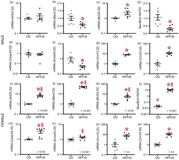
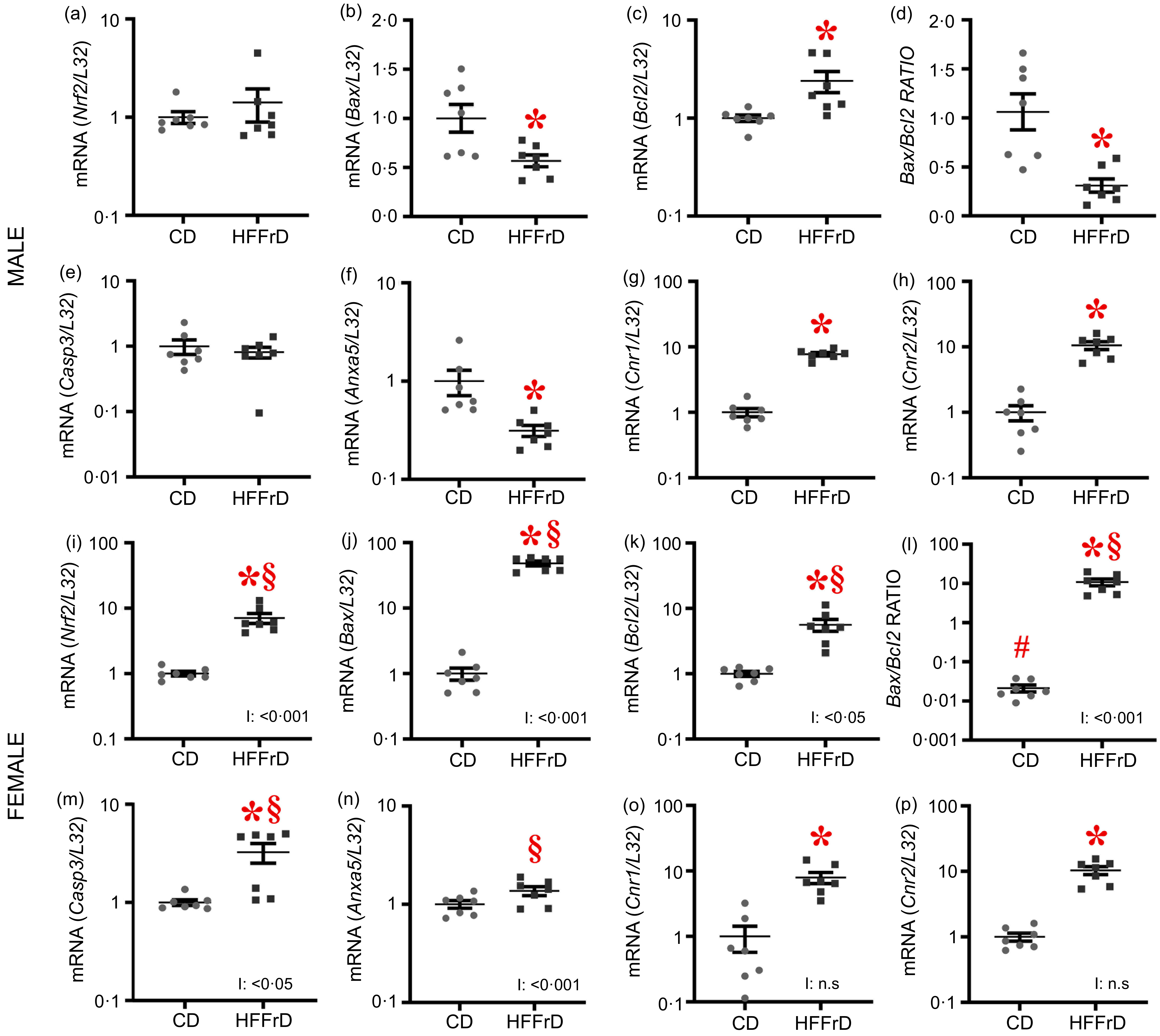
The mRNA expression of Nrf2 (nuclear factor erythroid 2-related factor 2) and Casp3 (caspase 3) was increased in females (Figure 3(i), (m)) but remained unchanged in males (Figure 3(a), 3(e)) from the HFFrD group compared with those in the CD group. Bcl2 (apoptosis regulator) expression was increased in both males (Figure 3(c)) and females (Figure 3(k)) from the HFFrD group compared with the CD group. Bax (Bcl2-associated X, apoptosis regulator) expression and the Bax/Bcl2 ratio were decreased in males (Figure 3(b), (d)) and increased in females (Figure 3(j), (l)) in the HFFrD group compared with those in the CD group. Anxa5 (annexin A5) expression decreased in males (Figure 3(f)) but remained unchanged in females (Figure 3(n)) from the HFFrD group compared with the CD group. A significant sex effect (P < 0·001) and a sex × diet interaction (P < 0·001) were detected for these genes. The mRNA expression of Cnr1 (cannabinoid receptor type 1) and Cnr2 (cannabinoid receptor type 2) was increased in both males (Figure 3(g), (h)) and females (Figure 3(o), (p)) from the HFFrD group compared with the CD group. No sex differences or sex × diet interactions were detected for these genes.
mRNA expression of genes involved in antioxidant response, apoptosis and endocannabinoid system in pancreatic islets. Male: (a) Nrf2, (b) Bax, (c) Bcl2, (d) Bax/Bcl2 ratio, (e) Casp3, (f) Anxa5, (g) Cnr1 and (h) Cnr2; female: (i) Nrf2, (j) Bax, (k) Bcl2, (l) Bax/Bcl2 ratio, (m) Casp3, (n) Anxa5, (o) Cnr1 and (p) Cnr2. Mean (sem), n 7 rats x sex and group. Control group (CD: 11 % kcal from fat and 0 % kcal from fructose) or a high-fat and high-fructose diet (HFFrD: 48 % kcal from fat and 33 % kcal from fructose). P < 0·05; * v. CD within male or female. § v. male HFFrD. I: interaction; n.s, non- significant.
Immunohistochemical staining for INS, GCG, CASP3, CNR1, CNR2 and TNFα and TUNEL staining in the pancreas
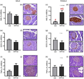
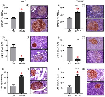
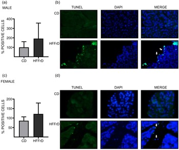
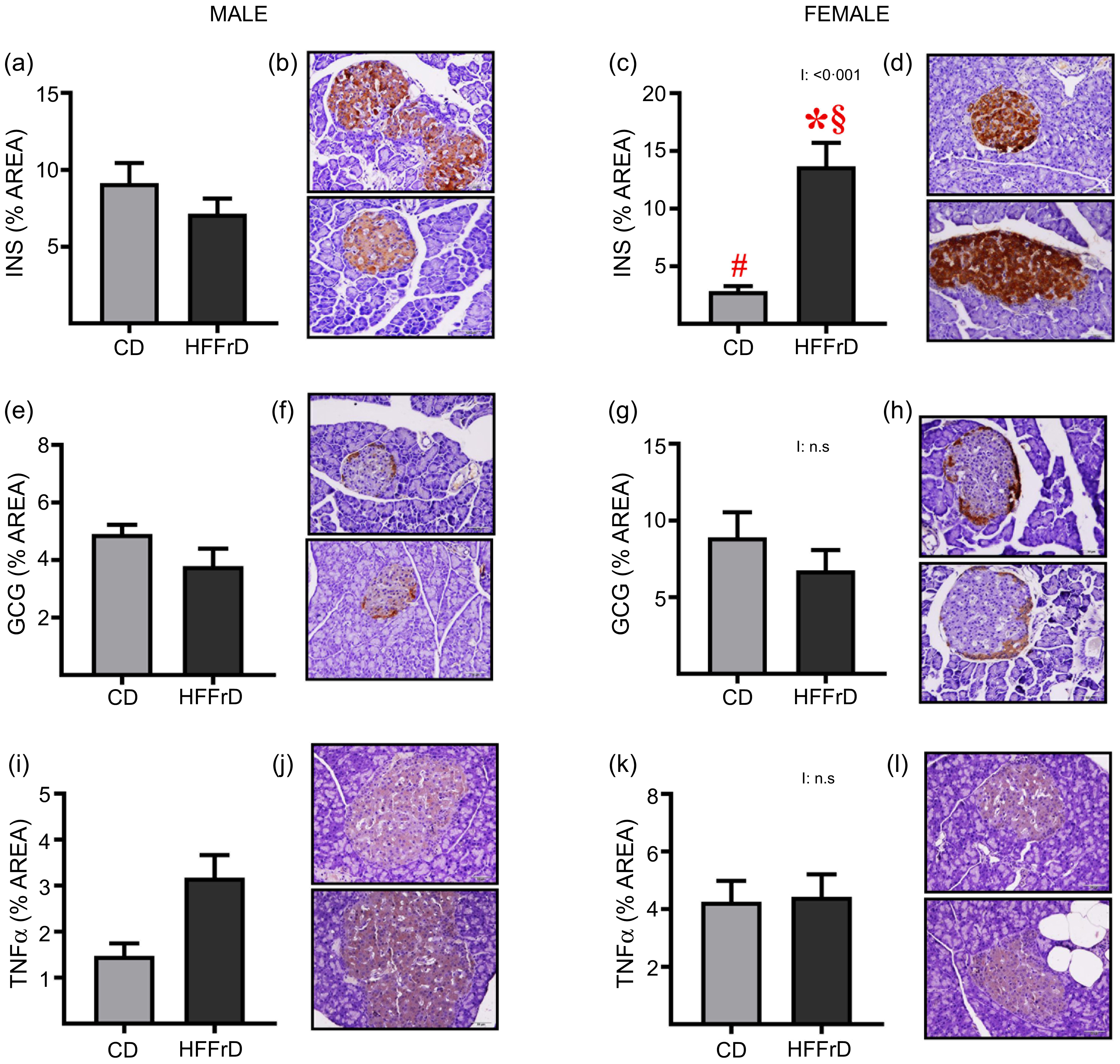
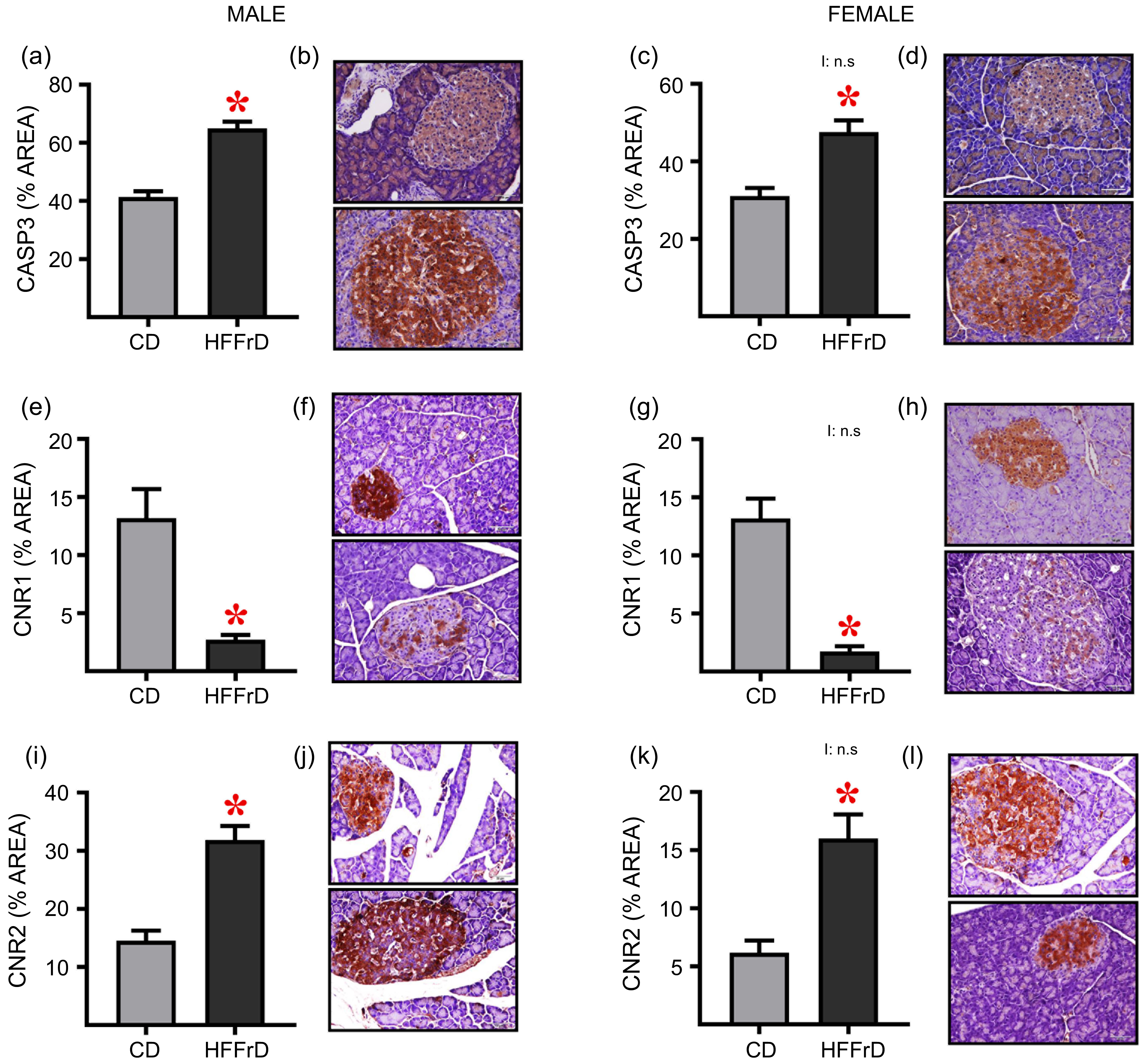
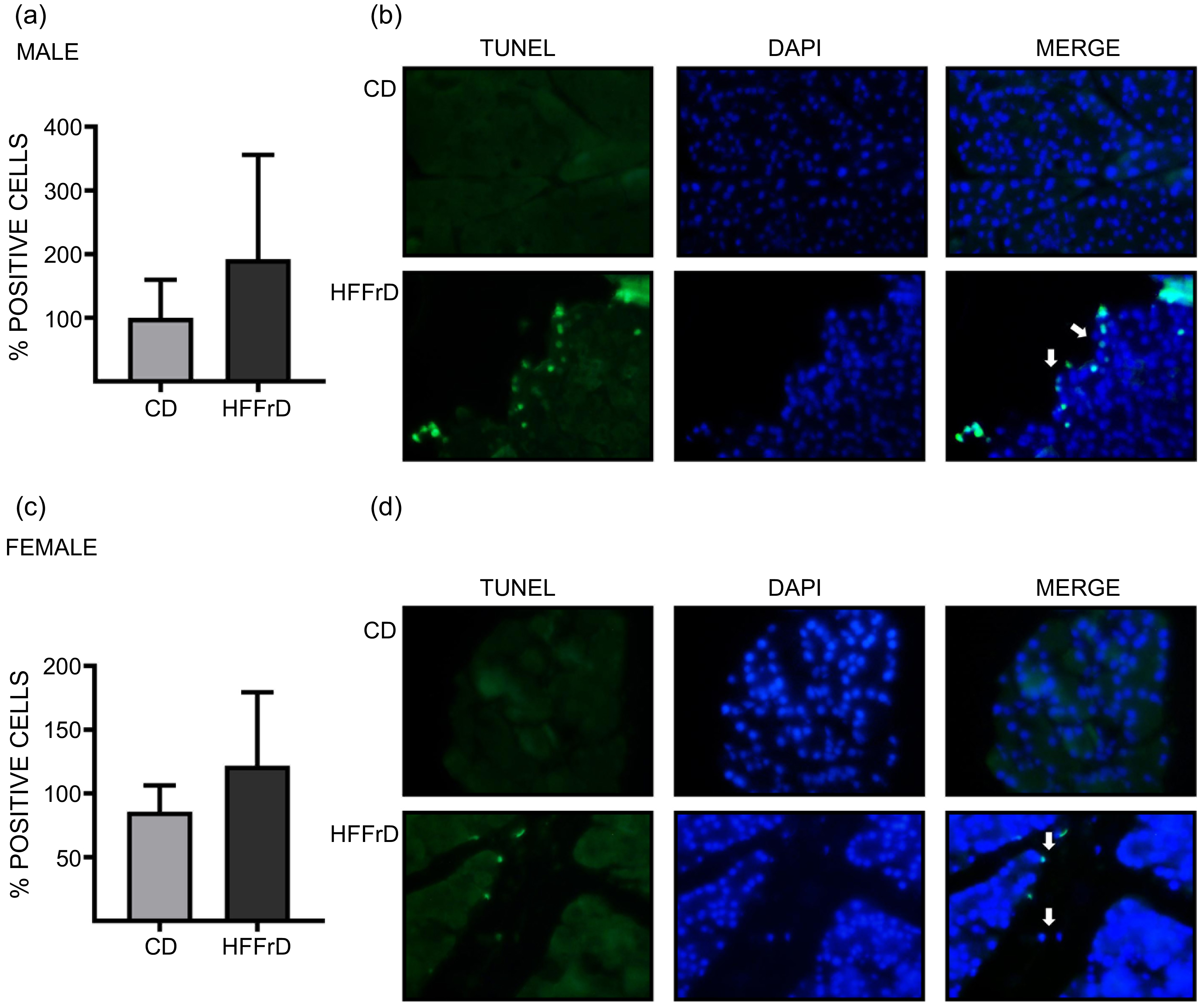
The percentage of the insulin-positive immunostained area was increased in females (Figure 4(c)) but remained similar in males (Figure 4(a)) from the HFFrD group compared with the CD group. Although no overall sex effect was detected, a significant sex × diet interaction was observed (P < 0·001). No diet-related differences in GCG or TNFα immunostaining were detected in males (Figure 4(e), (i)) or females (Figure 4(g), (k)). The percentages of the area of CASP3 and CNR2 immunostaining were increased in both males (Figure 5(a), (i)) and females (Figure 5(c), (k)) from the HFFrD group compared with the CD group. In contrast, CNR1 immunostaining decreased in both males (Figure 5(e)) and females (Figure 5(g)) from the HFFrD group. Although an overall sex effect was observed for CNR1 and CNR2 (P < 0·001) and for GCG and TNFα with a smaller magnitude (P < 0·05), no sex × diet interaction was detected for these markers. Additionally, the TUNEL assay showed no differences in apoptosis between the HFFrD and CD groups in either males (Figure 6(a)) or females (Figure 6(c)).
Pancreatic immunohistochemistry. Male: (a) INS, (e) GCG, (i) TNFα immunostained area (%), (b), (f) and (j) representative micrograph (40×); female: (c) INS (g) GCG (k) TNFα immunostained area (%), (d), (h) and (l) Representative micrograph (40×). Upper micrograph: CD group; lower micrograph: HFFrD group. Mean (sem), n 6 rats x sex and group. Control group (CD: 11 % kcal from fat and 0 % kcal from fructose) or a high-fat and high-fructose diet (HFFrD: 48 % kcal from fat and 33 % kcal from fructose). P < 0·05; * v. CD female. # v. male CD and § v. male HFFrD. I, interaction; n.s, non-significant.
Pancreatic immunohistochemistry. Male: (a) CASP3, (e) CNR1, (i) CNR2 immunostained area (%), (b), (f) and (j) representative micrograph (40×); female: (c) CASP3 (g) CNR1 K) CNR2 immunostained area (%), (d), (h) and (l) representative micrograph (40×). Upper micrograph: CD group; lower micrograph: HFFrD group. Mean (sem), n 6 rats x sex and group. Control group (CD: 11 % kcal from fat and 0 % kcal from fructose) or a high-fat and high-fructose diet (HFFrD: 48 % kcal from fat and 33 % kcal from fructose). P < 0·05; * v. CD within male or female. I, interaction; n.s, non-significant.
Pancreatic apoptosis by TUNEL. (a) Male and (c) female-positive cells (%), (b) and (d) representative micrograph (40×). Upper micrograph: CD group; lower micrograph: HFFrD group. Mean (sem), n 6 rats x sex and group. Control group (CD: 11 % kcal from fat and 0 % kcal from fructose) or a high-fat and high-fructose diet (HFFrD: 48 % kcal from fat and 33 % kcal from fructose).
Discussion
Obesity is a multifactorial disease, and its high prevalence is significantly influenced by poor dietary habits and a sedentary lifestyle. Foods high in fat and sugar have been identified as primary contributors to increased glucolipotoxicity in multiple organs, such as the brain, kidney, muscle, liver, adipose tissue and pancreas, because of altered carbohydrate and lipid metabolism, inflammation and oxidative damage(Reference Lama, Pirozzi and Severi31–Reference Melo, Zacarias and Oliveira35).
This study aimed to evaluate the effects of a HFFrD consumption on the expression of key genes that participate in insulin transcription and synthesis, major transcription factors, metabolic stress responses and genes related to inflammation and apoptosis in a rat model. Additionally, we explored whether HFFrD consumption induces changes in a sex-specific manner. The present study showed that 22 weeks (5·5 months) of HFFrD consumption induced multiple metabolic alterations in both male and female rats. These findings included increased body weight and total fat gain, hypertriglyceridaemia, insulin resistance, the accumulation of AGE, the dysregulation of proinflammatory cytokines and elevated serum concentrations of lipoperoxidation products such as malondialdehyde (MDA), resulting in oxidative stress and the formation of glyoxal-derived AGE, which are associated with increased oxidative lipid degradation(Reference Luevano-Contreras and Chapman-Novakofski36,Reference Son, Hwang and Han37) . Elevated concentrations of glyoxal-derived AGE, such as carboxymethyl-lysine (toxic AGE), bind to specific receptor for AGE (RAGE), triggering inflammatory responses and oxidative stress, and thereby generating a harmful cycle. This result was verified by the higher concentrations of TNFα, one of the main proinflammatory cytokines, in the serum of females fed HFFrD than in that of males. In fact, compared with males, females presented 110 % higher serum concentrations of glyoxal-derived AGE; nevertheless, the serum concentrations of MDA, an indicator of lipid peroxidation, increased in both males and females in the HFFrD group, with no significant difference between sexes. These findings in the present study, suggest that, in animals fed HFFrD, serum concentrations of these toxic AGE species may be a better marker of oxidative stress from lipid peroxidation than MDA. Additionally, serum concentrations of glucose-derived AGE (nontoxic AGE) increased in both males and females in the HFFrD group. These nontoxic AGE species are excellent markers of long-term glycaemic control(Reference Jimenez, Diaz-Diaz and Castro38). The lack of significant differences between the sexes could be because both groups fed the HFFrD produced maximal concentrations of glucose-derived AGE during the study period.
However, the serum concentrations of glucose-derived AGE observed in this experimental model are significantly higher than those observed in another rat model of metabolic syndrome induced by chronic consumption of 30 % sucrose, which is associated with similar levels of insulin resistance(Reference Perez-Torres, Torres-Narvaez and Pedraza-Chaverri39,Reference Perez-Torres, Gutierrez-Alvarez and Guarner-Lans40) . This evidence reinforces the concept that measuring these nontoxic AGE can reveal underlying metabolic alterations that a measurement of insulin resistance cannot detect. Taken together, these results show that, in contrast to the marked insulin resistance observed in males, female rats displayed lower HOMA-IR and more regulated glycemic control. However, this apparent advantage might be challenged by a significant increase in systemic glucose oxidation. Additionally, greater fat accumulation, along with higher levels of glyoxal-derived AGE and lipid peroxidation in females, suggests increased susceptibility to lipotoxic stress under HFFrD, leading to a pro-oxidant systemic environment that could affect islet integrity.
Research conducted in rodents has shown that the consumption of a Western diet(Reference Canova, Sipkova and Arora41,Reference Mazzoli, Spagnuolo and De Palma42) , cafeteria diet(Reference Sagae, Paz and Zanardini43,Reference Velasco-Gutierrez, de Alvarez-Buylla and Montero44) , high-fat diet(Reference Babaeenezhad, Farahmandian and Sotoudeheian45,Reference Pekand, Gholami and Abednatanzi46) and water enriched with sugars, specifically sucrose(Reference Osman, Laczko and Vagvolgyi47,Reference Suhaimi, Azemi and Mokhtar48) and fructose(Reference Tayfur, Palabiyik and Meric49,Reference Campanher, Andrade and Lopes50) , significantly contributes to the alarming incidence of overweight/obesity and its associated comorbidities, such as type 2 diabetes mellitus, dyslipidaemia, nonalcoholic fatty liver disease, systemic inflammation and oxidative dysfunction. These studies are consistent with our aforementioned results; however, some differences exist with respect to our experimental model. Most of the subjects being studied were male, with dietary exposure ranging from 2 to 16 weeks, and the studies focused mainly on the liver and adipose tissue.
Few studies have addressed the pancreas, mostly examining the morphological changes in pancreatic islets and the expression of certain genes involved in carbohydrate and lipid metabolism. Therefore, the results of this study allow us to elucidate the effects of HFFrD consumption on the expression of genes associated with insulin synthesis and to determine whether sex-related modifications occur.
In our study, the HFFrD-induced dysregulation of glucose sensing was involved in sex-dependent insulin expression. In male rats, we observed decreased expression of genes associated with glucose sensing (Glut2 and Gck) and metabolism (Khk), as well as the Ins2 gene. We also observed decreased expression of the Mafa gene and, conversely, overexpression of the NeuroD1 gene. These findings suggest that β-cells exhibit an adaptive response to increase and stimulate insulin synthesis, as evidenced by increased serum insulin concentrations. In contrast, females presented increased expression of the Glut2, Gck, Khk, Ins2, Irs-2 and Insr mRNAs, as well as increased expression of the transcription factors Mafa and Pdx1; these differential expression patterns support the idea that glucose metabolism exhibits a sex-specific pattern, as females fed the HFFrD also presented higher serum insulin concentrations and insulin resistance than those in the CD group.
Experimental studies have shown a decrease in Mafa mRNA in males fed an HFFrD. This factor is associated with the expression of the insulin gene and the modulation of transcription factors involved in the insulin signalling pathway(Reference Liang, Chirikjian and Pajvani51). Elevated glucose concentrations have been shown to decrease Mafa gene expression(Reference Kataoka, Han and Shioda52), as observed in our data. Another study demonstrated that insulin transcription and the insulin content decreased in Mafa-deficient mice; consequently, Ins2, Pdx1, Beta2 and Glut2 transcript levels decreased markedly(Reference Zhang, Moriguchi and Kajihara53). These observations support our findings from the HFFrD groups, as lower expression of genes involved in the insulin signalling pathway was observed in males. In contrast, higher expression of Mafa and genes involved in insulin signalling was observed in females.
The released insulin can exert autocrine effects on the insulin receptor, causing its autophosphorylation and catalysing the subsequent phosphorylation of Irs1–2. This process leads to the activation of different signalling pathways, particularly the PI3K–Akt and Ras–ERK pathways, which are activated by transcription factors such as Mafa, Pdx1 and NeuroD1, respectively, which are directly involved in insulin gene transcription(Reference Lazo-de-la-Vega and Fernandez-Mejia54,Reference Khoo, Griffen and Xia55) . Based on the results of these investigations, we hypothesise that the low expression of the Mafa gene in males fed the HFFrD negatively affects the PI3K–Akt pathway, in which this factor is involved; however, the overexpression of NeuroD1 observed in our study directly compensates for insulin transcription through the Ras–ERK pathway(Reference Lazo-de-la-Vega and Fernandez-Mejia54,Reference Khoo, Griffen and Xia55) , which supports the findings of higher blood insulin concentrations in this group than in the control group. On the other hand, in females, the PI3K–Akt pathway could be involved, since an increase in the expression of the transcription factors Mafa and Pdx1 and, consequently, an increase in blood insulin concentrations and protein levels detected by immunohistochemistry were observed compared with those in the CD group.
Few studies in the literature have investigated these transcription factors in female rats. To our knowledge, only one publication has evaluated the effects of a high-fat diet on the expression of Pax6 and its target genes Pdx1, Mafa and Glut2 in the pancreas of pregnant rats, reporting that the consumption of a high-fat diet can affect glucose tolerance and insulin resistance and reduce insulin secretion and sensitivity in the late stage of pregnancy, most likely due to reduced expression of Pax6 and its target genes. In our study, despite the observation that females showed insulin resistance, discrepancies in the expression of these genes were observed, which could be attributed to the type of diet and hormonal changes that occur during pregnancy(Reference Wu, Liu and Wang14).
On the other hand, obesity caused by the consumption of high-fat diets and/or sugar-rich products has been shown to induce chronic low-grade inflammation, leading to a variety of metabolic diseases. During obesity, TNF family cytokine signalling mediates cell death (apoptosis) and inflammation in adipose tissue, leading to lipid efflux, glucotoxicity and insulin resistance. These events ultimately lead to ectopic lipid accumulation, glucose intolerance and other metabolic complications with life-threatening consequences(Reference Hildebrandt, Ibrahim and Peltzer56). With respect to proinflammatory cytokines, in our study, we observed that in both sexes, HFFrD consumption increased the serum concentrations of TNFα and IL-6 and mRNA expression in pancreatic islets. Similar effects were observed in rodents fed a high-fat(Reference Abderrazak, El Hadri and Bosc57,Reference Flanagan, Brown and Santiago58) and high-fructose diet(Reference Prince, Lanzi and Toblli59,Reference Wang, Qi and Song60) .
A study investigated the effects of a standard American diet (16·7 % protein, 52·9 % carbohydrate and 17·1 % fat) on serum IL-10 levels in rats over 20 weeks. Serum IL-10 levels increased only in female rats, with no changes observed in males(Reference Totsch, Quinn and Strath61). Human studies have reported that in metabolically healthy obese individuals, IL-10 concentrations tend to be normal or slightly increased, indicating that the inflammatory state is attenuated(Reference Esposito, Pontillo and Giugliano62). Limited studies on animals have compared Il-10 levels between sexes; Albogami et al.(Reference Albogami, Hassan and Abdel-Aziem63) reported that in rats, the consumption of a high-fat diet decreased Il-10 expression in pancreatic tissue in both sexes. Our study showed that males presented a decrease in Il-10 mRNA expression and serum protein levels, whereas females presented an increase in mRNA expression but showed no alterations in serum IL-10 concentrations compared to the control group. However, males in the control group presented higher serum IL-10 concentrations than females. These results suggest that females exhibit a compensatory response, reflecting the immune system’s effort to restore homeostasis by counterbalancing the excessive inflammatory response triggered by the diet, whereas in males, this finding may imply the pre-existence of metabolic syndrome(Reference Sapan, Paturusi and Jusuf64). Similarly, only females fed the HFFrD showed an increase in Nrf2 mRNA expression, with no changes in males. These sex-related changes may be associated with the activation of antioxidant enzymes such as glutathione peroxidase, superoxide dismutase and glutathione reductase, which constitute the first line in the antioxidant defence system and reduce vulnerability to oxidative damage caused by the excessive production of reactive oxygen species(Reference Abebe, Mahadevan and Bogachus65).
Regarding the apoptotic process, oxidative stress and the chronic toxicity of high serum glucose levels contribute to the increase in caspase levels and the decrease in Bcl-2 expression in pancreatic β-cells(Reference Poitout and Robertson66,Reference Mandrup-Poulsen67) . In our study, we observed decreased expression of the Bax and Anxa5 mRNAs (markers of early apoptosis) in males fed an HFFrD, whereas the expression of the Bax and Casp3 mRNAs increased in females. Importantly, the Bax/Bcl2 ratio showed a dimorphic pattern, with females fed an HFFrD showing an increased susceptibility to apoptosis compared with males, and this ratio is increased in females(Reference Samm, Werner and Ruckert68), which is associated with an increase in the expression of the Casp3 mRNA, which plays a key role in the execution phase of early apoptosis(Reference Tomita69).
In our study, immunohistochemical staining showed increased levels of cleaved CASP3 in both males and females, but no differences in apoptosis were detected using TUNEL staining (a late apoptosis marker). This apparent discrepancy could be explained by the timing of activation of the markers of apoptosis. Since TUNEL staining detects DNA fragmentation in the terminal stages of apoptosis, the detection of TUNEL-positive cells may not accurately reflect the level of apoptosis, which may explain why the levels of the apoptosis markers reported in the present study are inconsistent. Our results are supported by those obtained by Nishimura et al. (Reference Nishimura, Iwashita and Hayashi70), who studied a mouse model fed a 60 % high-fat diet and observed greater numbers of CASP3-positive cells than in the control group; however, fewer β-positive cells were detected by TUNEL staining.
Cannabinoid receptors play key roles in energy metabolism and glucose and lipid homeostasis and participate in the modulation of insulin and glucagon secretion(Reference Cortes-Justo, Garfias-Ramirez and Vilches-Flores71). Our findings showed that HFFrD consumption increased the mRNA expression of Cnr1 and Cnr2 in pancreatic islets from both male and female rats. These results are consistent with those reported by Barajas-Martínez et al. (Reference Barajas-Martínez, Bermeo and de la Cruz72), who observed similar results in a rat model of diet-induced metabolic syndrome. Endogenous stimulation of the Cnr1 receptor plays a major role in the development of obesity, while Cnr2 plays a role in the inflammatory response induced by a high-fat diet(Reference Cortes-Justo, Garfias-Ramirez and Vilches-Flores71,Reference Ravinet Trillou, Delgorge and Menet73–Reference Bermudez-Silva, Sanchez-Vera and Suarez75) , which is consistent with the weight gain, fat accumulation and increased levels of inflammatory biomarkers observed in our study. However, when CNR1 and CNR2 were analysed using immunohistochemistry, we observed a negative regulation of CNR1 expression and a positive regulation of CNR2 protein levels by the HFFrD in both sexes. One possible explanation for this apparent discrepancy is that short-term administration of an obesogenic diet differentially affects the mRNA and protein expression of cannabinoid receptors, and long-term administration can cause a maladaptation(Reference Barajas-Martínez, Bermeo and de la Cruz72) that produce a compensatory mechanism counteracting the negative regulatory effect that the endocannabinoid system exerts on metabolic control during the onset of more serious dysfunctions, such as the development of type 2 diabetes and metabolic syndrome, in response to obesogenic diets(Reference Dörnyei, Vass and Juhász76).
Previous studies have demonstrated the harmful effects of an obesogenic diet on males(Reference Tramunt, Smati and Grandgeorge77,Reference Maric, Krieger and van der Velden78) . Our data revealed that compared with female rats, male rats were more vulnerable to developing metabolic abnormalities. Nevertheless, females exhibited adaptive responses to mitigate the adverse metabolic effects of the HFFrD diet. One explanation may be the role of oestrogens previously described by Chansela et al.(Reference Chansela, Potip and Weerachayaphorn79) who determined that the protective role of oestradiol in β-cells is to prevent ectopic pancreatic fat accumulation, thereby restoring and maintaining β-cell function. The preservation of β-cell survival mitigates endoplasmic reticulum stress and oxidative damage(Reference Zhou, Ribas and Rajbhandari80).
Recent advances in noninvasive imaging have enabled the assessment of pancreatic β-cell function in vivo. Mn2+ has been used as a calcium analogue and has shown high sensitivity in detecting glucose-stimulated β-cell activity in the rat pancreas, indicating its promising translational potential(Reference Kershaw, Lilburn and Jansen81). In the future, these imaging approaches could complement molecular analyses, providing a more comprehensive understanding of β-cell dysfunction during diet-induced metabolic stress.
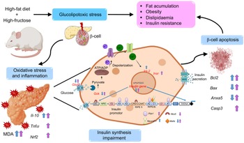
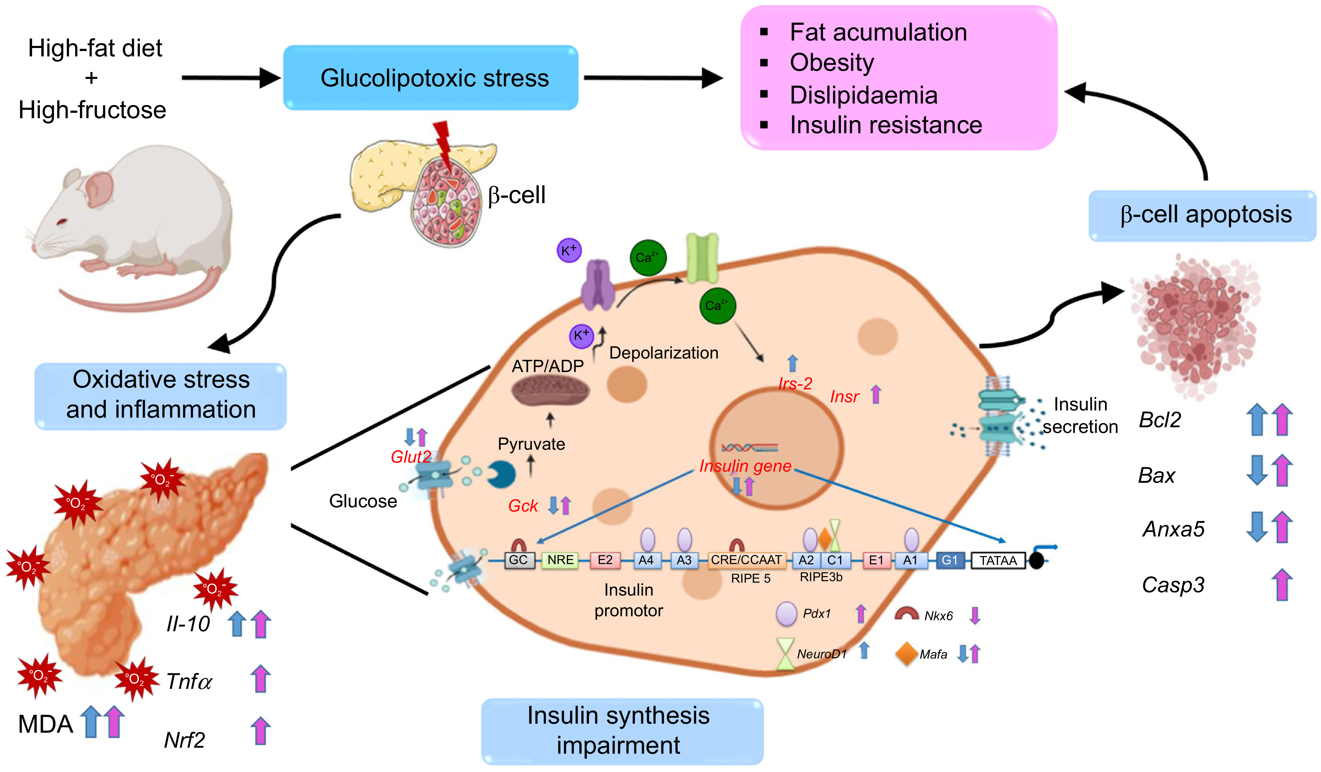
Our results indicate that consuming a HFFrD leads to glucolipotoxicity, which promotes excessive reactive oxygen species production and pancreatic inflammation. These changes disrupt β-cell insulin synthesis by altering the expression of genes and transcription factors that are essential for insulin synthesis signalling in a sex-dependent manner and activating genes related to inflammatory and apoptotic pathways. The resulting pancreatic dysregulation leads to fat accumulation, obesity and dyslipidaemia, all of which contribute to insulin resistance (Figure 7).
Summary of findings. A diet high-fat and high-fructose leads to glucolipotoxicity through sex-specific alterations in the expression of key genes associated with insulin secretion, oxidative stress, inflammation, and apoptosis. Male effects = blue arrow, Female effects = pink arrow. This figure was partially created with Biorender.
In conclusion, females fed an HFFrD exhibited lower vulnerability than males, showing a reduced disruption of insulin synthesis in β-cells, likely due to adaptive responses in insulin signalling, which are most likely associated with sex hormones. These findings highlight not only the importance of considering sex as a biological variable in metabolic research, particularly in terms of the mechanisms involved in insulin synthesis, but also provide the opportunity for timely interventions or appropriate treatments to prevent or mitigate the long-term consequences of dietary glucolipotoxicity while considering the relevance of sex-dependent effects.
Supplementary material
For supplementary material/s referred to in this article, please visit https://doi.org/10.1017/S0007114526106904
Acknowledgements
T. C. S-L. is a graduate student from Escuela Nacional de Ciencias Biológicas, Ciencias en Alimentos (IPN) and appreciates the financial support by CONAHCyT with the scholarship (252852) for her doctoral studies and BEIFI-IPN, grant numbers 20160499.
MVZ Mariela Contreras Escamilla provided support for animal facilities, and Msc David A. Olivas-Chairez and T.Q.I. Gimena Juárez-Pilares for their assistance in animal care and maintenance, as well as PhD. Carlos A. Ibáñez (Facultad de Química, UNAM) for his technical support
This work was partially supported by financial resources from INCMNSZ, Sociedad Mexicana de Nutrición y Endocrinología AC. (SMNE P E003909) and Instituto Politécnico Nacional, grant number SIP-20240629.
Conceptualisation: T. C. S-L., S. M. and L. A. R-C.; methodology and data analysis: T. C. S-L., A. M-M., C. J. B., V. R., E. D-D., L. V-H., P. C., J. A. S-R., S. M., and L. A. R-C.; preparation of manuscript: T. C. S-L., G. L. R-G.; S. M. and L. A. R-C.; review of manuscript: T. C. S-L., G. L. R-G., P. C., J. A. S-R., S. M., M.E. J-F. and L. A. R-C.; final approval of the manuscript: G. L. R-G., S. M., M. E. J-F., and L. A. R-C. All authors agree to the published version of the manuscript.
The authors declare no conflicts of interest.
The original contributions presented in this study are included in the article. Further inquiries can be directed to the corresponding author.
The nomenclature of genes and proteins was assigned in accordance with the guidelines for genetic nomenclature, markers, alleles and mutations in mice and rats, as set out in the manuscript.