Transthoracic two-dimensional echocardiography has been evaluated in the context of mass pre-participation cardiovascular evaluation of athletes because it is non-invasive, non-ionising, portable, and with acceptable costs. Reference Mont, Pelliccia and Piepoli1 Congenital or acquired coronary artery abnormalities are the second most common cause for sudden cardiac death in young athletic populations during training or competition. Reference Maron, Doerer, Haas, Tierney and Mueller2 It is well documented that coronary artery abnormalities can be detected by transthoracic echocardiography in a paediatric population using high-resolution transducers. Reference Gerling, Michel and Krutsch3
German elite soccer players in early adolescence (10–14 years of age) train between 8 and 14 hours and play one competitive match per week, for 11 ± 1 months per year. In this regard, the cardiovascular system has to increase its work significantly, compared with moderate intensity levels. Reference Wrigley, Drust, Stratton, Scott and Gregson4 In soccer player, intense and regular exercise does increase left ventricular wall thicknesses and left ventricular myocardial mass, as measured by echocardiography or MRI. Reference McClean, Riding and Wilson5,Reference Gerling, Pollinger, Michel, Dechant, Melter and Krutsch6 In adults training-induced myocardial hypertrophy involves a proportionate increase of coronary artery dimensions, Reference Pelliccia, Spataro, Granata, Biffi, Caselli and Alabiso7 but for adolescent athletes there is no sufficient data about coronary artery remodelling due to high-intensity exercise. Long-term high-intensity training stimuli might cause negative adaptations of the cardiovascular system. As high-intensity exercise causes relevant peaks in blood pressure, this could be a possible trigger for pathological adaptations of blood vessels. Reference Green, Spence, Rowley, Thijssen and Naylor8
Aims
With the increased professionalisation and subsequent increased training loads at early ages in elite soccer, there is an interest to analyse coronary artery adaptation. The aim of our prospective study was to compare cardiovascular z score values in adolescent elite non-symptomatic male soccer players and healthy matched controls using transthoracic echocardiography.
Methods
Study population
This prospective study was performed over 2 years at a tertiary children’s university hospital. A total of 259 consecutive adolescent male elite soccer player and 48 healthy matched controls has been enrolled. The top-level junior players (10–14 years old) of the Bavarian Football Association, the largest regional football association in Germany with 1.7 million players, participated in our study. As controls, we selected age- and sex-matched active voluntary scholars.
Inclusion criteria were male sex, age 10–14 years, no history of cardiovascular disease, Caucasian ethnicity, and field player. The soccer player training profiles were as follows: training load of 8.5 ± 3.5 hours per week, one competitive match per week, 11 ± 1 months per year training, and 3.5 ± 1.5 years of training. Controls were all recreationally active but were not taking part in any regular training scheme and exercised 2.5 ± 0.5 hours per week. All participants underwent a standardised cardiovascular screening protocol with a medical history, a physical examination, a 12-lead resting electrocardiogram, and a transthoracic echocardiography at rest. The participants should have a break from sports directly before the cardiovascular examination. A pause of 30 minutes was considered to be sufficient.
The study is in compliance with the Declaration of Helsinki and was approved by the institutional review board of the University of Regensburg (15-101-0079). Informed consent was obtained of all children and their legal guardians.
Echocardiography
Two-dimensional, M-mode and Doppler echocardiography was performed using a portable cardiovascular ultrasound system (Vivid i, General Electric’s Healthcare) with a 6S-RS (2.7–8.0 MHz) or a 3S-RS (1.5–3.6 MHz) transducer. All patients were examined in supine or left/right lateral decubitus position by well-experienced paediatric cardiologists (SG, HM).
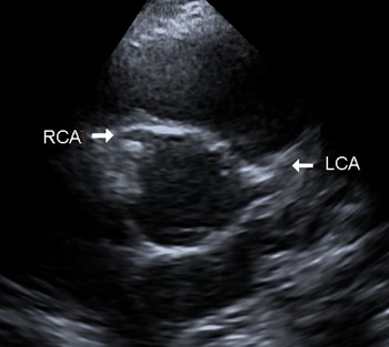
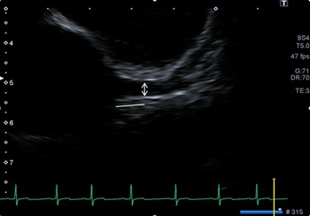
The recent guideline for chamber quantification from the American Society of Echocardiography and the European Association of Cardiovascular Imaging was followed for echocardiographic measurements. Reference Lang, Badano and Voigt9 The method for measuring the end diastolic coronary arterial luminal diameter in children was recommended by the Scientific Committee of the Japanese Society of Kawasaki disease. Reference Fuse, Kobayashi and Saji10 In accordance with these guidelines, both proximal main coronary arteries were examined in the parasternal short- or long-axis view (Fig 1). The distance from the trailing edge of the near wall to the leading edge of the far wall was measured as the coronary artery diameter 5 mm distal from the ostium (Fig 2). If there was a short stem of the main coronary artery (<5 mm), the diameter was measured in the middle between the ostia and the branching.
Regular aortic origin of the main coronary arteries. Parasternal short-axis view in transthoracic echocardiography. The main right coronary artery (RCA) originates at eleven o’ clock from the right sinus of Valsalva and the main left coronary artery (LCA) at three o’ clock from the left sinus of Valsalva.

Coronary artery dimensions were calculated as the average of two or three successive cardiac cycles. We assessed luminal dimensions normalised for body surface area as z scores according to Dallaire et al.Reference Dallaire and Dahdah11 A mean z score < 2.0 standard deviation was determined as normal, whereas a mean z score between 2.0 and < 2.5 standard deviation was declared as dilation. Small aneurysm of coronary arteries were defined as a mean z score 2.5–< 5.0, medium aneurysm 5.0–< 10, and large aneurysm from ≥ 10 or absolute dimension ≥ 8 mm. All studies were digitally stored for off-line analysis, retrospective re-evaluation, and follow-up examinations.
Statistical analysis
The body surface area was calculated with Haycock’s formula as follows: body surface area = 0.024,265 × height (cm) 0.3964 × weight (kg) 0.5378. Reference Haycock, Schwartz and Wisotsky12 As children undergo rapid changes in their physical development with resultant variations in their body size, a standardised score (z score) for assessing cardiovascular structures has been developed for use in paediatric cohorts. By definition, the z score values must be normally distributed, and the mean value should converge to zero and the standard deviation to 1. Reference Epstein, Goldberg, Allen, Konecke and Wood13 A Student’s independent t-test was used to compare coronary artery internal diameter in soccer player and controls. Statistical significance was defined as p < 0.05. All analyses were conducted using IBM SPSS Statistics, version 25.
Results
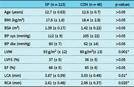
Two patients with minor cardiovascular disease and 42 goalkeepers had to be excluded due to position-specific training which might cause different cardiovascular adaptation compared to field players. A total of 215 soccer players and 48 controls were eligible for data interpretation. HM performed 41 and SG 222 echocardiographies. The mean age of soccer players was 12.7 ± 0.63 years compared with 12.6 ± 0.7 years in controls (p > 0.05). Soccer players had a slightly lower mean body mass index (17.6 ± 1.8 versus 18.4 ± 2.8 kg/ m2, p > 0.05) and body surface area (1.39 ± 0.17 versus 1.42 ± 0.22 m2, p > 0.05). Both groups had regular mean systolic (112 ± 9 versus 105 ± 22 mmHg, p > 0.05) and mean diastolic blood pressure values (60 ± 7 versus 62 ± 14 mmHg, p > 0.05) at rest. Soccer players had significant greater left ventricular mass indexed for body surface area (93 ± 13 versus 79 ± 12 g/m2, p = 0.001). There was no significant difference in conventional left ventricular function measurements (Table 1).
Demographic characteristics, left ventricular and coronary artery dimensions

Data from 215 adolescent elite male soccer players (SP) compared to 48 matched controls (CON)
BMI, body mass index (kg/m2); BP, blood pressure; BSA, body surface area; LCA, left main coronary artery; LVEF, left ventricular ejection fraction; LVMI, left ventricular mass index (g/m2); LVFS, left ventricular fraction of shortening; RCA, right main coronary artery
Values are presented as mean ± SD, percent, or number
* Significant p-values marked by Student t-test
Coronary arteries
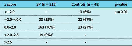
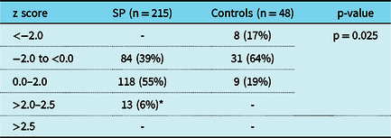
The origin of both coronary arteries could be identified in every participant. There was normal anatomy of the ostia, no pathologic ectopic origin and no stenosis or aneurysm of the proximal coronary arteries in our study cohort. In soccer players, the mean internal diameter of the left main coronary artery was 3.67 mm (SD ± 0.59, range 2.4–4.9 mm) and 2.61 mm (SD ± 0.48, range 1.4–3.85 mm) for the right main coronary artery. Controls showed a smaller mean luminal diameter of 3.03 mm (SD ± 0.49, range 2.3–4.7 mm) in the left coronary artery (p = 0.01) and 2.08 mm (SD ± 0.37, range 1.47–3.17 mm) in the right coronary artery (p = 0.025), respectively (Table 1). In soccer player, left main coronary artery and right main coronary artery mean internal diameter z scores were significantly increased compared to controls (Tables 2 and 3). In soccer players, a total of 91% (n = 196) and in controls a total of 94% (n = 45) showed left main coronary artery z scores within the normal range (−2.0 to 2.0) (Table 2). In the right main coronary artery z score values distribution in both groups were quite comparable (soccer players 94%, n = 202 versus controls 83%, n = 40 within the normal range) (Table 3). A subgroup of soccer players had supernormal (>2.0–2.5) z score values for left main coronary artery (9%, n = 19, p = 0.01) and right main coronary artery (6%, n = 10, p = 0.025), respectively. No coronary artery aneurysm or ectasia were found in both groups defined as a z score >2.5.
Left main coronary artery (LCA) z score values

Results for coronary artery (CA) z score values in adolescent male elite soccer players (SP, n = 215) and matched controls (n = 48)
z score values for left main CA
A mean z score −2.0 to 2.0 was determined as normal, whereas a mean z score < 2.0–2.5 in SP was declared as supernormal
Small aneurysm or CA ectasia were defined as a mean z score < 2.5–5.0
* Supernormal CA z score values marked by Student t-test
Right main coronary artery (RCA) z score values

Results for coronary artery (CA) z score values in adolescent male elite soccer players (SP, n = 215) and matched controls (n = 48)
z score values for right main CA
A mean z score −2.0 to 2.0 was determined as normal, whereas a mean z score < 2.0–2.5 in SP was declared as supernormal
Small aneurysm or CA ectasia were defined as a mean z score < 2.5–5.0
* Supernormal CA z score values marked by Student t-test
Discussion
The origin of both coronary arteries could be identified in every participant by transthoracic conventional echocardiography. There was normal anatomy of the ostia, no pathologic ectopic origin, and no stenosis of the proximal epicardial coronary arteries in our study cohort. Soccer players showed a significant increase in mean diameters of both coronary arteries compared to controls (Table 1). A small subgroup of soccer players showed even supernormal z score values (>2.0–2.5) for both main coronary arteries. No coronary aneurysm or ectasia were found in both groups defined as a z score >2.5 (Tables 2 and 3).
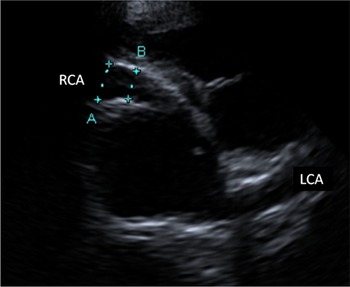
The incidence of sudden cardiac death in competitive athletes is significantly increased compared with their nonathletic counterparts. The most common etiologies include cardiomyopathies and coronary artery abnormalities. Reference Mont, Pelliccia and Piepoli1,Reference Maron, Doerer, Haas, Tierney and Mueller2 Coronary artery abnormalities most often regarded as predisposing to sudden cardiac death in athletes are a left coronary artery originating from the right sinus of Valsalva or a right coronary artery from the left sinus of Valsalva. Reference Cheitlin and MacGregor14,Reference Davis, Cecchin, Jones and Portmann15 But also ectasia or aneurysm of the coronary arteries bears a risk for myocardial ischaemia and consecutively life-threatening ventricular tachyarrhythmia. Coronary artery ectasia and aneurysm are seen in a wide variety of underlying etiologies like Kawasaki disease, genetic disorders like Noonan-Syndrome (Fig 3), Marfan-Syndrome, Ehler-Danlos-Syndrome, vasculitis, sickle cell disease, and chronic inflammatory bowel disease. Reference Dahhan16 In our adolescent Caucasian cohort, congenital coronary artery abnormalities or residuals from Kawasaki disease seem to be the most likely one.
Luminal diameter of the left main coronary artery (LCA). The distance from the trailing edge of the near wall to the leading edge of the far wall was measured as the coronary artery luminal diameter (arrow) 5 mm distal the ostium (line) in diastole.

Bilateral coronary artery ectasia in a 13-year-old boy with genetically confirmed Noonan syndrome. The main right coronary artery (RCA) originates at one o’ clock, distal the aortic sinotubular junction line. The luminal diameter 5 mm distal the ostium (distance B) measured 5.5 mm (4.87z) for RCA and 5.6 mm (4.79z) for the main left coronary artery (LCA).

The morphologic cardiac adaptation to exercise has been well studied in different populations of athletes and depends on the type, duration, and intensity of training. Reference Schuler, Adams and Goto17 Left ventricular work during exercise increases in proportion to the increased systolic arterial pressure. Therefore, chronic high intensity exercise leads to myocardial hypertrophy that can produce up to a 30% increase of relative LV mass in adults. Reference Poliner, Dehmer, Lewis, Parkey, Blomqvist and Willerson18 Mitchell et al defined adult soccer as a low-static and high-dynamic type of sport therefore predisposing more to endurance remodelling. Reference Mitchell, Haskell, Snell and Van Camp19 The significant left ventricular mass increase in our soccer player cohort may result from specificity of training in elite-level adolescent soccer, which differs significantly from training plans in adults. Reference McClean, Riding and Wilson5,Reference Gerling, Pollinger, Michel, Dechant, Melter and Krutsch6
Coronary vascular adaptations in response to exercise can be divided into functional (alterations in vasomotor control) and structural (angiogenesis and vascular remodelling) adaptations. Reference Laughlin and Joseph20 The increase in myocardial blood flow results from a combination of coronary vasodilation, with a decrease of coronary vascular resistance during heavy exercise to 20%–30% of the resting level. Reference Nelson, Gobel, Jorgensen, Wang, Wang and Taylor21
Hildick-Smith et al assessed coronary flow reserve in 29 male adult endurance athletes and 23 matched healthy sedentary controls, using adenosine infusion in transthoracic echocardiography. In addition to an increased proximal coronary artery diameter coronary flow reserve in endurance athletes was supernormal, and endothelium independent vasodilatation was enhanced. Reference Hildick-Smith, Johnson and Cooper22
Recently Ho et al studied the proximal diameters of the left main and right coronary arteries by gated multidetector CT scans in 500 men. They were able to show that larger coronary artery diameters after adjustment for body surface area were associated with higher physical fitness. Reference Ho, Cannaday and FitzGerald23
Pellicia et al evaluated 125 healthy, adult male top-level athletes by echocardiography and found a significant correlation between right and left main coronary artery diameter and left ventricular mass. They concluded that training-induced myocardial hypertrophy involves a proportionate increase of coronary artery dimension. Reference Pelliccia, Spataro, Granata, Biffi, Caselli and Alabiso7 These findings could be confirmed in our cohort of adolescent elite soccer players. They developed significant higher z score values for both main coronary artery diameters compared to controls. A small subgroup in soccer players showed even supernormal coronary artery z score values (>2.0–2.5), which might reflect a physiologic response induced by multiannual high-intensity exercise.
But long-term high-intensity training stimuli might cause negative adaptations of the cardiovascular system. As high-intensity exercise causes relevant peaks in blood pressure, this could be a possible trigger for pathological vascular adaptations. Green et al hypothesise that persistently increased exercise leads to an increase in arterial diameter and reduction in wall thickness. Reference Green, Spence, Rowley, Thijssen and Naylor8 Sharma et al refer to the relationship between increased arterial shear stress and increased oxidative stress in adults, which may cause a premature onset of atherosclerosis. Reference Sharma, Merghani and Mont24 In our cohort of elite SP in early adolescence, no coronary ectasia or aneurysm were found defined as a z score value >2.5, but it might be of great interest to follow-up coronary artery z score values in elite soccer player during their athletic career.
Limitations
There are several limitations of the present study. First of all, we did not test interobserver variability. Second, sonography results are strongly dependent on the examiner’s experience. Especially in imaging coronary arteries by transthoracic echocardiography, time is required for familiarisation with that topic. Both echocardiographers (HM, SG) are very experienced in imaging coronary arteries by transthoracic echocardiography. As coronary arterial dimensions were calculated as the average of two or three successive cardiac cycles, we believe that there is a very low risk for bias. Finally, our study was based on a male Caucasian population. Therefore, our results should not be extended to the general population.
Conclusions
The present study highlights that elite-level soccer training in early adolescence may be a stimulus strong enough to induce structural coronary artery adaptations. A small subgroup of soccer players developed supernormal coronary artery z score values, but did not exceed +2.5z. A longitudinal observation of structural and functional coronary artery adaptation to high-intensity exercise would be of interest.
Acknowledgements
None.
Authors’ contributions
Study design: SG, MM, HM
Data collection: SG, TP, MJD, HM
Statistical analysis and data interpretation: SG, HM, MM
Manuscript writing: SG, HM, WK
Financial support
This research received no specific grant from any funding agency, commercial, or not-for-profit sectors.
Conflict of interest
None.