




No CrossRef data available.
Article contents
Congenital cardiac malformations in Iceland from 1990 through 1999
Published online by Cambridge University Press: 21 January 2005
Abstract
Introduction and Background: About 1% of live-born children have congenital malformations of the heart. The aim of our study was to investigate the incidence of such defects in children born in Iceland during a period of 10 years, extending from 1990 through 1999. Materials and Methods: Information about the patients was obtained from medical records from two hospitals that cover the whole country, a private clinic of pediatric cardiologists, an echocardiography database, autopsy reports, and death certificates. We investigated the distribution of specific malformations, the age at diagnosis, the symptoms leading to the diagnosis, the source of referral, and treatment and quality of life. Results: Between 1990 and 1999, there were 44,013 live births in Iceland, of which 740 patients were diagnosed with congenital cardiac malformations, accounting for 1.7% of the live-born children. The distribution was made up of 338 patients with ventricular septal defect (45.7%), 90 with atrial septal defect (12.2%), 85 with patency of the arterial duct (11.5%), 48 with pulmonary valvar stenosis (6.5%), 38 with a bicuspid aortic valve (5.1%), 28 with aortic coarctation (3.8%), 22 with tetralogy of Fallot (3.0%), 14 with transposed great arteries (1.9%), 11 with aortic stenosis (1.5%), 10 with atrioventricular septal defect and common atrioventricular orifice (1.4%), 9 with mitral valvar regurgitation (1.2%), 7 with sub-aortic stenosis (0.9%), and 5 with hypoplasia of the left heart (0.7%). Extracardiac anomalies were seen in 89 patients (12.0%). Chromosomal defects were seen in 36 patients, of whom 28 had Down's syndrome. Discussion: The annual incidence of diagnosis of patients with congenital cardiac malformations increased during the period of study. This was noted for minor defects, but the incidence of the major anomalies did not alter. Our observed yearly incidence, at 1.7%, was higher than noted in a previous study covering the years 1985 through 1989, and is also higher than in other population-based studies. The most likely explanation is the fact that access to pediatric cardiologists in Iceland is very good. Diagnosis, registration, and follow-up are conducted by only a few cardiologists, and take place at a single center for pediatric cardiology.
Information
- Type
- Original Article
- Information
- Copyright
- © 2004 Cambridge University Press
Cardiac anomalies are amongst the most common congenital malformations, and are said to afflict about 1% of all live-born children.1–12 The cause of the lesions is unknown, but genetic and environmental factors, such as maternal diseases or the use of medications and drug abuse during pregnancy, are thought to play a role.13 The defects are often a component of a number of syndromes, such as Marfan's, as well as some chromosomal anomalies such as Down's syndrome.13 To obtain better understanding of the etiology of the lesions, it is important to know their precise incidence, as well as their distribution in the population. These factors are likely to become increasingly significant with the ongoing increase in genetic knowledge. The purpose of our study, therefore, was to document the incidence and type of congenital cardiac malformations occurring in Iceland between 1990 and 1999. A similar study had been performed in our country between 1985 and 1989,1 which gave us the added opportunity to compare the incidence of the anomalies over the two periods of time. Iceland is a small nation, with a relatively small genetic pool. The medical system is socialized, and follow-up of patients is centralized. For these reasons, we think the Icelandic population can provide accurate information on the incidence of congenital cardiac malformations, as well as the age at diagnosis and mode of presentation.
Materials and methods
Information on patients born in the period 1990 through 1999, and with the diagnoses of congenital heart defect of 745 through 747 according to the ninth revision, Q20 through Q28 of the tenth revision, of the International Classification of Disease, was obtained from medical records in two hospitals, and an echocardiography database and medical records held in a private clinic of three pediatric cardiologists. We also obtained access to autopsy reports and death certificates of those patients who had died. We collected the data over the period September 10th 1997 to May 17th 2000, obtaining approval for the study from the ethical committee of the University Hospital of Iceland.
As minor defects, we defined those anomalies for which no treatment was needed during the period of the study. Major defects were defined as defects that needed surgical correction or other invasive intervention, along with defects considered inoperable and those requiring medical treatment. When a patient had more than one lesion, we considered the defect that required treatment, or the one that caused the greatest hemodynamic effect, to be the main malformation. Diagnoses were confirmed with echocardiography, cardiac catheterization, or autopsy. Patency of the arterial duct in babies of <37 weeks gestational age, along with patency of the oval foramen or atrial septal defects less than 4.0 mm in diameter, were not considered cardiac anomalies for the purpose of our study. In contrast to the previous study, we did chart the incidence of bicuspid aortic valves.
All pregnant women in Icleand are offered prenatal ultrasonic screening in the nineteenth week of gestation, and they are referred for specialized fetal echocardiography if an abnormality is suspected from the four chamber view obtained at that visit, or if there are other fetal or maternal factors for increased risk. We studied the number of pregnancies terminated because of severe cardiac lesions detected at fetal echocardiography. Fetal echocardiography has become more advanced in the last decade, and we tried to evaluate the effect of these diagnostic methods on the incidence of the congenital cardiac anomalies.
We also studied the distribution of the cardiac anomalies relative to other congenital anomalies, chromosomal defects, and comorbid diseases. We studied the symptoms leading to the diagnosis, the age at diagnosis, and the pattern of referral, for the latter features establishing whether patients were referred from those responsible for the birth, from those organizing screening of the well child, from the general practitioner or pediatrician, from hospital, or because of fetal ultrasound. We also investigated treatment and quality of life. We then divided the patients into four groups based on symptoms and the need for treatment. The first group was made up of patients in whom we deem the problem created by the lesion to be solved, and who do not need further follow-up. The second group consisted of patients having no symptoms, but who require regular follow-up. The third group consisted of patients having daily symptoms, and/or needing medical treatment. The final group consisted of patients limited in their daily activities because of the cardiac anomaly.
Results
From January 1st 1990, through December 31st 1999, there were 44,013 live births in Iceland, producing 22,554 boys (51.2%) and 21,459 girls (48.8%). By May, 2000, congenital cardiac anomalies had been diagnosed in 740 patients, 363 boys (49%) and 377 girls (51%), giving an incidence of 1.7%. The yearly incidence varied from 1.04% of live-born infants in 1991 to 2.34% in 1997. Of the defects, 225 (30.4%) were deemed to be major, the remaining 515 (69.6%) being considered minor defects within our system. Figure 1 shows the distribution of major and minor defects, according to the year of birth of the patient, and the number of elective abortions based on antenatal diagnosis of severe malformations.

Figure 1. Number of patients with major and minor defects, born in each year of the period of study, and the yearly number of elective abortions due to severe defects diagnosed prenatally.
Ventricular septal defect was the most common heart defect, accounting for almost half of the total number (338 patients – 45.7%, or 0.77% of live-born infants). Of the number, 176 were girls and 162 were boys. An atrial septal defect was diagnosed in 90 patients, and patency of the arterial duct in 85 patients. Table 1 lists the distribution of the specific lesions and the gender of the patients.
Figure 2 shows the source of referral for the suspected cardiac anomalies, while Figure 3 shows the age at diagnosis. Nearly half of the patients (323, or 44%), were diagnosed during the first week of life, and 70% (523 patients) had been diagnosed by 6 months of age. Table 2 shows the nature of the anomalies that were diagnosed after 6 months of age. Of these patients, 190 (87.6%) were diagnosed because of a cardiac murmur detected on routine physical examination. Table 3 lists the symptoms leading to the diagnosis.
Table 1. Distribution of congenital cardiac defects, and the ratio of males to females, in children born from 1990 through 1999.


Figure 2. The sources of referral of our patients diagnosed with congenital cardiac defects.

Figure 3. The age at diagnosis of the patients with congenital cardiac malformations.
Table 2. Number of the most common congenital cardiac defects diagnosed after the age of 6 months.

Table 3. Symptoms leading to the diagnosis of congenital cardiac defects.

We diagnosed 225 (30.4%) major defects during the period of study, accounting for 0.51% of live-born infants, and with proportions varying from 0.74% in 1993 to 0.30% in 1996. Significantly fewer incidences of major lesions were found in the latter half of the period of study (p = 0.04). We found 98 boys with major defects, as opposed to 127 girls, giving a ratio of 1 to 1.3. About half of the patients were diagnosed before discharge from their site of birth, but 114 patients were diagnosed later. Of live-born children with major defects, 102 were diagnosed in the newborn intensive care unit or the newborn nursery, and 9 were diagnosed during routine fetal ultrasound. We had 58 patients referred from clinics proving “well care” services, while 19 were referred from offices of general practitioners, 9 from pediatricians' offices, and 28 patients with major defects were diagnosed when they were admitted to a hospital for some other reason. During the period of study, 19 fetuses were diagnosed with a major cardiac malformation considered to have poor prognosis despite intervention, and justifying termination of pregnancy (Fig. 1).
Of all 740 patients with congenital cardiac malformations, 89 (12.0%) had chromosomal defects, syndromes and/or other congenital anomalies (Table 4). Of the patients deemed to have major cardiac defects, 41 (18.2%) had other defects or anomalies.
Table 4. Distribution of congenital heart defects accompanied by syndromes, chromosomal anomalies and other congenital defects.

The majority of patients, specifically 499, has no symptoms, and is followed regularly by a pediatric cardiologist, but 194 patients have been discharged from follow-up. No patient is considered to be significantly limited in daily activities because of the cardiac disease, but 20 patients have daily symptoms and/or need daily medical treatment.
Of the 44,013 infants born during the period of study, 27 have died because of the cardiac anomaly or other related problems. Mortality related to congenital cardiac was thus 0.06% over the period of study. Of those dying, 10 died in the first week of life, 2 in the second week, and 15 between 2 months and 4 years of age. Other chromosomal anomalies and multi-system problems were found in 7 patients who died. Of these, 2 had trisomy 18, one had trisomy 8, one had trisomy of both chromosomes 9 and 18, and one had incomplete trisomy 22. Table 5 lists the causes of death.
Table 5. Cause of death of patients with congential heart defects.

Discussion
According to our current study, the yearly incidence of congenital cardiac malformations in Iceland over the period 1990 through 1999 was 1.7%. Within the period, the incidence varied from 1.04% in 1991 to 2.34% in 1997. This incidence is higher than that found in the previous study done in Iceland for the years 1985 through 1989, when it was 1.1%.1 The increase is largely produced by the greater number of patients found to have minor defects. The incidence of major defects, at 0.50%, was similar to the figure of 0.47% found during the earlier study.1 Since, however, the number of major defects diagnosed over the last 5 years of the period was significantly lower, the incidence of major defects might well be decreasing. One explanation for this trend could be the improved prenatal detection of the malformations. With improved fetal echocardiography, severe lesions, such as hypoplasia of the left heart, are detected earlier in pregnancy, which might lead to increased number of elective abortions, thus decreasing the number of live-born infants with major defects. Although the number of elective abortions almost doubled from the first to the second half of the study, this can only partially explain the decrease in the incidence of major cardiac defects. It is likely related to overall improvement in antenatal diagnosis of malformations and chromosomal abnormalities.
We counted bicuspid aortic valves in our current analysis, since we now have diagnostic tools with sufficient resolution to detect this malformation, which in later life can lead to valvar complications such as calcification, prolapse, or endocarditis, and thus warrants lifelong follow-up.14 Since the bicuspid aortic valve also constitutes a significant risk for sub-acute bacterial endocarditis, prophylactic antibiotics are needed whenever there is a risk of bacteremia. The increased incidence of defects deemed minor in our study can partly be related to the inclusion of bicuspid aortic valves, since these were not counted in the previous study. We assume that, with improving quality of echocardiography, such bicuspid aortic valves will be accounted for in all future studies on congenital cardiac malformations.
If we look only at the incidence of congenital cardiac malformations over the last three years, from 1997 through 1999, we find a figure of 2.11%, which is significantly higher than that found over the first years of the study, and also higher than in the previous study. Since all the children born in our current period of study with congenital cardiac malformations have likely not yet been diagnosed, this number might still rise, increasing the incidence even further. It is almost certainly the better quality of echocardiography, better access to pediatric cardiologists, and the increased number of referrals to their service, that are the causes for the increase in the diagnosis of the minor defects. It is also likely that such defects resolved in previous eras without ever having been detected, like small ventricular septal defects. These are now being diagnosed earlier with the help of echocardiography.
The incidence of congenital cardiac anomalies in Iceland is, according to our experience, higher than that has been reported in similar studies elsewhere. The incidence found in other countries has ranged from 0.28% to 1.22%,3–12 but the incidence in older studies is generally lower than those performed more recently. When we compare our own findings to those from the earlier study performed in our country from 1985 through 1989, it seems that the number of congenital anomalies could truly be increasing in Iceland. The definitions of anomalies used for the studies performed in the other countries are generally compatible with our own, apart for bicuspid aortic valves, but their incidence is still markedly lower.3–12 Other studies, nonetheless, have reported an increased number of small ventricular septal defects diagnosed with improved ultrasonography,15 as we found in our own study.
The most likely reason for the high incidence of congenital cardiac anomalies in Iceland is the fact that diagnosis, follow-up, and documentation are done by a few physicians. The center for diagnosis is located in the University Hospital of Iceland, and only a few pediatric cardiologists diagnose congenital cardiac anomalies. The Icelandic people are fairly homogenous, both genetically and socioeconomically, and their access to medical specialists is good.16–18
In our study, almost half of the patients with cardiac malformations were diagnosed prior to discharge from the hospital in which they were born.
This number is lower than the findings reported in a recent study from Norway,11 where three-quarters of afflicted patients were diagnosed in the hospital where delivery occurred. When these numbers were studied as a proportion of live-born infants, we found that, in both studies, 0.8% of live-born infants with cardiac defects were diagnosed before discharge from the institution providing delivery. These are often major defects, where symptoms become evident shortly after birth. The number of congenital cardiac defects diagnosed after discharge from institution responsible for delivery, nonetheless, seems to be higher in Iceland, since 0.9% of live-born infants were thus diagnosed as opposed to only 0.24% in the Norwegian study.11
When looking at the age of diagnosis, we found that 323 patients, or 43.6%, were diagnosed during the first week of life. At 6 months of age 70% of the patients with congenital heart defects had been diagnosed, rising to 86% at 2 years of age, results that are comparable to a similar study from Qatar.6 The majority of patients with bicuspid aortic valves (81.5%), two-thirds of those with patency of the arterial duct, and almost half of those with atrial septal defects, were diagnosed after the age of 6 months. All of these malformations continued to be diagnosed in patients up until the age of twelve. In contrast, only one-sixth of the patients with ventricular septal defects were diagnosed after the age of 6 months, and the oldest patient was eleven years of age. Almost nine-tenths of the patients diagnosed after the age of 6 months were asymptomatic, being referred to a pediatric cardiologist because of discovery of a heart murmur.
Patients with cardiac defects often have other congenital anomalies, chromosomal defects, and syndromes. The proportion of comorbidity in our current patients with major cardiac lesions was lower than in the previous study.1 Our patients with co-existing chromosomal defects, at 4.9%, was also lower than the proportion of 13% encountered in a Swedish study carried out from 1981 through 1986.8 The proportion of our current patients with Down's syndrome was 3.8%, compared to 4.2% in the previous one.1 This number is generally between 6 and 8%.19 The low percentage of chromosomal defects and other anomalies in our study can be explained by the increased number of minor defects detected, thus diluting the proportion of other anomalies, that more often follow major cardiac defects. It is also possible that the general improvement in prenatal diagnoses, as discussed earlier, might play a role.
Of the patients identified in our study, 27 have died (Table 5). The rate of mortality is thus 0.06% of live-born infants. This rate has decreased from the period 1985 through 1989, when it was 0.08%. We did not include, however, those incidents when pregnancy was terminated because of a severe defect diagnosed prenatally.
When we examine the outcome of the patients born with congenital cardiac malformations, it is interesting to see that only 20 patients have daily symptoms or need medical therapy, and no one is considered to have significant limitations in their daily activities because of a cardiac disease. The majority of patients is symptom-free according to the pediatric cardiologists. These results suggest that the majority of children born with congenital heart defects has a good chance of recovery, and will lead a normal life.
Acknowledgements
Grants: The Science Fund of the University Hospital of Iceland and the Memorial Fund of Magnus Benjaminsson and his wife Sigridur Einarsdottir.
References
Sigfusson G, Helgason H. Incidence and diagnosis of congenital heart defects in Iceland. Icelandic Med J 1993; 79: 107–114.Google Scholar
Hoffman JIE. Incidence of congenital heart disease: I. Postnatal incidence. Pediatr Cardiol 1995; 16: 103–113.Google Scholar
Meberg A, Otterstad JE, Frøland G, Sørland S. Barn med medfødt hjertefejl i Vestfold 1982–1988. Tidsskr Nor Laegeforen 1990; 110: 354–357.Google Scholar
Anand R, Mehta AV. Incidence of cardiovascular malformation in the Tennessee and southwest Virginia area. Am J Cardiol 1996; 78: 610–611.Google Scholar
Bosi G, Scorrano M, Tosaro G, Forini E, Chakrokh R. Working Party of the Italian Society of Pediatric Cardiology. The Italian Multicentric Study on Epidemiology of Congenital Heart Disease: first step of the analysis. Cardiol Young 1999; 9: 291–299.Google Scholar
Robida A, Folger GM, Hajar HA. Incidence of congenital heart disease in Qatari children. Int J Cardiol 1997; 60: 19–22.Google Scholar
Samánek M, Zdenek S, Bozena Z, Hrobonová V, Vorísková M, Skovránek J. Prevalence, treatment and outcome of heart disease in live-born children: A prospective analysis of 91, 823 live-born children. Pediatr Cardiol 1989; 10: 205–211.Google Scholar
Pradat P. Epidemiology of major congenital heart defects in Sweden, 1981–1986. J Epidemiol Community Health 1992; 46: 211–215.Google Scholar
Grech V, Gatt M. Syndromes and malformations associated with congenital heart diseases in a population-based study. Int J Cardiol 1999; 68: 151–156.Google Scholar
Samánek M, Vorísková M. Congenital heart disease among 818, 569 children born between 1980 and 1990 and their 15-year survival: A prospective Bohemia survival study. Pediatr Cardiol 1999; 20: 411–417.Google Scholar
Meberg A, Otterstad JE, Frøland G, Hals J, Sørland SJ. Early clinical screening of neonates for congenital heart defects: the cases we miss. Cardiology in the Young 1999; 9: 169–174.Google Scholar
Abdulla R. What is the prevalence of congenital heart diseases? Pediatr Cardiol 1997; 18: 269.Google Scholar
Clark EB. Epidemiology of Congenital Cardiovascular Malformations. In: Moss AJ, Adam FJ (eds). Heart Disease in Infants, Children and Aolescents Including the Fetus and Young Adult, 5th edn. Williams and Wilkins, Baltimore, 1995, pp 60–69.
Beppu S, Suzuki S, Matsuda H, Ohmori F, Nagata S, Miyatake K. Rapidity of progression of aortic stenosis in patients with congenital bicuspid aortic valves. Am J Cardiol 1993; 71: 322–327.Google Scholar
Martin GR, Perry LW, Ferencz C. Increased prevalence of ventricular septal defect: epidemic or improved diagnosis. Pediatrics 1989; 83: 200–204.Google Scholar
Fixler DE, Pastor P, Sigman E, Eifler CW. Ethnicity and socioeconomic status: impact on the diagnosis of congenital heart disease. J Am Coll Cardiol 1993; 21: 1722–1726.Google Scholar
Newacheck PW, Stoddard JJ, McManus M. Ethnocultural variations in the prevalence and impact of childhood chronic conditions 1993. Pediatrics 1993; 91 (Suppl): 1031–1039.Google Scholar
Allen HD, Taubert KA, Deckelbaum RJ, Driscoll D, Dunningan , Gidding SS, et al. Poverty and cardiac disease in children. Am J Dis Child 1991; 145: 550–553.Google Scholar
Moller JH, Taubert KA, Allen HD, Clark EB, Lauer RM. Cardiovascular health and disease in children: current status. Circulation 199; 89: 923–930.

Number of patients with major and minor defects, born in each year of the period of study, and the yearly number of elective abortions due to severe defects diagnosed prenatally.
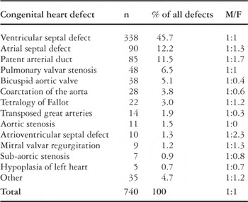
Table 1.

The sources of referral of our patients diagnosed with congenital cardiac defects.

The age at diagnosis of the patients with congenital cardiac malformations.
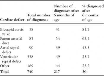
Table 2.
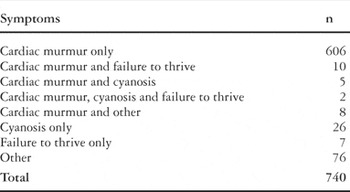
Table 3.
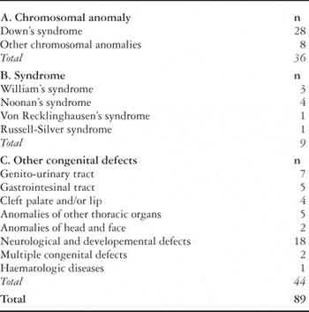
Table 4.
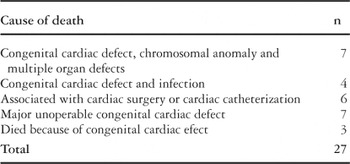
Table 5.
 You have
Access
- 38
- Cited by