Introduction
When the mountain pine beetle (MPB), Dendroctonus ponderosae Hopkins (Coleoptera: Curculionidae), overwhelms the defenses of a pine tree (Pinaceae) and successfully colonises the phloem, it creates a habitat that is favourable for itself and associated species, many of which arrive on or in the bodies of the beetles. Soon a microcosm is established that includes numerous fungi (Lee et al. Reference Lee, Kim and Breuil2006b), bacteria (Winder et al. Reference Winder, Macey and Cortese2010), mites (Acari) (Mori et al. Reference Mori, Proctor, Walter and Evenden2011), Nematoda (Massey Reference Massey1974), insect predators and parasitoids (Dahlsten and Stephen Reference Dahlsten and Stephen1974), and phloeophages that interact in diverse ways, mostly undocumented (Gibson and Hunter Reference Gibson and Hunter2009). However, it is the relationships between tree-killing bark beetles and symbiotic Ophiostomatales (Fungi) that has garnered the most scientific attention (e.g., Paine et al. Reference Paine, Raffa and Harrington1997; Klepzig and Six Reference Klepzig and Six2004; Six and Klepzig Reference Six and Bentz2004).
Despite the importance of bark beetle-fungus systems, it has proven challenging to elucidate interspecific interactions as the system is difficult to manipulate experimentally (Six Reference Six and Paine2012). Several approaches have been employed to gain insight into bark beetle-fungus interactions, all with some disadvantages. Inferences can be made based on correlations of occurrence of species in situ (e.g., Adams and Six Reference Adams and Six2007), but the co-occurrence of other microorganisms within the beetle’s environment could confound these interactions. Phloem sandwiches, constructed by placing phloem between glass plates and introducing insects and fungi, have been used to explore interactions, but it is impossible to eliminate contaminating microbes (Bleiker and Six Reference Bleiker, Potter, Lauzon and Six2007). Six and Paine (Reference Six1998) acquired fungus-free MPB adults by sterilising pupae and rearing them in pseudo-pupal chambers where teneral adults were then allowed to feed on individual fungal species (and thus acquire the fungi in their mycangia) or no fungi before introduction into fresh bolts; however, this labour-intensive method resulted in low rearing success and few replicates. Clearly, there is a need for a controlled rearing technique that is simple, economical in labour requirements, minimises contamination, and yields high rearing success. Herein we describe the novel “rearing-tube” technique that meets these criteria, allowing controlled rearing with modest space requirements, and permitting observations of growth patterns and behaviour. This method was applied to investigate the influence of three associated symbiotic fungi on MPB development, survival, and fitness.
Two ophiostomatalean fungi, Grosmannia clavigera (Robinson-Jeffrey and Davidson) Zipfel, de Beer, and Wingfield (Ophiostomataceae) and Ophiostoma montium (Rumbold) von Arx (Ophiostomataceae), are commonly associated with MPB throughout its range (Six and Paine Reference Six1998). Both are carried in the maxillary mycangia of the MPB (Whitney and Farris Reference Whitney1970), and also on the exoskeleton (Bleiker et al. Reference Bleiker and Six2009). In lodgepole pine, Pinus contorta Douglas var. latifolia Engelmann (Pinaceae), both of these fungi increased total nitrogen content of phloem and provided nutritional benefits to developing MPB larvae (Bleiker and Six Reference Bleiker, Potter, Lauzon and Six2007). They also provide a source of sterols, especially ergosterol, which is important for development, metamorphosis, and reproduction (Bentz and Six Reference Bentz and Six2006). In general, G. clavigera is the most beneficial of the two species, (Cook et al. Reference Cook, Shirley and Zambino2010), but there is adaptive value for the MPB to maintain associations with both species as they have different temperature optima for growth (Six and Paine Reference Six1998; Solheim and Krokene Reference Solheim and Krokene1998; Addison et al. Reference Addison, Powell, Six, Moore and Bentz2013), different water potential optima (Bleiker and Six Reference Bleiker and Six2009b), and may use different carbon sources (Bleiker and Six Reference Bleiker and Six2009b). There is also evidence that the relative prevalence of each symbiotic fungus varies predictably with temperature, influencing which fungus dominates in a particular population and which instars develop with each fungus (Adams and Six Reference Adams and Six2007; Six and Bentz Reference Six and Klepzig2007; Bleiker and Six Reference Bleiker and Six2009a). The fungi may also play roles in conditioning host tree tissues for insect development by altering phloem chemistry and moisture content (e.g., Paine et al. Reference Paine, Raffa and Harrington1997).
Since 2003, the MPB has invaded the Canadian boreal forest, starting in northeastern British Columbia and spreading throughout most of north-central Alberta (western Canada), where it is infesting lodgepole pine, jack pine (Pinus banksiana Lambert (Pinaceae)), and their hybrids (Safranyik et al. Reference Safranyik and Linton2010; Cullingham et al. Reference Cullingham, Cooke, Dang, Davis, Cooke and Coltman2011). The beetle threatens to continue spreading eastward across Canada’s boreal ecozone (Safranyik et al. Reference Safranyik and Linton2010). The boreal forest is a novel ecosystem and jack pine a new host for MPB. Environmental and biological selection pressures imposed on MPB in the boreal forest may change the relationships between MPB and associated microorganisms, potentially altering the beetle’s success in this newly invaded ecosystem, and consequently the ecosystem itself (Safranyik et al. Reference Safranyik and Linton2010).
Grosmannia clavigera and O. montium are commonly associated with MPB across its range. However, a third MPB-associated ophiostomatalean species, Leptographium longiclavatum Lee, Kim, and Breuil (Ophiostomataceae), was described in 2005 (Lee et al. Reference Lee, Kim and Breuil2005). Although much rarer than G. clavigera and O. montium in British Columbia (Kim et al. Reference Kim, Allen, Humble and Breuil2005; Lee et al. Reference Lee, Kim and Breuil2006b) and California, United States of America (Lee et al. Reference Lee, Kim and Breuil2005), L. longiclavatum is a much more prevalent associate of the MPB in the boreal (Rice and Langor Reference Rice, Thormann and Langor2009) where it appears to be dominating G. clavigera (Roe et al. Reference Roe, James, Rice, Cooke and Sperling2011). Furthermore, in montane British Columbia L. longiclavatum is less pathogenic than G. clavigera to lodgepole pine (Lee et al. Reference Lee, Kim and Breuil2006a), whereas in the boreal both species were equally virulent on lodgepole pine, jack pine, and hybrids (Rice et al. Reference Rice, Thormann and Langor2007).
The nutritional benefits of these three fungus species to MPB have not been investigated in jack pine, or in any host for L. longiclavatum. We describe a novel “rearing-tube” technique and apply it to assess the impacts of these three fungi on the development and survival of MPB progeny using a ground phloem diet of jack pine.
Materials and methods
Rearing tube preparation
Bolts were cut from healthy jack pine (~30 cm diameter) growing near Lac La Biche, Alberta, Canada (55.022°N, 111.985°W) and were brought to the laboratory. Trees from which bolts were cut were deemed to have predominantly jack pine characteristics based on tree and cone morphology; however, it is likely that there was some lodgepole pine gene introgression in that population (Cullingham et al. 2011). In the laboratory, bark was removed and phloem separated from the outer bark using a large knife. Some outer sapwood (1.0–1.5 mm in thickness) was also shaved from bolts using a knife. Sapwood and phloem were dried at 60 °C for three to four days and ground separately in a Wiley mill with a 3 mm mesh followed by a 1 mm mesh. Ground phloem and sapwood were mixed at a ratio of 9:1, which was experimentally determined through earlier trials (not reported) as optimal for texture, aeration, and moisture-holding capacity. Dried medium was stored at room temperature in sealed plastic containers.
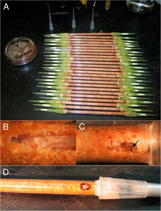
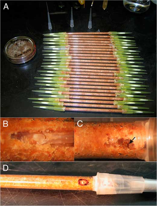
Rearing tubes (10 cm long) were constructed from Kimax™ (Gerresheimer Glass Inc., Vineland, New Jersey, United States of America) glass tubing (5 mm outer diameter, 3.6 mm inner diameter). Each tube was plugged on one end with cotton wool, filled with ~0.4 g of medium that was lightly compressed with a tamping rod, and plugged in the second end with cotton wool. Around each end of the tube, a ~2-mm-wide strip of Parafilm™ (Bemis Co. Inc., Oshkosh, Wisconsin, United States of America) was wrapped, over which a 200 µL pipette filter tip was tightly fitted to function as a gas permeable seal while preventing contamination. The assembled tubes (Fig. 1A) were dry-autoclaved twice at 121 °C for 30 minutes and stored at room temperature until required.
Photographs of rearing tubes and mountain pine beetle eggs: A. Medium-filled rearing tubes before eggs were added; B. Egg added using a pipette tip; C. Close-up of egg and fungal inoculum (arrow); D. Completed rearing tube.
Before use, the medium in each tube was hydrated in a laminar flow hood by removing the pipette tip and cotton from one end and adding 0.6 mL of sterile distilled water with a sterile syringe, and replacing the pipette tip and cotton. Hydration resulted in a gravimetric moisture content of 60% (=150% oven-dry-weight), comparable to that of the fresh phloem (~66% as measured in the laboratory before and after drying). Hydrated tubes were stored up to a week at −20 °C until used.
Beetle egg source and sterilisation
Bolts of MPB-infested lodgepole pine were cut in April 2010 at two boreal locations near Peace River, Alberta, Canada (56.480°N, 118.518°W and 56.178°N, 116.800°W), transported to the laboratory, and stored at 4 °C. After approximately two weeks of storage, bolts were moved to ambient temperatures and placed in rearing cages. Emerged adults were collected daily, sexed, segregated by sex, and stored at 4 °C in Petri dishes containing rehydrated ground phloem until used. In October 2009, fresh uninfested jack pine bolts (~1 m long, ~30 cm diameter) were cut near Lac La Biche, Alberta, brought to the laboratory where cut ends were sealed with paraffin, and stored at −20 °C until needed. Upon removal from cold storage, each bolt was inoculated with four pairs of MPB adults spaced equidistantly around the circumference. Females were first introduced through short transparent straws pinned to the bolts and subsequently plugged. After boring dust was observed, one male beetle was placed in each straw. One to two weeks after male beetle introductions, bark was stripped from the bolts and eggs were gently transferred from galleries to sterile moistened filter paper in Petri dishes. A 200 µL gel-loading pipette tip (see end of tubes in Fig. 1B) dipped in water generated hydroscopic forces to transfer eggs without damaging them. Eggs were stored at 4 °C for up to two days until used. To accomplish surface sterilisation, eggs were placed in fine mesh baskets, rinsed for one minute in sterile water, transferred into modified White’s solution (Barras Reference Barras1972) for four minutes, and rinsed three times (one minute each) in sterile water. Sterilised eggs were transferred to sterile moistened filter paper and stored at 4 °C for ⩽three days.
Egg placement and fungal inoculation
Eggs are fragile and can easily be damaged. To reduce damage we waited until embryo development advanced to the point where the centre of the egg became opaque leaving the periphery relatively clear (Stages II–IV of egg development; Reid and Gates Reference Reid and Gates1970). Because of attrition due to damage or lack of fertilisation we found it important to collect and sterilise 50% more eggs than required for an experiment. Eggs with discoloration or evidence of deflation were avoided.
After the filter tip and cotton plug were removed from one end of a hydrated tube, a depression ~5 mm deep was made in the medium down one side of the tube using a sterile 200 µL pipette tip. Under a dissecting microscope, an egg was gently placed into the depression as close to the bottom as possible and resting on the medium (Fig. 1B). Placement of the egg was done using a 200 µL gel-loading pipette tip with a slight fork cut into the end (Fig. 1B) and dipped in sterile water. Eggs were placed to avoid contact with the tube as eggs tended to suffocate in the condensation on the glass.
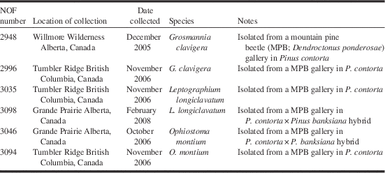
The fungal cultures used in this study were from the Northern Forestry Centre culture collection (Edmonton, Alberta, Canada) (NOF). Two isolates of each fungus species were used: O. montium (NOF3046, NOF3094), G. clavigera (NOF2948, NOF2996), and L. longiclavatum (NOF3035, NOF3098) (Table 1). Inoculum was added to sterile hydrated phloem medium (from the same source used to prepare tubes) placed in sterile plastic Petri plates and incubated at room temperature for three weeks before introduction to rearing tubes.
Detailed source information about isolates of mountain pine beetle-associated ophiostomatalean fungi used in rearing tube experiments.

Note: NOF, Northern Forestry Centre Culture Collection, Edmonton, Alberta, Canada.
The fungal treatment was added to the tube after egg placement by filling the depression with fungal culture (or sterile medium for controls) without putting pressure on the egg (Figs. 1C, 1D), and cotton plugs and pipette tips were replaced. All work was done under a laminar flow hood using sterile technique. Inoculated rearing tubes were placed into sterile covered trays and incubated at 24 °C in the dark.
Experimental design and data collection
There were 50 replicates for each of the six fungus isolates and 100 replicates of the controls. Each tube was examined daily, except on weekends, using a stereomicroscope and the following events/data recorded: egg hatch, larval moulting, pupation, eclosion, and mortality. When a egg-hatch or larval-moulting event occurred on a weekend, it was relatively easy, with experience, to project on which day the event occurred based on the colour of the head capsule, size of the larva and distance the larva had moved since moulting/hatching. Newly moulted larvae had pale head capsules and the moulted cuticle was found nearby making them easy to recognise; however, this could only be observed for less than a day after moulting before the head capsule darkened and the exuvium, except for the head capsule, was consumed. Whenever visible, larval head capsules were measured using the ocular micrometer of the stereomicroscope. After four weeks of incubation, larval gallery length and the distance of fungal advancement (fungal hyphae could be directly observed under the stereomicroscope) from the inoculation point were measured with a ruler. Daily observations continued until all individuals were either dead or had matured to adults. Two weeks after eclosion, each adult was removed from its tube by cutting the tube open near the adult and teasing it out with sterile forceps. Adults were allowed to walk around for several minutes on 1.5% malt extract agar in Petri plates which were then incubated at room temperature for three to four weeks and later checked for presence of fungi. After removal from agar plates, beetles were rinsed in sterile distilled water to remove debris, measured for maximum pronotum width and body length, and sexed before freezing at −20 °C.
Adults designated for fat extraction were removed from the freezer and dried at 47 °C for four days in individual 9 mL glass vials that were placed without lids in a desiccator containing phosphorus pentoxide, and the dry weights were recorded to the nearest 0.1 mg using a Mettler AC100 balance (Mettler-Toledo International Inc., Columbus, Ohio, United States of America). To extract fat, 5 mL of petroleum ether was added to each vial which were then capped and incubated for 24 hours at 47 °C. This step was repeated four times, each time decanting the old petroleum ether and replacing with fresh solution. This was followed by two rounds of immersion and incubation in 95% ethanol to remove any residue adhering to the specimens and tubes. Extracted beetles were dried and reweighed as previously described.
Nutrient analyses
To assess N and C concentration changes in the sterilised rearing medium due to fungal treatment, rearing tubes without beetles were inoculated with each of the six fungal isolates or left as un-inoculated controls (five replicates of each). Tubes of medium were left for 30 days at 24 °C after which medium was removed, placed in separate glass vials, and dried at 60 °C for 18 hours. Vials of medium were stoppered and stored at −20 °C until subjected to nutrient analyses. At the Northern Forestry Centre, total carbon and nitrogen were estimated through combustion and Infra-Red detection using a Leco® C/N Analyzer (Leco Corporation, St. Joseph, Michigan, United States of America).
Statistical analysis
Analysis of variance (ANOVA, Proc GLM; SAS Institute Inc., Cary, North Carolina, United States of America) was used to assess treatment effects on most measured parameters (exceptions noted below). All data were normally distributed so no transformations were applied. When significant effects were observed, post hoc least-squares-means tests (Tukey–Kramer adjustment for comparisons) were applied to compare means. χ 2 tests (Proc Freq; SAS) were employed to assess treatment effects on MPB hatch rates, larval survival and number of supernumerary instars. As there were no significant differences between isolates within a fungal species for most measured parameters, data were pooled by species for subsequent analyses. The only exception was that there was an O. montium isolate effect on larval survival.
Results
Rearing protocol
The amount of time required to check each tube varied according to the amount of data to be collected. Only a few seconds per tube were needed to ascertain if hatch had occurred and whether fungi were growing. About 30 seconds per tube were needed to measure larval gallery length and distance of mycelium advancement, and up to a minute to ascertain larval health (active, lethargic, or dead) and measure head capsule widths (if possible). Observations of behaviour of MPB larvae, pupae, and adults required additional time.
Only 17 of 400 rearing tubes (4.25%) became contaminated during the rearing process, all by a Penicillium Link (Trichocomaceae) species, and these were excluded from analyses. Nine of the 17 contaminated tubes likely resulted from a contaminated inoculum source. None of the control tubes (no-fungus) exhibited any growth of fungi, indicating that surface sterilisation of the eggs was successful and that eggs contained no internal fungi.
Effects of fungi on MPB success and development
Hatch
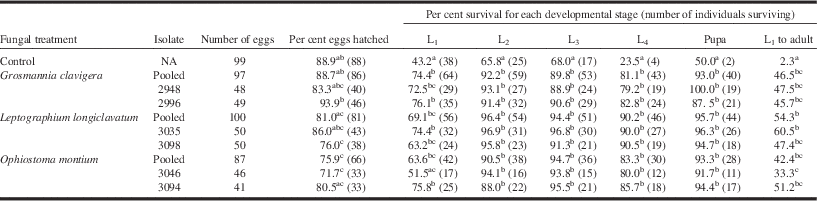
Per cent egg hatch was 89% for controls and the G. clavigera treatment, 81% for L. longiclavatum, and 76% for O. montium. Hatch success for the O. montium treatment (especially isolate NOF3046) was significantly lower than for the G. clavigera treatment (χ 2 1, 184=5.2285, P=0.0222) and the control treatment (χ 2 1, 186=5.166, P=0.0188) (Table 2). Sometimes eggs were overgrown by the fungus prior to hatching, likely resulting in their mortality.
Per cent survival (number of individuals that survived) by development stage of mountain pine beetle in rearing tubes inoculated with each of three fungi or with no fungi (controls).
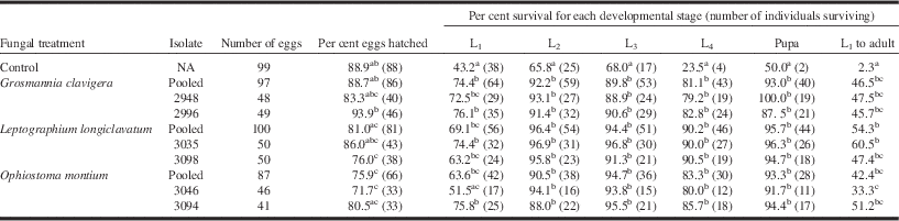
Note: Within columns, the values followed by the same superscript letters are not significantly different as determined by χ 2 tests (α=0.05). Rearing tubes contaminated by other fungi were eliminated from analysis.
Number and size of larval instars
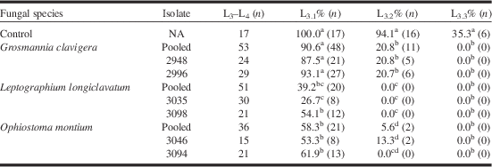
Although the MPB is documented to have four instars (Reid Reference Reid1962a), some larvae in this study had up to seven instars, based on signs of moulting (Table 3). The head-capsule sizes of the three extra instars were intermediary between those of third (L3) and final (L4) instars and were designated L3.1, L3.2, and L3.3. Of the 157 larvae that reached L4, 106 exhibited a fifth instar (L3.1), 29 exhibited a sixth (L3.2), and six (all in controls) exhibited a seventh (L3.3). The propensity for additional instars was highest in controls (Table 3). Significantly more larvae in the G. clavigera treatment exhibited extra instars than in the other treatments, and the L. longiclavatum treatment exhibited a lower occurrence of L3.2 than the O. montium treatment. Extra instar frequencies did not vary significantly by sex.
Per cent (number) of individual mountain pine beetles that had supernumerary instars.

Note: Within columns, the values followed by the same superscript letters are not significantly different as determined by χ 2 tests (α=0.05).
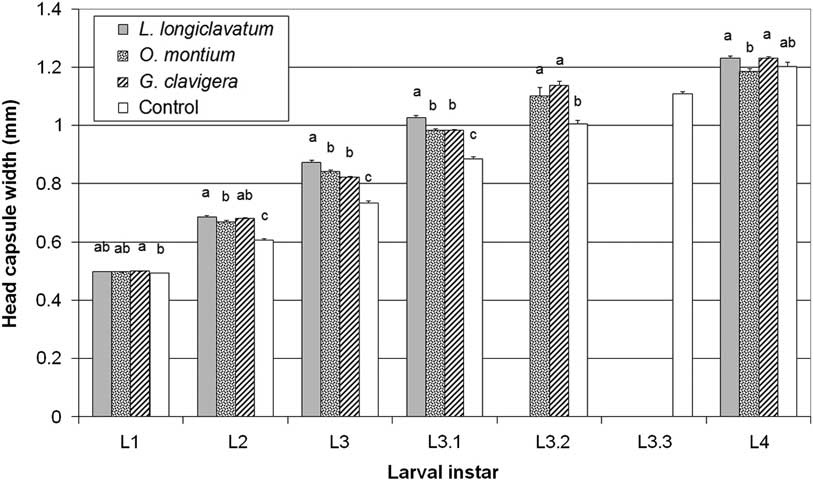
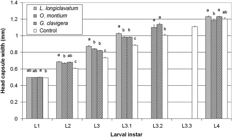
Larval head capsule width (a measure of larval size) was affected by treatment for all larval instars (F 3, 321=4.80, P=0.0028; F 3 , 200=75.85, P<0.0001; F 3, 176=75.14, P<0.0001; and F 3, 157=5.86, P=0.0008 for instars L1 to L4, respectively; Fig. 2). Larvae reared with L. longiclavatum generally had the widest head capsules and larvae in controls the narrowest. The greatest treatment effects on head-capsule width occurred for L3 as those reared with L. longiclavatum were larger (P<0.001) and control larvae were smaller (P<0.0001) than for all other treatments. However, through the addition of extra instars between L3 and L4, larvae achieving L4 were mostly equivalent in head-capsule width among treatments and controls, except that L4 larvae reared on O. montium were smaller than for those reared on L. longiclavatum (P=0.0028) or G. clavigera (P=0.0032), but this did not result in a significantly smaller adult size for this treatment. Head-capsule width did not vary by sex for most instars, but L4 females (1.237 mm±0.0072 SE) were significantly larger (F 1, 112=23.23, P<0.0001) than males (1.187 mm±0.0075).
Mean maximum head capsule width for each mountain pine beetle larval instar reared in association with three ophiostomatalean fungal species (Grosmannia clavigera, Leptographium longiclavatum, Ophiostoma montium) and with no fungus (control). Lines associated with each bar indicate one SE above the mean. Within an instar, bars containing the same lowercase letters do not have significantly different mean values as determined by post hoc Tukey–Kramer comparisons of means (α=0.05). Instars L3.1, L3.2, and L3.3 represent extra instars between L3 and L4, which occurred in some individuals.
Feeding behaviour and development
Larvae were observed to feed mainly on fungal hyphae and spores. L1 larvae in controls seemed “directionally” challenged and meandered short distances in all directions, eventually becoming lethargic and usually dying. However, in the presence of fungi, L1 larvae fed for a short time on fungal inoculum but soon constructed a gallery away from the inoculum. During gallery extension, larvae moved back and forth and groomed galleries by packing frass and rearing medium tightly into the sides and ends of galleries, and frequently moved these materials from the leading end of the gallery to the rear. While continually moving back and forth in galleries, larvae frequently fed on mycelium and spores by scraping gallery walls for fungi using their mouthparts. These actions kept the active part of the galleries relatively free of fungal growth. Little phloem was ingested by larvae, but was rather moved around and packed within the galleries. New adults did not extend galleries but fed on fungus spores in and near the pupal chamber.
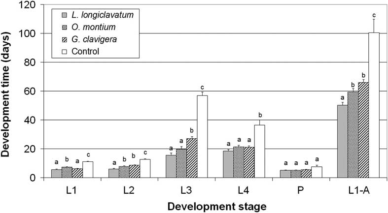
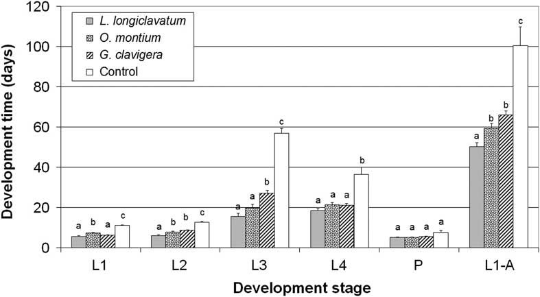
Development rate was calculated for each instar (L3 includes supernumerary instars), pupae, and summed for L1 to adult eclosion. Development for all instars and stages except pupae were significantly affected by treatment F 3, 200=58.7, F 3, 176=41.6, F 3, 157=76.3, F 3, 127=9.1, and F 3, 114=18.7 for L1, L2, L3, L4, and L1 to adult, respectively; P<0.0001 for all tests; Fig. 3). The slowest development occurred in controls for all instars and for L1-adult; this was particularly pronounced for L3 and L4. In general, development for each instar was fastest, sometimes significantly so, for individuals associated with L. longiclavatum, and total development from L1 to adult was fastest for that treatment (P=0.0272). Third instar development was faster for the O. montium treatment than for the G. clavigera treatment (P=0.0071), although the total development time from L1 to adult was not. There was no significant difference in development time between isolate treatments within a fungal species. Overall, males and females developed at the same rate; however, females developed slower than males at the L4 instar F 1, 112=9.98, P=0.0021; 22.2 days±0.87 SE and 18.19 days±0.91 SE for females and males, respectively).
Mean development time for each instar and pupal stage and total development time from egg hatch to adult eclosion for mountain pine beetles reared from the egg stage in association with three ophiostomatalean fungus species (Grosmannia clavigera, Leptographium longiclavatum, Ophiostoma montium) and with no fungus (control). Lines associated with each bar indicate one SE above the mean. Within an instar or stage, bars containing the same lowercase letters do not have significantly different mean values as determined by post hoc Tukey–Kramer comparisons of means (α=0.05). Instar L3 includes the extra instars L3.1, L3.2, and L3.3 where they occur.
Larval gallery length
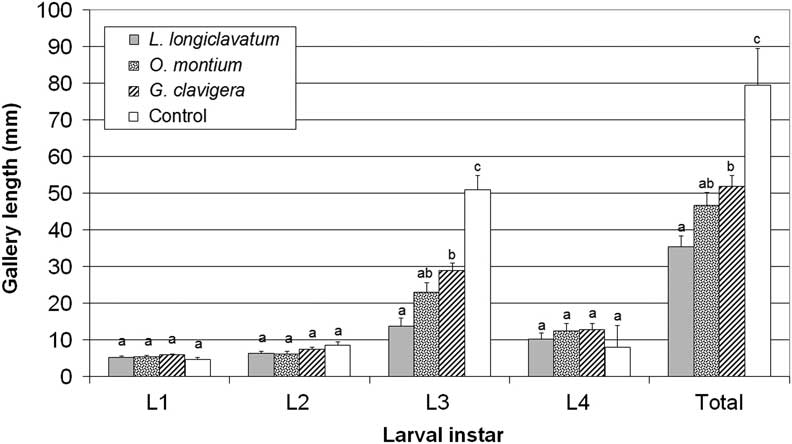
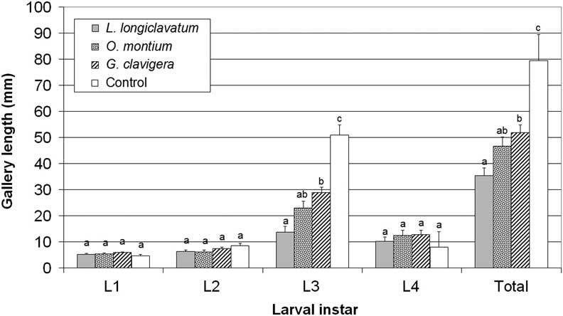
Gallery extension was predominantly accomplished by the L3, including extra instars (Fig. 4). During this period, there was a significant effect of treatment on gallery length F 3, 157=25.48, P<0.0001). Galleries were significantly longest in controls. Galleries associated with G. clavigera were longer than those associated with L. longiclavatum, that were shorter (NS) than those associated with O. montium. Total gallery length was affected by treatment (F 3, 124=9.17, P<0.0001) similar to that described for L3. The only gender effects were that females constructed longer galleries than males in the L4 instar F 1, 112=10.91, P=0.0013; 15.9 mm±1.58 SE for females, 8.4 mm±1.64 for males) and overall F 1, 112=8.37, P=0.0046; 49.4 mm±2.64 for females, 38.4 mm±2.74 for males).
Mean length of gallery excavated by each larval instar and total gallery length summed over all instars for mountain pine beetles reared in association with three ophiostomatalean fungus (Grosmannia clavigera, Leptographium longiclavatum, Ophiostoma montium) species and with no fungus (control). Lines associated with each bar indicate one SE above the mean. Within an instar, bars containing the same lowercase letters do not have significantly different mean values as determined by post hoc Tukey–Kramer comparisons of means (α=0.05). Instar L3 includes the extra instars L3.1, L3.2, and L3.3 where they occur.
Survival
Survival was determined for each instar, pupae, and totalled from egg hatch to adult eclosion. For all fungal treatments, the lowest survival occurred for L1, ranging from 52% to 76% among isolates (Table 2). For other instars and pupae, survival was much better at 79–100%, and generally was >90%. First instar survival varied among the two O. montium isolates (Table 2). Survival was lower in controls than in fungal treatments for all instars and stages (Table 2; χ 2 1=7.2654 to 49.331, P=0.007 to P<0.0001) except that L1 survival was similar for isolate NOF3046 (O. montium) and controls. Significant treatment effects occurred only for L1 survival, which was lower for O. montium isolate NOF3046 than for G. clavigera (pooled), and for total survival, which was lower for isolate NOFC3046 than for L. longiclavatum (pooled). Although not significant, the trend was for better survival in association with L. longiclavatum than with other species. Larvae typically died just before or during moulting, especially in controls.
Adult size and fat content
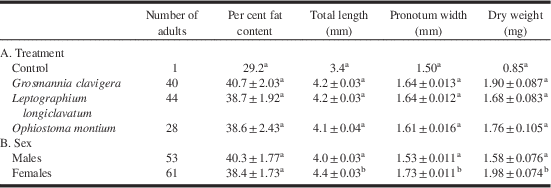
Only two small adults were produced in controls. These died before maturation and were excluded from analyses. There were no treatment effects on adult length, pronotum width, dry weight, and per cent fat content, although adults from the O. montium treatment tended to be smaller than those from the other treatments (Table 4). Females were significantly longer, wider and heavier F 1, 106=119.51, 176.23, 19.60, respectively, P<0.0001) than males, but there was no significant effect of gender on fat content (Table 4).
Mean±SE per cent fat content, total length, maximum pronotum width, and dry weight of mountain pine beetle adults.

Notes: A. Reared without fungi (control) or with one of three ophiostomatalean fungus species; B. By sex (due to low numbers of beetles reared, data from controls were excluded). Within columns of each section of the table, the values followed by the same superscript letters are not significantly different as determined by Tukey–Kramer comparison of least square means (α=0.05).
Fungal growth and interactions with larvae
Four weeks after fungal inoculation, treatment effects on hyphal growth were evident F 2, 282=16.21, P<0.0001). Hyphal advancement by L. longiclavatum (39 mm±2.3 SE) was greater than for G. clavigera (31 mm±2.3), which was greater than for O. montium (21 mm±2.4). Conidia were observed in >90% of replicates for each fungus.
The fungal species differed in growth patterns and dynamics. Hyphal growth of O. montium seemed limited to galleries and did not advance much beyond the end of the gallery into unmined and water-saturated phloem, possibly indicating limitation by oxygen availability. However, once the phloem was exposed to air through gallery extension or drying of medium over time, it exhibited the most aggressive growth of all fungi. Larvae were affected most by O. montium as the thick mycelial growth was observed to overwhelm and apparently kill some eggs and young, inactive. The rearing medium was also observed to dry out most quickly with O. montium.
Leptographium longiclavatum penetrated hydrated and unmined medium faster than other species and appeared least limited by low oxygen. Its mycelium was much less dense than that of O. montium. Leptographium longiclavatum sporulated more abundantly than the other fungi. Larvae were most active and vigorous in the L. longiclavatum treatment, and tended to spend more time “back-feeding” and less time extending galleries than in other treatments.
Grosmannia clavigera tended to have the least dense fungal growth, and larvae easily kept galleries free of excessive mycelium. All fungi degraded dead larvae and eggs (as evidenced by the darkening and eventual disappearance of bodies which did not occur in the no-fungus control); however, G. clavigera seemed to be the least and O. montium the most saprophytic.
Nutrient analyses
The concentration of carbon in the rearing medium was not affected (F 3, 35=1.72, P=0.18) by fungal inoculation, and ranged from 48.7% to 49.0% among treatments and the sterilised control. Nitrogen concentration in the rearing medium was significantly affected by treatment (F 3, 35=5.94, P=0.0025); the concentration in the L. longiclavatum treatment (0.41%±0.012 SE) was significantly lower compared to sterilised controls (0.49%±0.018) and the O. montium (0.46%±0.012) and G. clavigera (0.47±0.012%) treatments.
Discussion
Rearing protocol
This novel protocol is useful for rearing MPB from egg to adult in a controlled fashion. Some advantages of this protocol are that it: is easy, relatively inexpensive and takes up little space; allows individual rearing; provides an opportunity to view bark beetle development and behaviour; minimises contamination; allows easy control of environmental conditions (moisture, light, temperature); and can be adapted to examine many kinds of interspecific interactions. We offer some suggestions to guide future users of this method:
-
1. Phloem used for our experiments was taken from trees in October after they had hardened-off, but it is not known if this has an impact on fungus and MPB performance. It is preferable to use phloem taken from trees in July–August, the period when trees are normally attacked by MPB.
-
2. The protocol for adding inoculum will need to be refined if combinations of fungi or bacteria are used. It is recommended that species that are less aggressive and slower growing be added sometime before more aggressive species to reduce the chance of competitive exclusion. As bacteria may be slow to colonise phloem from a single inoculation site, we recommend inoculation using a liquid suspension.
-
3. This method could be used to examine behaviour of adult MPB by adding a female in one end of the tube followed by a male one day later. Wider tubes (~7 mm) are recommended to allow beetles room to turn around, and longer tubes (30–40 cm) will accommodate the extensive boring of the adults. The end of the tube where adults are added should have 2–3 cm of empty space to accommodate frass expulsion from egg galleries.
-
4. This method could also be used to examine interactions between other organisms (mites, nematodes, microorganisms, beetle eggs/larvae). As these organisms are small, use of narrow tubes (e.g., 3 mm inner diameter) are recommended so that behaviours are more easily observed.
Ophiostomatalean fungus growth and effects on phloem nutrients
The relative commonness of L. longiclavatum in boreal Alberta compared to elsewhere (Rice and Langor Reference Rice, Thormann and Langor2009; Roe et al. Reference Roe, James, Rice, Cooke and Sperling2011) suggests that it is better adapted to the boreal environment. This fungus is as pathogenic to boreal pines as are G. clavigera and O. montium (Rice et al. Reference Rice, Thormann and Langor2007) and is adapted to growth and survival at cooler boreal temperatures (Rice et al. Reference Rice and Langor2008). We have now demonstrated that it is the fastest growing, in terms of hyphal extension at room temperature, of the three ophiostomatalean species on jack pine phloem; however, its mycelial growth did not appear to be as dense as that of O. montium. Much of the growth capacity of L. longiclavatum likely comes from its apparent ability to tolerate lower oxygen levels, which enabled it to penetrate unmined and water-saturated phloem more readily than the other species. This apparent tolerance to low oxygen may provide L. longiclavatum with the means to outcompete O. montium in fresh phloem, while O. montium seems to have faster growth in mined and drier phloem.
Grosmannia clavigera is also more tolerant of low oxygen conditions than is O. montium (Solheim and Krokene Reference Solheim and Krokene1998), and in previous studies was consistently at the leading edge of fungal penetration of sapwood of MPB-colonised pines (Solheim Reference Solheim1995). It also grew faster than O. montium in the current study and was able to penetrate unmined medium (although not as rapidly as L. longiclavatum). The superior growth performance of L. longiclavatum in low oxygen conditions may explain why this species seems to be dominating G. clavigera in Alberta where they commonly co-occur (Roe et al. Reference Roe, James, Rice, Cooke and Sperling2011), have similar physiological requirements (Rice et al. Reference Rice, Thormann and Langor2007, Reference Rice and Langor2008), and are therefore likely in direct competition.
Our results corroborate earlier work (Six and Paine Reference Six1998) that suggests that O. montium is not as beneficial to MPB as G. clavigera. Although MPB seems to benefit nutritionally from consuming mycelium and spores of O. montium, the aggressive growth of this species in mined areas and its capacity to dry out phloem and apparently overcome eggs and small larvae suggests that it may sometimes be detrimental to MPB. Beetles that carry all three species may have advantages over individuals with one or two species in terms of capacity to successfully colonise a wider variety of hosts and habitats across a wider range of environmental conditions (Six and Bentz Reference Six and Klepzig2007).
Interactions between MPB and ophiostomatalean fungi
Since Whitney (Reference Whitney and Farris1971) first suggested that MPB gained nutritional benefit from associated ophiostomatalean fungi, the evidence for the importance of fungi in the nutritional ecology of the MPB has mounted (Six Reference Six and Paine2012 and references therein). However, as the MPB has many associated fungi and other microorganisms (Lee et al. Reference Lee, Kim and Breuil2006b; Winder et al. Reference Winder, Macey and Cortese2010), the attribution of beetle benefits (or detriments) to individual fungus species is difficult and necessitates empirical approaches to control MPB-microbe interactions. This has proven difficult to achieve, but the one successful attempt previous to this study has provided new insights into MPB-fungus interactions and has demonstrated that different ophiostomatalean species do not have equivalent benefits to the MPB (Six and Paine Reference Six1998).
Our study builds significantly on earlier knowledge and, for the first time, compares the effects of L. longiclavatum to the two more commonly studied species, G. clavigera and O. montium. Clearly, ophiostomatalean fungi benefit MPB, as progeny reared in the absence of fungi fared significantly worse in terms of survival and development rate. Furthermore, larval galleries were longest in controls, indicating a poor larval environment (Six and Paine Reference Six1998; Bleiker and Six Reference Bleiker, Potter, Lauzon and Six2007). Our observations of axenically reared L1 larvae indicate that they wander, frequently becoming lethargic and dying. The frequent active consumption of hyphae and spores of all three fungus species by all instars, and the indication that fungal tissue is frequently consumed more than phloem suggests that these fungi contribute directly to the MPB nutrition. This is not surprising, as mycophagy confers many benefits to insects (Martin Reference Martin1979). Our observations corroborate those of Adams and Six (Reference Adams and Six2007) by showing that MPB larvae are associated with and ingest the symbiotic fungi and move back in galleries to feed on areas colonised by the fungi. This contrasts somewhat to the observations of Whitney (Reference Whitney and Farris1971) who reported that late instars became separated from fungi as they mined; however, his observations were in experimental logs under conditions very different than those seen in nature where beetles consistently are found feeding in fungus-colonised phloem. Whitney (Reference Whitney and Farris1971) did report that early instars consumed fungi, possibly deriving nutritional benefits.
Different ophiostomatalean species had different effects on MPB success on jack pine phloem. Mountain pine beetle fared best in association with L. longiclavatum as indicated by faster development. Faster development times will help larvae achieve the fourth instar before onset of winter which has implications for survival because larger larvae are more cold tolerant than small larvae (Safranyik and Linton Reference Safranyik, Carroll, Régnière, Langor, Riel and Shore1998). Galleries were also shortest and there were fewer supernumerary instars in association with L. longiclavatum, suggesting a better larval nutritional content (Six and Paine Reference Six1998). However, final adult size, weight and fat content, all indicators of better fitness (Reid Reference Reid1962b; Amman Reference Amman1972; Barras and Hodges Reference Barras and Hodges1974; Safranyik Reference Safranyik1976), did not vary significantly between fungus treatments.
Previous work suggested that MPB benefitted more from G. clavigera than O. montium in lodgepole pine logs (Six and Paine Reference Six1998; Bleiker and Six Reference Bleiker, Potter, Lauzon and Six2007; Cook et al. Reference Cook, Shirley and Zambino2010). Our work corroborates this as L4 head-capsule width was smaller for the O. montium treatment than for the G. clavigera treatment. Adult size and fat content also tended to be lower for the O. montium treatment than for the G. clavigera treatment, though the results were not significant. We suggest additional experiments using lodgepole pine and jack pine phloem and all three fungi, to discern whether pine species mediates the interactions between MPB and associated ophiostomatalean fungi.
Little is known about underlying processes responsible for the responses of MPB to different fungi. As the carbon and nitrogen nutrient analysis was performed on the entire contents of rearing tubes (including fungus-colonised and uncolonised portions), the analysis was not structured to compare how well each fungus concentrated nutrients. Low nitrogen availability is limiting to insect growth in phloem and it is known that ophiostomatalean fungi can concentrate nitrogen (Ayers et al. Reference Ayers, Wilkens, Ruel, Lombardero and Vallery2000). Grosmannia clavigera and O. montium serve to mobilise nitrogen concentration, and make it available to MPB larvae, and G. clavigera is better at this role than O. montium at 20–25 ºC and at a range of medium nitrogen concentrations (Cook et al. Reference Cook, Shirley and Zambino2010). Furthermore, fungi provide MPB larvae with sterols, especially ergosterol and possibly vitamins, important for development (Bentz and Six Reference Bentz and Six2006; Douglas Reference Douglas2009). The fact that mortality in controls usually occurred just before or during moulting may suggest a limited access to sterols required for hormone production. The superior success of MPB larvae with L. longiclavatum may be related to a better capacity to concentrate nitrogen in a form used by beetles or produce sterols and vitamins.
It is interesting that all three fungi tended to reduce overall nitrogen, although this reduction was significant only for L. longiclavatum. This suggests that fungi may metabolise nitrogen and release some as gas, possibly nitrous oxide (Bollag and Tung Reference Bollag and Tung1972) or ammonia (Aldarf et al. Reference Aldarf, Amrane and Prigent2002). In nature, these fungi have access to large pools of nitrogen in the sapwood and concentrate it in the phloem (Bleiker and Six Reference Bleiker, Potter, Lauzon and Six2007; Goodsman et al. Reference Goodsman, Erbilgen and Lieffers2012) so availability of nitrogen to beetles in the phloem is not likely to be limited even if fungi are metabolising a portion of it.
Supernumerary instars
Our work is the first report of supernumerary instars in Scolytinae. Supernumerary instars are reported from several insect orders (Heming Reference Heming2003; Esperk et al. Reference Esperk, Tammaru and Nylin2007) and a few wood-boring Coleoptera in the families Cerambycidae and Bostrichidae (Lyctinae) (Iwata and Nishimoto Reference Iwata and Nishimoto1984; Shintani et al. Reference Shintani, Munyiri and Ishikawa2003). Extra instars are frequently associated with starvation or suboptimal nutritional intake, short day lengths, high temperature, and high rearing density (Esperk et al. Reference Esperk, Tammaru and Nylin2007). As MPB larvae were reared solitarily at room temperature and in darkness, the most reasonable explanation for extra instars is inadequate nutrition. In MPB, the likelihood of supernumerary instars and the average number of extra instars was directly related to developmental time. Thus the existence and number of extra MPB instars likely reflects the quality of the nutritional environment, with the likelihood and number of extra instars lowest in association with L. longiclavatum.
In MPB, pupation was not elicited by number of larval moults (i.e., four) but was rather determined by the larval stage achieving some size threshold (L4 head capsule size was roughly the same regardless of number of supernumerary instars) or possibly a threshold nutritional intake. This is similar to the situation reported for Manduca sexta (Linnaeus) (Lepidoptera: Sphingidae), wherein larvae must achieve a size threshold between the penultimate and ultimate instars before pupation will occur; if the threshold is not achieved, additional instars occur (Nijhout Reference Nijhout1994). This means that in a situation where MPB encounters suboptimal nutritional conditions, evolution has favoured a longer developmental time and extra instars in order to achieve a threshold size and ultimately larger adults versus the alternative of shorter developmental times, a limit of four instars, no threshold size, and smaller adults. This may indicate that small adults have low probability of reproductive success and that it is better to risk a longer development time and later dispersal period with larger adults than a shorter development time and earlier dispersal time with smaller adults.
Conclusions
The rearing tube method offers a relatively easy means of individually rearing large numbers of MPB from egg to adult in association with various fungal associates to investigate beetle-microorganism interactions. While not natural, this method can allow insights into interactions and behaviours that may be explored in additional experiments. Furthermore, this method should be easily adapted to examine other interspecific relationships.
Using this method, we have gained new insights into MPB-ophiostomatalean fungus interactions. Rearing of MPB in jack pine phloem in combination with each of its three most commonly associated ophiostomatalean fungi reveals that beetles do not benefit equivalently from all fungi. As larval galleries were shortest and there were fewer supernumerary instars in association with L. longiclavatum, this suggests that this fungus provides more nutritional benefits than the other two species. This allows MPB to develop faster in association with L. longiclavatum, which is postulated to increase overwinter survival in boreal climates. Furthermore, the superior capacity of L. longiclavatum to penetrate hydrated, unmined phloem compared to the other two fungus species may explain why it is dominant in the boreal. Clearly, interspecific interactions between MPB and ophiostomatalean fungi are different in the boreal than elsewhere and may represent an adaptation to boreal environments. Understanding the changing natural history of the MPB in the boreal is critical to helping predict the persistence, success, spread, and potential impact of this species in this newly invaded ecosystem.
Acknowledgements
The authors thank Daryl Williams for field assistance, Joel Crumbaugh and Curtis Olive for doing nutrient analyses, Tod Ramsfield for helpful advice, and Tod Ramsfield, Kathy Bleiker, and two anonymous reviewers for reviewing an advanced draft of the manuscript. Funding was provided by Natural Resources Canada, Saskatchewan Ministry of Forests, Alberta Environment and Sustainable Resource Development, Foothills Research Institute (Mountain Pine Beetle Ecology Program), and the Spray Efficacy Research Group International.