Implications
Escherichia coli O157:H7 is causing, every year, deaths and costly recalls worldwide, yet little is known of the relationship between E. coli O157:H7 and the gastrointestinal colonization of cattle. These studies offer insight regarding the potential of E. coli O157:H7 to utilize intestinal mucus and its components as substrates for growth in the gastrointestinal tracts of cattle. Factors influencing intestinal mucus secretion in cattle may be important determinants of E. coli O157:H7 colonization rates and, thus, important components in the development of innovative and efficient preharvest interventions to prevent food chain contaminations.
Introduction
Escherichia coli are commensal copiotroph bacteria found in the intestinal mucus layer (Montagne et al., Reference Montagne, Toullec and Lalles2000; Moller et al., Reference Moller, Leatham, Conway, Nuijten, de Haan, Krogfelt and Cohen2003; Naylor et al., Reference Naylor, Low, Besser, Mahajan, Gunn, Pearce, McKendrick, Smith and Gally2003) of mammalian digestive tracts (Ihssen and Egli, Reference Ihssen and Egli2005). Some strains of E. coli are pathogenic to humans, such as the well known Enterohemorrhagic (EHEC) E. coli O157:H7. Infection with EHEC often originates from the ingestion of contaminated food products. E. coli O157:H7 is non-pathogenic to cattle, and cattle are thus recognized as an important reservoir for the pathogen. Exposure in human populations can occur either directly through contact with cattle or their waste products, or indirectly through water, meat or other food products that have been contaminated by cattle feces. To control contamination in the food chain, it is essential to understand how this pathogen is able to grow and compete with other bacteria in the bovine gastrointestinal tract. Previous studies have shown that bovine intestinal mucus supports bacterial colonization and can selectively influence composition of the bacterial population (Deplancke and Gaskins, Reference Deplancke and Gaskins2001), yet little is known of the nutrients and metabolic pathways used by E. coli O157:H7 (Miranda et al., Reference Miranda, Conway, Leatham, Chang, Norris, Allen, Stevenson, Laux and Cohen2004). Intestinal mucus is comprised of mucins, glycoproteins, glycolipids, epithelial cell debris and electrolytes (Conway et al., Reference Conway, Krogfelt and Cohen2006). Degradation of the complex mucin components to simpler, more readily fermentable substrates requires multiple enzymes. Proteases convert the mucus from viscous to fluid state and endoglycosidases act at internal sites to release oligosaccharide fragments. Sialic acid is then removed from these fragments by neuraminidases, allowing the degradation of the remaining chain by exo-glycosidases and β-galactosidases (Corfield et al., Reference Corfield, Wagner, Clamp, Kriaris and Hoskins1992). E. coli does not produce polysaccharide-degrading enzyme (Chang et al., Reference Chang, Smalley, Tucker, Leatham, Norris, Stevenson, Anderson, Grissom, Laux, Cohen and Conway2004), thus restricting its capacity for carbohydrate utilization to mono- or disaccharides (Mayer and Boos, Reference Mayer and Boos2005). Moreover, E. coli O157:H7 lacks neuraminidase activity (Hoskins et al., Reference Hoskins, Agustines, McKee, Boulding, Kriaris and Niedermeyer1985), and therefore has limited ability to degrade complex mucin molecules. The organism is thus dependent upon other anaerobes present within the gastrointestinal tract to degrade mucin polysaccharides and release fragments beneficial for their growth (Jones et al., Reference Jones, Jorgensen, Chowdhury, Rodgers, Hartline, Leatham, Struve, Krogfelt, Cohen and Conway2008). Analysis of bovine small intestinal contents revealed the presence of free mucus-derived carbohydrates, including galactose (1.43 mM), GlcNAc (0.89 mM), GalNAc (0.72 mM), fucose (0.64 mM), mannose (0.50 mM) and N-acetyl neuraminic acid (0.09 mM; Bertin et al., Reference Bertin, Chaucheyras-Durand, Robbe-Masselot, Durand, de la Foye, Harel, Cohen, Conway, Forano and Martin2013), all of which can be metabolized by E. coli O157:H7. Nevertheless, to be maintained in the gastrointestinal tract of cattle, E. coli O157:H7 needs to compete with other bacteria to colonize the mucus layer. In the 1980s, it was believed that bacteria have a single preferred nutrient as a substrate for growth (Freter, Reference Freter1988); however, recent studies have illustrated that E. coli relies on a diverse range of nutrients for its growth (Chang et al., Reference Chang, Smalley, Tucker, Leatham, Norris, Stevenson, Anderson, Grissom, Laux, Cohen and Conway2004), allowing EHEC to proliferate in cattle and shed in their feces, thus providing opportunity for contamination of carcasses at harvest. Fox et al. (Reference Fox, Drouillard, Shi and Nagaraja2009) demonstrated that galactose, gluconic acid, glucuronic acid, galacturonic acid, glucosamine and porcine mucin increased growth of NalR E. coli in fecal fermentations. In addition, Bertin et al. (Reference Bertin, Chaucheyras-Durand, Robbe-Masselot, Durand, de la Foye, Harel, Cohen, Conway, Forano and Martin2013) showed that EDL933, an EHEC strain, consumed the free mucus-derived carbohydrates present in bovine small intestinal content more rapidly than resident microflora. The following in vitro experiments were conducted to gain further insight on the mechanisms used by E. coli O157:H7 to thrive and compete with other bacteria of the bovine gastrointestinal tract. Fermentation assays were performed on small or large bovine intestinal mucus, or their constituents, with or without various enzymes, and in the presence or absence of fecal inoculum.
Material and methods
Intestinal mucus harvest
Intestinal tissues were collected from freshly harvested cattle and, immediately, transported to the Preharvest Food Safety Laboratory (Kansas State University, Manhattan, KS, USA). Sections of the ileum and colon were excised with sterile scissors and washed with a HEPES (N-2-hydroxyethylpiperazine-N'-2-ethanesulfonic acid)-Hanks buffer (8.0 g of NaCl, 0.4 g of KCl, 0.185 g of CaCl2∙2H2O, 0.2 g of MgSO4∙7H2O, 0.05 g of Na2HPO4, 0.35 g of KH2PO4, 2.6 g of HEPES; adjusted to pH 7.4) to remove digesta. Mucus was collected from each of the sections by gently scraping the epithelium with a sterile microscope slide. Harvested mucus was centrifuged twice at 27 000×g for 20 min to remove cellular debris and impurities. Supernatant containing the crude mucus was dialyzed overnight at 4°C in HEPES-Hanks buffer, lyophilized and stored at −20°C.
Bacterial strains
Five Shiga toxin-producing E. coli O157:H7 strains (STEC; 01-2-1863, 01-2-7443, 01-2-10004, 01-2-10530 and 01-2-12329), isolated from feedlot pen fecal samples by Sargeant et al. (Reference Sargeant, Sanderson, Smith and Griffin2003), were used in this experiment. Mutants resistant to nalidixic acid (NalR) were obtained by serial transfer into Luria-Bertani broth (Neogen Inc. Baltimore, MD, USA) containing increasing concentrations of nalidixic acid (from 0.2 to 50 mg/l; Sigma-Aldrich, St. Louis, MO, USA) following the procedure outlined by Schamberger et al. (Reference Schamberger, Phillips, Jacobs and Diez-Gonzalez2004).
Bacterial inoculum preparation
All five NalR STEC were plated onto tryptic soy agar containing 5% sheep’s blood (Blood agar; Thermo Fisher Scientific, Waltham, MA, USA) and were incubated overnight at 37°C. A single colony was picked from each plate and grown overnight at 37°C in 10 ml tryptic soy broth (TSB; Thermo Fisher Scientific) to be transferred to five other 9-ml TSB tubes. After incubation at 37°C for 18 h, the five broths were combined in equal proportions to create a five-strain cocktail of NalR STEC.
Fecal inoculum preparation
Feces were collected by rectal palpation from a steer fed a concentrate-based diet and transported to the Preharvest Food Safety Laboratory in a pre-warmed thermos. Feces were blended in an Osterizer blender for 60 s with McDougall’s buffer (pH 6.8; McDougall, Reference McDougall1948) in a 1 to 6 ratio (weight/volume) under a stream of CO2, strained through two layers of cheese cloth, and the resulting fecal suspension was collected.
In vitro fermentation assay
Fecal inoculum or McDougall’s buffer were added to tubes containing substrates to be tested in a 1 : 2 ratio (volume/volume) under a stream of CO2 to maintain anaerobic environment. Tubes were inoculated with the five-strain bacterial inoculum to achieve a final concentration of 103 CFU/ml, gassed with CO2, capped with butyl stoppers fitted with Bunsen valves, and incubated on a shaker at 40°C. A volume of 100 µl was extracted from each fermentation tube after 0, 6, 8, 12 and/or 24 h and diluted into 900 µl of Butterfield’s phosphate buffer (26.22 g of KH2PO4/l; pH 7.2). Subsequent dilutions (100 µl) were plated in triplicate onto sorbitol MacConkey agar (Becton Dickinson, Franklin Lakes, NJ, USA), supplemented with cefixime (0.05 mg/l), potassium tellurite (2.5 mg/l), and nalidixic acid (50 mg/l; CTN-SMAC). Plates were incubated at 37°C for 24 h and non-sorbitol fermenting colonies were enumerated to determine NalR STEC concentrations.
Nitrogen and organic matter (OM) content of mucus
OM contents of mucus harvested from the small and large intestine were determined by ashing the samples (Undersander et al., Reference Undersander, Mertens and Thiex1993). Briefly, mucus samples were dried in aluminum pan overnight at 105°C to determine dry matter content. Pans were then heated to 450°C overnight, slowly cooled and transferred to a desiccator to be weighted. OM percentage was obtained by subtracting ash content from a hundred. A bicinchoninic acid assay (Thermo Fisher Scientific) was performed to assess CP content of samples. In vitro fermentations performed in this study were carried out in tubes containing mucin from the small and large intestine at equal levels of OM (4.4 mg of OM/ml; 7.5 and 9.2 µg of protein/ml for small and large intestinal mucus, respectively) unless stated otherwise.
Increasing concentrations of mucus
Increasing concentration of mucus from the small intestine (0, 0.5, 1.0, 2.0, 4.4, 10 and 15 mg OM per ml of inoculum) were tested in an in vitro fermentation assay as described previously. In addition, aliquots obtained at different sampling times were plated in triplicate onto tryptic soy agar (TSA; Thermo Fisher Scientific) to allow enumeration of total anaerobic bacteria. The TSA plates were inoculated with 100 µl of the cultures and incubated at 40°C in a Coy rigid anaerobic chamber (Coy Laboratory Products, Grass Lake, MI, USA) containing 90% nitrogen, 5% CO2 and 5% hydrogen.
Mucus and mucus components
Mucin from the ileum and the colon (4.4 mg of OM/ml), as well as mucus components such as the lipid l-α-phosphatidylserine, (1 mg/ml; CAS number: 840032P, Avanti Polar Lipids Inc., Alabaster, AL, USA), and the carbohydrates: d-gluconic acid (CAS number: G9005), d-glucuronic acid (CAS number: 6556-12-3), N-acetyl-d-glucosamine (CAS number: 7512-17-6), d-galacturonic acid (CAS number: 91510-62-2), sialic acid (CAS number: 131-48-6), galactose (CAS number: G5388) and mannose (10 mg/ml; CAS number: 3458-28-4,Sigma-Aldrich) were tested as growth substrate in the in vitro fermentation assays.
Enzymes and enzyme inhibitors
All enzymes and inhibitors were added to tubes containing small or large intestinal mucin and McDougall’s buffer to be subjected to a fermentation assay. A protease from bovine pancreas (CAS number: 9001-92-7), an endoglycosidase from Elizabethkingia meningoseptica (PNGase F; CAS number: 83534-39-8), a neuraminidase from Clostridium perfringens (CAS number: 9001-67-6) and a lipase from Candida antartica (CAS number: 9001-62-1) all were purchased from Sigma-Aldrich and tested at a concentration of one unit per milliliter of McDougall’s buffer. β-Galactosidase (CAS number: 9031-11-2; Sigma-Aldrich) and phenylethyl β-d-thiogalactopyranoside (PETG; CAS number: P-1692; Invitrogen, Grand Island, NY, USA) were inoculated at a concentration of 100 and 200 µM, respectively. Finally, bacterial protease inhibitor cocktail containing 4-(2-aminoethyl)benzenesulfonyl fluoride, EDTA disodium salt, bestatin, pepstatin A, and E-64 (P8465; Sigma-Aldrich) was added at two different concentrations, 0.25 and 2.5 ml/g E. coli O157 culture.
Statistical analysis
Colony forming units (CFU) counts were transformed to the log10 scale. Statistical analysis of fermentation assays were performed using the MIXED procedure of SAS (SAS 9.2, Cary, NC, USA). Sampling times, substrate types, presence or absence of fecal inoculum and their interactions were included in the model as fixed effects. Effect of increasing level of small intestinal mucus on E. coli O157:H7 and total anaerobes were analyzed using the MIXED procedure of SAS and linear contrasts. Sampling time, small intestinal mucus concentration, and the interaction of sampling time×small intestinal mucus concentration were included as fixed effects in the model. Differences were considered different at P<0.01 and P<0.05.
Results
Mucus anatomical origin and composition
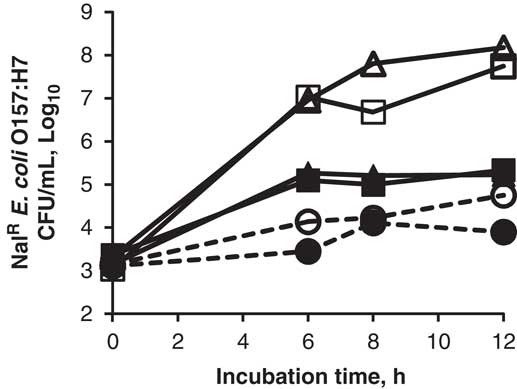
The analysis of the nitrogen and OM content of the mucus collected at the ileum and colon revealed slight differences in composition with 1.068 µg of protein and 627 µg of OM per milligram of small intestinal mucus v. 0.910 µg of protein and 442.6 µg of OM per milligram of large intestinal mucus. Figure 1 depicts the growth of NalR E. coli O157:H7 in presence or absence of a fecal inoculum, with small intestinal mucus, large intestinal mucus, or no substrate overtime. It reveals no significant differences (P>0.10) in growth when mucus from the ileum and colon were used as substrate. The bacteria increased from 103 CFU/ml of culture, at hour 0, to between 107 and 108 CFU/ml at hour 8. An overall time effect was observed on the growth of the bacteria (P<0.01); however, there was no significant difference between hour 8 and hour 12. The presence or absence of fecal inocula in the culture did affect growth of E. coli O157:H7 (P<0.01). Final concentration of the pathogen was 107 CFU/ml in tubes without fecal inoculum, compared with 105 CFU/ml in tubes containing fecal inoculum.
Growth of NalR E. coli O157:H7, with buffer (○), small (▵) or large (□) intestinal mucus as a substrate, in presence (bold symbols) or absence of fecal inoculum (open symbols). s.e.m.=0.21, effect of time, P<0.01; effect of mucus origin, P>0.01; effect of addition of fecal inoculum, P<0.01.

Increasing concentrations of mucus
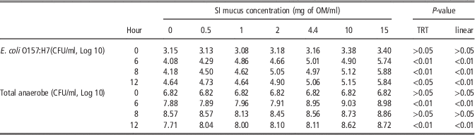
Growth of total anaerobic bacteria and NalR E. coli O157:H7 was tested with increasing levels of small intestinal mucus. Growth of total anaerobes was significantly influenced by the levels of substrate addition at hours 6 and 12 (Table 1; SI concentration effect, P<0.01). A linear increase in anaerobe concentration was observed with increasing concentration of small intestinal mucus (linear effect, P<0.01). Cultures containing 15 mg of mucus OM/ml supported a 28% increase in anaerobe growth after 12 h incubation compared with cultures without mucus. Growth of NalR E. coli O157:H7 increased at every time point in response to increased concentrations of mucus (linear effect, P<0.01). Counts of NalR STEC counts were increased by 72% in the presence of 15 mg of mucus OM/ml of culture compared with tubes containing 0 mg of mucus/ml after 12 h of fermentation.
Growth of NalR Escherichia coli O157:H7 and total anaerobes with increasing levels of small intestinal (SI) mucus over a 12 h fermentation at 40°C

s.e.m.=0.3.
Mucus and mucus components
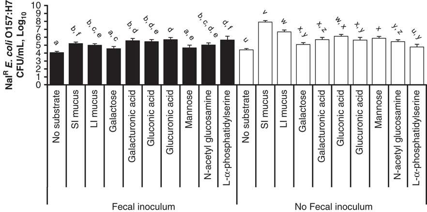
Figure 2 depicts the growth of E. coli O157:H7 after 8 h of anaerobic incubation with or without fecal inoculum and either whole mucus or selected components of mucus as substrates. In presence of a fecal inoculum, all substrates increased the growth of E. coli O157:H7 (P<0.05), with the notable exceptions of mannose and galactose. Growth was numerically greatest with l-α-phostatidylserine and glucuronic acid as substrates, but was not different from that obtained with whole mucus from the small or large intestines (P>0.179). In the absence of fecal inoculum, all of the mucus components tested, with the exception of l-α-phosphatidylserine, increased growth of the bacteria in comparison to the batch containing no substrate (P<0.05). Mucus originating from the large and small intestines supported greater growth than individual mucus fractions (P<0.05), which is in contrast to results observed in the presence of fecal inoculum. Gluconic acid was the only single compound to yield growth similar to that obtained with whole large intestinal mucus (P>0.09), but still was less than that observed for whole small intestinal mucus (P<0.0001).
Growth of NalR E. coli O157:H7 after 8 h incubation at 40°C with mucus or single mucus components as a substrate for growth, in presence or absence of fecal inoculum. Means are compared within fecal inoculum groups. Means without a common superscript letter are different, P<0.05.
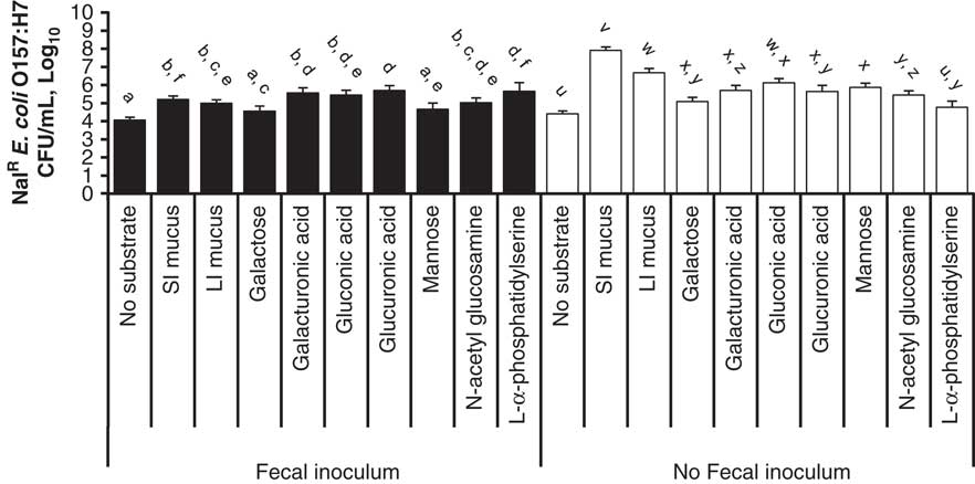
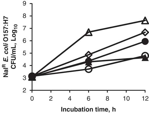
Figure 3 illustrates growth of E. coli O157:H7 in response to small intestinal mucus or sialic acid substrates, in presence or absence of a fecal inoculum. NalR STEC growth was significantly lower in sialic acid than in mucus (P<0.01) in absence of fecal inoculum. When fecal inoculum was added to cultures with small intestinal mucus, there was a decrease in growth of NalR E. coli compared with similar cultures without fecal inoculum (P<0.0001). When fecal inoculum was added to cultures containing sialic acid, growth of E. coli O157:H7 increased in comparison to similar cultures without fecal inoculum (P<0.0002). The addition of neuraminidase to cultures containing sialic acid fermentation increased growth of the bacteria compared with tubes containing sialic acid with (P<0.025) or without fecal inoculum (P<0.0001), but growth still was less than that of cultures containing small intestinal mucus as substrate (P<0.0001).
Growth of NalR E. coli O157:H7 in response to small intestinal mucus (▵) and sialic acid (○), in presence (bold symbols) or absence of fecal inoculum (open symbols) and in response to sialic acid with neuraminidase (◊). Effect of time, P<0.01; effect of substrate, P<0.01; effect of addition of fecal inoculum, P<0.01; interaction between substrate and fecal inoculum, P<0.01.

Enzymes and enzyme inhibitors
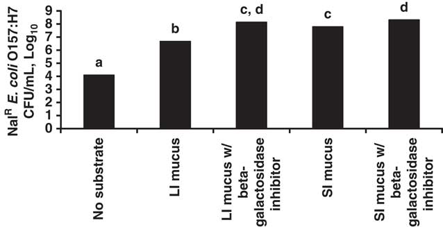
Figure 4 illustrates our attempt to evaluate the stimulatory effect of mucus degrading enzymes on growth of E. coli O157:H7. There were no effects of protease, endoglycosidase, lipase, β-galactosidase, neuraminidase or protease inhibitor addition to cultures (data not shown; P>0.05). Conversely, addition of β-galactosidase enzyme inhibitor increased growth of NalR E. coli O157:H7 cultured with either small or large intestinal mucus (P<0.05).
Growth of NalR E. coli O157:H7 after 8 h incubation at 40°C with no substrate, small (SI) or large (LI) intestinal mucus and in presence or absence of β-galactosidase inhibitor. Means without a common superscript letter are different, P<0.05.

Discussion
Previous studies have shown heterogeneity in mucus composition and thickness across the human and rat gastrointestinal tracts (Atuma et al., Reference Atuma, Strugala, Allen and Holm2001; Freitas et al., Reference Freitas, Axelsson, Cayuela, Midtvedt and Trugnan2002; Robbe et al., Reference Robbe, Capon, Coddeville and Michalski2004). Bovine mucus is believed to display a certain level of heterogeneity between the different sections of the intestine, and this diversity is considered partly responsible for the bacterial tropism (Robbe et al., Reference Robbe, Capon, Coddeville and Michalski2004; Snider et al., Reference Snider, Fabich, Conway and Clinkenbeard2009). Our analysis of the ileum and colon mucus composition revealed no major differences in nitrogen or OM content. Moreover, pathogenic E. coli was equally capable of growing on large and small intestinal mucus. These observations led us to postulate that heterogeneity in bovine mucus is either less important than in other species or that differences in composition do not appreciably influence bacterial growth.
Growth of the pathogen was attenuated by the presence or absence of fecal inoculum in the assay. In presence of fecal inoculum, NalR E. coli growth was reduced by at least 2 log units, which likely is due to competition for nutrients between fecal bacteria and our introduced strains of NalR E. coli O157:H7. These observations are consistent with the principle of competitive exclusion (Tkalcic et al., Reference Tkalcic, Zhao, Harmon, Doyle, Brown and Zhao2003), by which the presence of other bacteria in the medium limits substrate availability, thus reducing pathogen growth.
Another interesting observation was the linear increase in NalR E. coli O157:H7 and total anaerobes with increasing level of small intestinal mucus. Small intestinal mucus at 15 mg of OM/ml supported a 2 log increase in growth of both NalR E. coli O157:H7 and total anaerobes after 12 h incubation. Our results suggest that intestinal mucus stimulated pathogenic E. coli and total anaerobe.
NalR E. coli O157:H7 was able to grow on all mucus components tested in the absence of fecal inoculum, indicating that the bacteria were able to metabolize all compounds evaluated. Whole mucus resulted in the greatest degree of growth, despite the ability of the pathogen to grow on all substrates. Whole mucus may represent a combination of substrates that more closely meet requirements of the pathogen or, alternatively, a component of mucus not tested in this experiment may be a key ingredient for optimal growth of NalR E. coli O157:H7. For example, an E. coli mutant deficient in the catabolic pathway for l-fucose demonstrated a marked decrease in colonization of the rectal mucus (Snider et al., Reference Snider, Fabich, Conway and Clinkenbeard2009). Similar results were observed in mice and in vitro with E. coli MG1655 (Chang et al., Reference Chang, Smalley, Tucker, Leatham, Norris, Stevenson, Anderson, Grissom, Laux, Cohen and Conway2004; Fabich et al., Reference Fabich, Jones, Chowdhury, Cernosek, Anderson, Smalley, McHargue, Hightower, Smith, Autieri, Leatham, Lins, Allen, Laux, Cohen and Conway2008). Fucose is only a minor component of calf ileal mucus (Montagne et al., Reference Montagne, Toullec and Lalles2000), and thus was not evaluated in this study. In retrospect, it could be a key constituent supporting optimal growth of NalR E. coli O157:H7. Fox et al. (Reference Fox, Drouillard, Shi and Nagaraja2009) indicated that, in presence of fecal inoculum, addition of galactose, gluconic acid, glucuronic acid, glucosamine, galacturonic acid, mannose, galactosamine, fucose, and porcine mucin, all supported growth of NalR E. coli O157:H7. However, mannose, fucose and galactosamine addition did not support growth greater than the control. In the current experiment, in the presence of fecal inoculum, mannose and galactose were the only two components tested that yielded growth not different from the control. In addition, growth observed with whole bovine mucus was similar to that observed by Fox et al. (Reference Fox, Drouillard, Shi and Nagaraja2009) using porcine mucin, suggesting that compositional differences may not appreciably influence bacterial growth.
Cultures of NalR E. coli O157:H7 alone with sialic acid as substrate resulted in modest bacterial growth, whereas in the presence of fecal inocula growth of the pathogen closely resembled that obtained with whole mucus. The E. coli strains used in our experiment appeared to have limited ability to use sialic acid, but seem capable of using intermediate products or metabolites synthesized by other fecal bacteria in the degradation of sialic acid. This observation could explain partly why feeding distiller’s grains to feedlot cattle stimulates E. coli O157:H7 shedding (Jacob et al., Reference Jacob, Parsons, Shelor, Fox, Drouillard, Thomson, Renter and Nagaraja2008, Reference Jacob, Fox, Drouillard, Renter and Nagaraja2009). Distiller’s grains are rich in yeasts, which contain up to 3% of their dry mass as sialic acid, (Malhotra and Singh, Reference Malhotra and Singh2006). In such conditions, it is conceivable that a derivative of sialic acid is the active component that stimulates proliferation of the pathogen in cattle fed dried distiller’s grains. In addition, the EHEC strain O157:H7 used by Bertin et al. (Reference Bertin, Chaucheyras-Durand, Robbe-Masselot, Durand, de la Foye, Harel, Cohen, Conway, Forano and Martin2013) was able to grow on N-acetyl neuraminic acid to level comparable to that obtained with N-acetyl glucosamine, which may indicate a strain specific ability to metabolize sialic acid.
One of our initial hypotheses was that E. coli O157:H7 would have little ability to use whole mucus to support its growth because the organism is not known to produce endoglycosidase (Chang et al., Reference Chang, Smalley, Tucker, Leatham, Norris, Stevenson, Anderson, Grissom, Laux, Cohen and Conway2004), but our results suggest otherwise. First, NalR E. coli O157:H7 grew best on whole mucus compared with individual compounds. It is possible that the mucus harvested for this experiment had already been partially cleaved by the gastrointestinal microflora and was readily available to NalR E. coli O157:H7. This may explain why addition of protease, endoglycosidase, lipase or neuraminidase to the medium did not improve growth of the pathogen. Only β-galactosidase inhibitor had an effect on the growth of the bacteria and, surprisingly, increased the NalR E. coli O157:H7. Presence of β-galactosidase activity is used in chromogenic medium as a means of distinguishing E. coli O157:H7 from other E. coli which are β-galactosidase negative and glucuronidase positive. Therefore, we were expecting the addition of β-galactosidase inhibitor to decrease growth of the organism. It is possible that galactosides are more stimulatory to growth of NalR E. coli O157:H7 than degradation products derived from galactosides, or that the inhibitor itself was used as a source of nutrient by the bacteria. The latter explanation seems unlikely, based on the very small amount of inhibitor (200 µM) added in this assay. Additional experiments are needed to further investigate this effect.
In conclusion, this series of experiments provided information regarding metabolism of mucus and mucus components by pathogenic E. coli O157. We were unable, however, to identify a single component as a key stimulator or inhibitor of growth of these bacteria. In order to develop innovative and efficient preharvest intervention measures, it is important to further investigate the relationship between E. coli O157:H7, or other STEC, and the gastrointestinal colonization of cattle.
Acknowledgment
This project was funded, in part, by beef and veal producers and importers through their $1-per-head checkoff and was produced for the Cattlemen’s Beef Board and state beef councils by the National Cattlemen’s Beef Association.