Brief report
A patient with neonatal diagnosis of cor triatriatum, unroofed coronary sinus with persistent left superior caval vein, and unrestrictive perimembranous ventricular septal defect underwent a single-stage repair at 2 months of age with ligation of the left superior caval vein. The postoperative course was complicated by thrombotic obstruction of the right superior caval vein. Surgical resection and balloon angioplasty restored partial patency of the right superior caval vein, and the patient was discharged home asymptomatic. After 2 years, diagnostic catheterisation demonstrated complete occlusion of the right superior caval vein. The mechanism of occlusion was not clearly understood at that juncture. Thrombophilia screening was reported as normal: protein C, protein S, and investigation for mutations, such as factor V Leiden (R506A), prothrombin (G20210A), and MTHFR (C667T), were all negative.
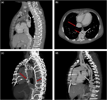
At 11 years of age, an epicardial pacemaker was implanted for sino-atrial nodal dysfunction and intermittent complete atrioventricular block. After 3 years, the patient was admitted to the emergency department because of facial swelling. Clinical examination was in keeping with superior caval vein syndrome, and there was also peripheral lower-limb oedema. Laboratory testing found hypoalbuminaemia and elevated levels of faecal α-1 antitrypsin, confirming protein-losing enteropathy. As MRI was contraindicated by the presence of the pacemaker, the patient underwent thoracic contrast-enhanced CT, which documented complete occlusion of the right superior caval vein, abnormal dilation of the inferior caval vein, and a significant discrepancy in diameter between the dilated inferior caval vein (25 mm) and the orifice of drainage at the veno-atrial junction (12 mm) (Fig 1a and b). The superior systemic venous return was connected to the inferior caval vein via three descending plexuses of tributary veins: on the anterior right side of the mediastinum, there was a tortuous vein descending on the diaphragm and draining into the inferior caval vein (Fig 1b and c, long red arrow); in addition, there was a perivertebral right posterior descending vein, mimicking the original azygos vein, with multiple connections with the perivertebral and the internal vertebral venous plexuses (Fig 1b and c, short red arrow). On the left side of the mediastinum, CT visualised an abnormal tortuous vein, mimicking a dilated accessory hemiazygos vein, draining the left jugular vein and descending into the abdomen in the left paravertebral position, also connected with the perivertebral and the internal vertebral venous plexuses (Fig 1b and d, white arrow).
( a ) Sagittal view passing through the inferior caval vein as a bicaval projection showing the occlusion of the right superior caval vein. Please note in a and b the dilation of the inferior caval vein. In b and c , the long red arrow indicates the anterior tortuous vein descending on the diaphragm and draining into the inferior caval vein; the short red arrow indicates the right paravertebral vein descending as a true azygos. In b and d , the white arrow indicates the left posterior descending vein draining the left jugular vein and descending as an accessory hemiazygos vein. In c and d , the descending veins are shown in maximum intensity projection reconstructions connected to the perivertebral and internal vertebral venous plexuses.

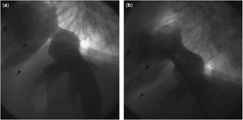
Peripheral injections from the arms confirmed the CT findings concerning the superior systemic venous drainage. Conventional angiography of the inferior caval vein assessed the stenosis at the veno-atrial junction (Fig 2a). The peak gradient between the dilated inferior caval vein and the right atrium was 2 mmHg. The stenosis was treated by using a 45-mm, uncovered Cheatham-Platinum stent (Numed, Hopkinton, New York, United States of America) that was dilated on a 22-mm BiB® balloon (NuMED, Hopkinton, New York, United States of America). There was compete resolution of the peak gradient and significant improvement in the calibre of the inferior caval vein–right atrial junction (Fig 2b). As both brachiocephalic veins were occluded, we did not pursue further interventional procedures to re-cannulate the superior caval vein.
( a and b ) The conventional angiography, respectively, before and after stenting of the veno-atrial junction.

The patient remains asymptomatic without any clinical signs of protein-losing enteropathy 4 years after the procedure; his blood albumin levels were normal.
Discussion
Protein-losing enteropathy is a rare and life-threatening condition that is characterised by protein loss in the gastrointestinal tract. It can be caused by different disorders, with protein leakage through either mucosal injury, as in the case of inflammatory bowel diseases and neoplasms, or through abnormalities of the lymphatic circulatory system, such as in patients with intestinal lymphangiectasia.Reference Braamskamp, Dolman and Tabbers 1 Lymphangiectasia may occur as a congenital disorder – primary intestinal lymphangiectasia – or as an acquired complication that may be secondary to different cardiovascular diseases.Reference Braamskamp, Dolman and Tabbers 1 Occasionally, lymphangiectasia and protein-losing enteropathy have been reported in patients with congestive heart failure, tricuspid regurgitation, and constrictive pericarditis, however, it is more often recognised as a devastating complication in up to 15% of patients with single-ventricle physiology after Fontan surgery.Reference Pundi, Johnson and Dearani 2 High pre-Fontan mean pulmonary pressure, postoperative high systemic atrial pressure, and low cardiac output are well-known risk factors for protein-losing enteropathy in these patients;Reference Pundi, Johnson and Dearani 2 moreover, abnormal abdominal vascular pressure, resistance and even flow pattern, and elevated levels of inflammatory markers are frequently found in patients with protein-losing enteropathy.Reference Pundi, Johnson and Dearani 2 Even if all the elements are not completely understood, these and other findings suggest that abnormal flow and resistances in the mesenteric artery in combination with elevated caval and portal venous pressures and persistent subclinical inflammation might play a key role in the pathophysiology, provoking impaired mesenteric perfusion, intestinal mucosal disruption, and protein leakage.Reference Rychik 3 Indeed protein-losing enteropathy may also develop in cases of stenotic Fontan conduits and even in the setting of turbulent flow,Reference Butera, Marini and MacDonald 4 and also after hepatic vein obstruction – Budd–Chiari syndrome – typically after liver transplantation.Reference Braamskamp, Dolman and Tabbers 1
Our case highlights that protein-losing enteropathy may also occur in the presence of relatively mild stenosis of the inferior caval vein when the superior caval vein is completely occluded. Total occlusion of the superior caval vein may occasionally complicate central vascular line positioning or surgical anastomosis, especially if genetic procoaugulant conditions exist.Reference Agnoletti, Marini, Bordese, Villar and Gabbarini 5 Superior systemic venous return is then forced to find an alternative route that usually involves hyper-development of the paravertebral/azygous/hemiazygous systems connecting with the inferior caval vein. Hence, when all of the systemic venous return is dependent upon the patency of the inferior caval vein, even a mild or moderate reduction in calibre may significantly increase portal venous pressure, potentiating protein-losing enteropathy. For this reason, in clinical practice, stenosis and occlusion of the main systemic veins should always be prevented, screened, and treated with all available interventional tools.Reference Agnoletti, Marini, Bordese, Villar and Gabbarini 5 In our patient, percutaneous stenting of the stenosis was effective as a definitive treatment for this potentially catastrophic complication.
Of note, contrast-enhanced CT provided an exhaustive evaluation of the systemic venous patency, elucidating the definitive diagnosis of total superior caval vein occlusion and mild inferior caval vein stenosis. This information guided the interventional strategy by excluding the practicality of interventional re-canalisation of the superior caval vein.
Acknowledgements
The authors thank Renato Prosio for the technical assistance.
Financial Support
This article received no specific grant from any funding agency or from commercial or not for profit sectors.
Conflicts of Interest
None.
Ethical Standard
The authors assert that all procedures contributing to this study comply with the ethical standards of the relevant guidelines on human experimentation and with the Helsinki Declaration of 1975, as revised in 2008, and has been approved by the institutional committees.