Introduction
Epstein–Barr virus (EBV) is a double-stranded deoxyribonucleic acid enveloped virus belonging to the herpes virus family. It is associated with lymphomas, nasopharyngeal carcinomas, human immunodeficiency virus-associated and post-transplant lymphoproliferative disorders.Reference Debriec-Rychter, Croes, De Vos, Marynen, Roskams and Hagemeijer1 Epstein–Barr virus-associated smooth muscle tumour is a smooth muscle tumour characterised by the presence of EBV-encoded ribonucleic acid (RNA) in smooth muscle cells.Reference Deyrup, Lee, Hill, Cheuk, Toh and Kesavan2 It usually exhibits little atypia and low levels of mitotic activity.Reference Deyrup, Lee, Hill, Cheuk, Toh and Kesavan2 Little is known of the natural history of this rare disorder, but the condition seems to improve with modification and reduction of the immunotherapy dose. The tumour has been reported to occur in the lung, kidney, liver, spleen, dura and skull base.Reference Deyrup, Lee, Hill, Cheuk, Toh and Kesavan2, Reference Cheuk, Li and Chan3 However, the current report is believed to be the first published case in the English literature of EBV-associated smooth muscle tumour affecting the vocal process.
Case report
A 36-year-old Chinese lady was diagnosed in September 1986 with end-stage renal failure as a result of Goodpasture's syndrome. She received continuous ambulatory peritoneal dialysis, but this was complicated by methicillin-resistant Staphylococcus aureus peritonitis in May 1990. As a result, she was placed on haemodialysis for one year before receiving a cadaveric renal transplant in March 1992. Post-transplantation, she received cyclosporine, azathioprine and prednisolone for immunosuppression.
The patient was referred to the Ear, Nose and Throat Centre at Singapore General Hospital in May 2005 with a one-year history of throat discomfort associated with regurgitation of acid into her throat at night. She denied dysphagia, odynophagia or hoarseness of voice.
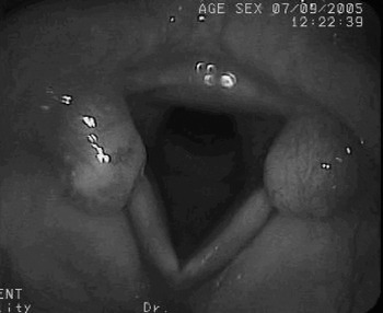
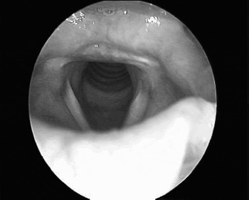
Videolaryngoscopy revealed what appeared to be bilateral vocal process granulomas (Figure 1). In view of the patient's history of acid regurgitation and coexisting signs of posterior laryngeal and subglottic oedema, these granulomas were presumed to be a result of gastroesophageal reflux. She received a four-week course of high dose proton pump inhibitor (40 mg omeprazole twice daily).
Videolaryngoscopic view of presumed bilateral vocal process granulomas (first presentation). Note also the presence of posterior laryngeal and subglottic oedema, suggestive of gastroesophageal reflux disease.

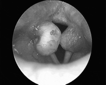
On follow up a month later, the patient's symptoms persisted and the vocal process lesions had increased in size (Figure 2). Subsequently, she underwent laser excision of the lesions.
Videolaryngoscopic view of bilateral vocal process ‘granulomas’ which have enlarged despite one month's treatment with high dose omeprazole.

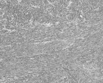
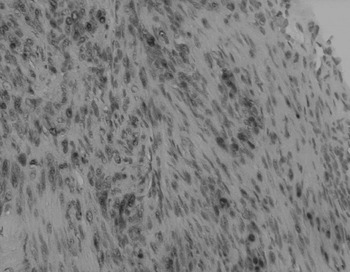
Histological examination was consistent with Epstein–Barr virus (EBV) associated smooth muscle tumour. The polypoid tissue was covered by squamous epithelium, associated with a proliferation of spindle cells resembling smooth muscle cells arranged in fascicles (Figure 3). On immunohistochemical analysis, the spindle cells stained for smooth muscle actin (Figure 4) and vimentin. Focal staining was seen with caldesmon. Staining for desmin, S-100, epithelial membrane antigen (EMA), leucocyte common antigen (LCA), CD34, CK19, AE1/3 and CD99 were negative. In situ hybridisation for EBV-encoded RNA was focally positive (Figure 5).
Medium power photomicrograph of vocal fold biopsy, revealing a proliferation of spindle cells within the stroma, accompanied by a lymphoplasmacytic infiltrate (H&E; original magnification ×100).

Immunohistochemical photomicrograph showing spindle cells with positive staining for antibodies against smooth muscle actin, indicating smooth muscle differentiation (Avidin biotin complex; original magnification ×200).

On in-situ hybridisation, spindle cell nuclei were labelled by probes against Epstein–Barr virus encoded ribonucleic acid (peptide nuclei acid (PNA) detection kit; original magnification ×400).

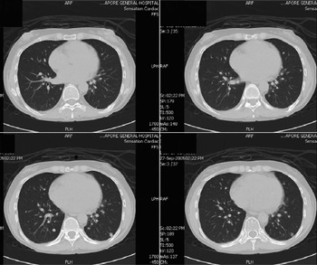
A computed tomography (CT) scan of the thorax was ordered in view of the patient's long-standing dry cough and suspicion of a lymphoproliferative disorder in the setting of immunosuppression. The CT showed bilateral, subcentimeter lung nodules suggestive of metastatic disease or lymphoproliferative disorder (Figure 6). The patient refused biopsies of the pulmonary nodules. Her azathioprine and cyclosporine prescriptions were discontinued and replaced with sirolimus.
Axial computed tomography scans of the thorax, showing multiple subcentimeter pulmonary nodules.

At out-patient follow up, two weeks following surgery, the patient's symptoms had subsided. Nasoendoscopy showed that the vocal process wounds were healing well. A follow-up CT scan of the thorax, three months after surgery, showed no progression of the lung lesions. Further nasoendoscopic evaluation five months after surgery showed no recurrence of the vocal process lesions (Figure 7).
Videolaryngoscopic view of the vocal processes five months after surgical resection, showing no recurrence of the lesions.

Discussion
Epstein–Barr virus-associated smooth muscle tumour is a recently identified entity that occurs in immunocompromised patients. The association between post-transplant spindle cell tumour and Epstein–Barr virus (EBV) was first described by Lee et al. in 1993.Reference Lee, Dickman, Jaffe, Alashari, Tzakis and Reyes4 In a 2002 literature review by Cheuk et al., 46 cases of EBV-associated smooth muscle tumour were identified from various sites. The liver, gastrointestinal tract and lung were the most commonly involved locations.Reference Cheuk, Li and Chan3 However, to date, EBV-associated smooth muscle tumour has not been reported in the vocal process or larynx. Epstein–Barr virus-associated smooth muscle tumour has been found to affect three groups of immunocompromised patients: those with acquired immunodeficiency syndrome (AIDS), organ transplantation recipients and those with severe congenital immunodeficiency.Reference Cheuk, Li and Chan3 In post-transplant patients, there seems to be a preponderance for the liver. The time of discovery of the tumour has varied from 1.25 to 66 months post-transplantation.Reference Cheuk, Li and Chan3 The majority of cases have occurred in children and adolescents, but this could be due to the age prevalence of the AIDS and transplant recipient patients in the series described.
The pathogenesis of EBV-associated smooth muscle tumour remains unclear due to the rarity of this condition. It has been hypothesised that high viral titres in immunocompromised patients allow tissues such as smooth muscle, which are normally resistant to infection under normal physiological conditions, to become infected.Reference Liebowitz5 Viral infection precedes clonal expansion of smooth muscle cells, and the presence of EBV monoclonality in these tumours suggests a primary role of EBV in tumorigenesis.Reference Lee, Locker, Nalesnik, Reyes, Jaffe and Alashari6 The current understanding of the ability of EBV to promote cell growth is derived mainly from in vitro studies of EBV-transformed B-lymphocyte cell lines.Reference Wang, Gregory, Sample, Rowe, Liebowitz and Murray7 Epstein–Barr virus gains entry into lymphoid cells via a transmembrane glycoprotein receptor known as CD21.Reference Morgello, Kotsianti, Gumprecht and Moore8 This receptor has been found in high levels in immunocompromised paediatric patients with leiomyomas and leiomyosarcomas.Reference McClain, Leach, Jenson, Joshi, Pollock and Parmley9 The route of EBV entry into smooth muscle cells is unknown, although up-regulation of CD21 receptors has been shown.Reference Lee, Locker, Nalesnik, Reyes, Jaffe and Alashari6 Once within the myocytes, EBV maintains itself in three latency states, depending on the type of antigens expressed.Reference Morgello, Kotsianti, Gumprecht and Moore8 Epstein–Barr virus-associated smooth muscle tumour and post-transplant lymphoproliferative disorder typically exhibit EBV type III latency with the expression of EBV nuclear antigen 2 and latent membrane protein 1.Reference Cheuk, Li and Chan3 On the other hand, EBV type I latency is seen in Burkitt's lymphoma, while EBV type II latency is seen in Hodgkin's disease and nasopharyngeal carcinoma.Reference Cheuk, Li and Chan3
The biological behaviour of EBV-associated smooth muscle tumour does not seem to correlate with its histological features. Tumours classified as benign or of uncertain malignant potential have been seen to behave aggressively, with multiorgan involvement and death in at least one case despite aggressive chemotherapy.Reference Lee, Locker, Nalesnik, Reyes, Jaffe and Alashari6 In our patient, multiple subcentimetric bilateral pulmonary nodules were seen. The patient refused biopsy, but, in the setting of immunosuppression, likely possible causes of the pulmonary nodules would be EBV-associated smooth muscle tumour or lymphoproliferative disorders. Epstein–Barr virus-associated smooth muscle tumour has been shown to involve more than one location in the same individual.Reference Deyrup, Lee, Hill, Cheuk, Toh and Kesavan2, Reference Cheuk, Li and Chan3, Reference Lee, Locker, Nalesnik, Reyes, Jaffe and Alashari6, Reference Boman, Gultekin and Dickman10 In a study of 19 patients by Deyrup et al. in 2006, 13 (68 per cent) were reported to have multiple tumours.Reference Deyrup, Lee, Hill, Cheuk, Toh and Kesavan2 These authors found that multiple tumours in the same patient were clonally distinct, and they concluded that these tumours were the result of multiple infection events rather than metastasis.Reference Deyrup, Lee, Hill, Cheuk, Toh and Kesavan2 There is no consensus on the standard treatment of EBV-associated smooth muscle tumour, although reduction or discontinuation of immunosuppression, with resultant tumour shrinkage, and surgical resection leading to cure have been reported in a few cases.Reference Debriec-Rychter, Croes, De Vos, Marynen, Roskams and Hagemeijer1, Reference Lee, Locker, Nalesnik, Reyes, Jaffe and Alashari6, Reference Creager, Maia and Funkhouser11
The clinical presentation of EBV-associated smooth muscle tumour depends on the site of involvement. In this patient, EBV-associated smooth muscle tumour mimicked the appearance of bilateral vocal process granulomas, which were presumed to be due to gastroesophageal reflux. Vocal process granulomas or contact granulomas are benign, inflammatory lesions located at the cartilaginous part of the posterior glottis.Reference Benjamin and Croxson12, Reference Holinger and Johnston13 Proposed mechanisms for their formation include mechanical causes (such as vocal abuse, intubation injury and surgical trauma) and inflammatory causes (such as gastroesophageal refux, infection, postnasal drainage and allergy).Reference Hoffmann, Overholt, Karnell and McCulloch14
The current management of contact granulomas is directed mainly at the multifactorial aetiology and includes speech therapy, lifestyle modification and antireflux measures.Reference Havas, Priestley and Lowinger15 Surgical resection of vocal process granuloma is usually avoided, as the recurrence rate after surgery is high (92 per cent).Reference Ylitalo and Lindestad16 Treatment should be aimed at removing chronic irritants leading to granuloma formation.Reference Havas, Priestley and Lowinger15 Indications for surgery include airway obstruction from large granulomas, diagnostic doubt and failed conservative treatment.Reference Havas, Priestley and Lowinger15 In our patient, failed response to high dose antireflux medication and an increase in size of the lesions led to surgical excision to obtain a histological diagnosis.
Other pathologies can mimic vocal process granulomas. In 1949, New and Devine reported three cases in which presumed vocal process granulomas were variously histologically identified as myxomatous tissue, haemangioendothelioma and squamous cell carcinoma.Reference New and Devine17 In addition, in 1990 Wenig and Heffner reported 105 cases that had been diagnosed histologically as contact ulcers and sent for review at the Armed Forces Institute of Pathology. Their review reported histological misdiagnoses including pyogenic granuloma, haemangioma, haemangiopericytoma, Kaposi's sarcoma, angiosarcoma, spindle cell carcinoma and granulomatous infectious diseases.Reference Wenig and Heffner18 The percentage of misdiagnoses was not available.
• This is a case report of an immunocompromised patient who presented with granulomas over the vocal processes of the vocal folds
• The masses were thought initially to be due to reflux
• Subsequent analysis showed that they were smooth muscle tumours due to Epstein–Barr virus
• The literature surrounding such lesions is reviewed
Bilateral vocal process granulomas are less prevalent than unilateral granulomas.Reference Ylitalo and Lindestad16, Reference Jaroma, Pakarinen and Nuutinen19 In Ylitalo and Lindestad's retrospective study of 120 patients with contact granulomas, only seven cases were bilateral.Reference Ylitalo and Lindestad16 Jaroma et al. reported 10 cases of bilateral vocal process granuloma in their series of 98 patients;Reference Jaroma, Pakarinen and Nuutinen19 the histology of these bilateral granulomas was not available. However, a search of the English literature (via the Pubmed and MEDLINE databases) revealed one reported case of bilateral vocal process granuloma with a histological diagnosis of squamous cell carcinoma.Reference Sidle, Haines and Altman20
Conclusion
Epstein–Barr virus-associated smooth muscle tumour is a rare condition that can mimic the appearance of vocal process granulomas in immunocompromised patients. It should be considered in the differential diagnosis when granulomas in such patients persist despite a period of medical and speech therapy. Bilateral granulomas are not common, and further studies would be needed to determine common aetiologies, so that a management algorithm can be formulated to optimise treatment.