Introduction
Stab wounds to the head and neck are unfortunately a fairly common occurrence, but a retained knife blade in this region is a rare event, with only isolated case reports and small series published previously.Reference Grobbelaar and Knottenbelt1–Reference Gardner, Righi and Shahbahrami5 Patients with retained knife blades in the head and neck region seldom present alive. When they do, they pose not only management challenges but also diagnostic problems.
We present three such cases, in two of which the diagnosis was delayed by more than eight weeks. We discuss the appropriate radiological investigations, method of removal and the inevitable association of alcohol consumption in violent trauma cases; we also present a literature review regarding this unusual phenomenon.
Case one
A 35-year-old man presented with a history of having been stabbed in the left eye two months previously. He had attended the local day hospital, where the wound had been cleaned but no further imaging studies had been performed. In the weeks after the incident, he had noted continuing blurring of vision and swelling of the left eye. During a subsequent one-month jail sentence, he had again sought medical attention, but, again, no further investigations had been performed. Three weeks prior to presentation, he had noted a knife blade extruding through the left upper eyelid.
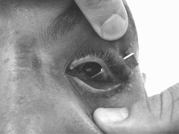
On examination, the tip of a knife blade was protruding through the left upper eyelid. This extended anterior to the globe and through the floor of the orbit (Figure 1). Eye movements were normal, except for restricted eye elevation. The patient had 6/24 vision in the left eye. The cornea and globe were normal.
Clinical photograph of case one, showing the visible, impacted knife blade extending from the left upper lid anterior to the globe.

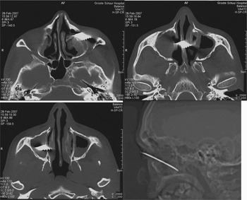
Plain X-rays and a computed tomography (CT) scan demonstrated the knife blade extending from the left upper eyelid anterior to the globe, through the floor of the orbit, the left maxillary sinus, septum and lodging within the right maxillary sinus (Figure 2). No angiography was performed, as it was clear that the blade was not closely related to important vascular structures.
Axial and survey computed tomography scans of case one, demonstrating the path of the knife blade from the left orbit, through the septum and extending to the right maxillary sinus.

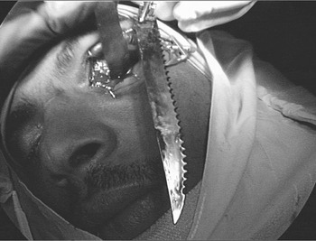
The blade was removed under general anaesthesia. A lateral canthotomy was first performed and the upper lid elevated over the blade. An inferior conjunctival incision was made and the orbit was dissected free from the blade, protecting it during removal. A 13 cm steak-knife blade was removed along the pathway of insertion (Figure 3). There were no sequelae following removal.
The knife blade in case one after removal.

The patient failed to keep his follow-up appointments.
Case two
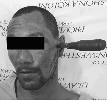
A 37-year-old man presented to our trauma unit four hours after an assault in which he reported being stabbed in the head. A knife remained embedded in the temporal bone on the left side of his head (Figure 4). The entrance wound was anterior to the superior attachment of the helix, with the blade directed medially and angled postero-inferiorly.
Clinical photograph of case two, showing the knife embedded in the patient's left temporal bone. Note the presence of a left-sided facial nerve palsy.

On examination, the patient was conscious and fully orientated. Clotted blood filled the left external auditory canal. There was no evidence of a cerebrospinal fluid leak. There was a conductive hearing loss on the affected side. The patient was not vertiginous, and no nystagmus was present. The patient had a left-sided, lower motor neurone facial nerve palsy.
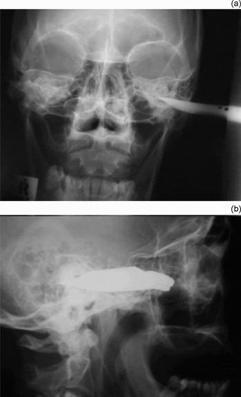
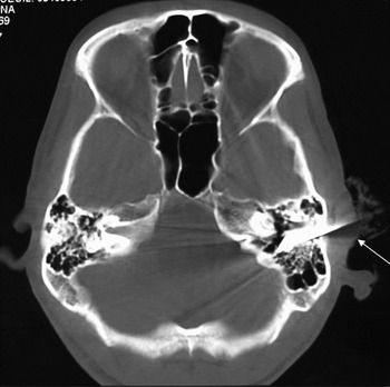
Plain skull X-rays (Figure 5) and CT scans (Figure 6) demonstrated the knife blade within the petrous temporal bone. The path of the blade passed posterior to the temporomandibular joint and the internal carotid artery. The blade tip could be seen within the petrous bone, with no extension into the posterior cranial fossa.
(a) Antero-posterior and (b) lateral X-rays of case two, showing the knife visible in the left temporal bone.

Axial computed tomography scan of case two, demonstrating the knife blade lodged within the left temporal bone (arrow).

Surgical exploration was urgently undertaken. The blade was removed along the pathway of insertion and the path of the knife explored through a mastoidectomy approach. The blade had traversed the external auditory canal, penetrating the anterior and posterior canal walls, and had transected the facial nerve at the junction of the upper third and lower two-thirds of its vertical segment. An interposition greater auricular nerve graft was used to repair the defect. The patient was discharged following an uncomplicated 48-hour post-operative stay.
Case three
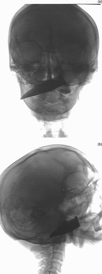
A 31-year-old man was referred to the ENT department on suspicion of an oesophageal malignancy. He had a three-month history of dysphagia, voice change and a right-sided neck lump. A barium swallow had been performed as part of his investigations at the referring hospital, and this had revealed the presence of a knife blade extending from the left nasopharynx, passing behind the soft palate and lying underneath the mandible (Figure 7).
Barium swallow of case three, showing the knife blade extending from the left posterior choanae to the right submandibular region.

On examination, the patient had a ‘hot-potato voice’ and numerous facial scars.
Flexible naso-endoscopy revealed a metal object in the left nasopharynx, depressing the soft palate inferiorly. The mass palpated in the right submandibular area was shown to be the tip of the knife blade on subsequent imaging.
Angiography was performed to exclude injuries to the carotid artery and its branches. This showed the blade to be clear of surrounding major vessels.
The patient denied any knowledge of how the knife blade might have lodged in his nasopharynx, but admitted to having been severely inebriated just prior to the onset of his symptoms.
The knife blade was removed under general anaesthesia. A transverse neck incision was made and dissection continued down to the blade. The knife blade was removed by pulling it away from the pathway of insertion, without complication. The patient was discharged after 48 hours, with no dysphagia or voice problems.
He too did not attend his follow-up appointments.
Discussion
A retained knife blade is an uncommon problem, although some of the largest published series of this unusual and spectacular event have come from South Africa.Reference Grobbelaar and Knottenbelt1, Reference Daya and Liversage2 In 1972, Jett et al. evaluated the socioeconomic pattern of 254 victims with penetrating knife and gunshot wounds, and found the typical patient to be male, 15 to 35 years old and from low socioeconomic strata.Reference Jett, van Hoy and Hamit6 The incidents were more frequent on a Friday and Saturday night between 2100 and 0200 hours.Reference Jett, van Hoy and Hamit6 In addition, other authors noted a high level of alcohol consumption associated with such trauma.Reference Daya and Liversage2 In the present series, the patient profile was found to be quite similar to that described above.
Daya and Liversage described a series of eight patients with retained knife blades, and Grobbelaar and Knottenbelt described 11 patients with similar injuries.Reference Grobbelaar and Knottenbelt1, Reference Daya and Liversage2 In both these series, the knife blades could be removed along the pathway of entrance, with no significant resultant sequelae. The authors of both series suggested that impacted knife blades could safely be removed in theatre with little fear of complication.Reference Grobbelaar and Knottenbelt1, Reference Daya and Liversage2 Scheepers and Lownie suggested that angiography be performed in cases in which the foreign body was close to major arteries. This would allow the surgeon to pre-empt the complication of acute perforation resulting from pulsation of the artery against the retained object, and the subsequent development of a pseudo-aneurysm.Reference Scheepers and Lownie3 In cases of excessive arterial bleeding, angiography can aid identification of the bleeding vessel, which can then be managed by selective embolisation or surgical ligation.Reference Scheepers and Lownie3 If the path of the knife is clear of the cranium and major vessels, angiography is not mandatory, as was found in cases one and two.
• This paper reports three cases in which patients presented with retained knife blades in the head and neck region. In two of these cases, the diagnosis was delayed by more than eight weeks
• All patients had the knife blades removed through the pathway of insertion
• These cases demonstrate the need for a high index of suspicion for retained knife blades in the head and neck, when stab wounds are anything more than superficial
• Routine radiographic examination is recommended for all deep facial stab wounds or where the history is suggestive of a possible retained knife blade
• In most cases, simple removal along the pathway of entry seems safe and effective
Following a facial stab wound, a high index of suspicion for a retained knife blade is required when the stab wound is anything more than superficial and the knife has not been recovered.Reference Gardner, Righi and Shahbahrami5 A careful history from paramedics or witnesses is required to determine if the knife blade was intact after the stabbing.Reference Gardner, Righi and Shahbahrami5 A complete head and neck physical examination should include an assessment of the extent and depth of the stab, as well as an assessment of relative neurovascular structures. All cranial nerves should be assessed, particularly the facial nerve. Pharyngeal injuries should be assessed using endoscopy or Gastrografin swallow. If there is any doubt that the knife was intact after the stabbing, or if examination reveals a deep wound, then appropriate radiographs should be performed to exclude a retained knife blade.Reference Gardner, Righi and Shahbahrami5
These cases demonstrate the need for a high index of suspicion for retained knife blades in the head and neck. In two of the cases, the knife blade eluded detection immediately after the event. We recommend routine radiographic examination in all cases of deep facial stab wounds or where the history is suggestive of a possible retained knife blade. In most cases, simple removal along the entry pathway seems safe and effective.
Conclusion
Radiography should be performed on all patients presenting with stab injuries to the head and neck area which are anything more than superficial. We further suggest that direct extraction of sharp objects via the pathway of insertion is safe if radiological studies show no risk of vascular injury.