Patients with β-thalassaemia major usually receive periodic blood transfusions to prevent complications of chronic anaemia; however, these transfusions could result in excessive iron loading, which in turn leads to endocrine, liver, and cardiac dysfunctions.Reference Olivieri 1 The survival in these patients has significantly improved as a result of chelation therapy. In addition to improved chelation regimens, new imaging methods also allowed better management of the iron overload.Reference Wood 2
Although these novelties have significantly reduced mortality and morbidity in these patients, cardiac complications are still the major cause of death in β-thalassaemia major,Reference Modell, Khan and Darlison 3 , Reference Borgna-Pignatti, Rugolotto and De Stefano 4 and regular evaluation of cardiac function is recommended for all patients with β-thalassaemia major. Cardiac involvement in β-thalassaemia major is generally characterised by iron-induced heart failure, cardiomyopathy, and arrhythmias.Reference Ehlers, Levin and Markenson 5 , Reference Olivieri, Nathan and MacMillan 6 Serum ferritin levels and transthoracic echocardiography have been used previously for predicting iron-induced cardiac involvement.
According to the latest data, T2* cardiac magnetic resonance imaging has been approved as a quantitative method for evaluating myocardial iron overload, and magnetic resonance imaging parameter T2* was used to determine cardiac iron loading in patients with β-thalassaemia major.Reference Meloni, Positano and Pepe 7 However, once iron-induced cardiac damage is demonstrated by magnetic resonance imaging (T2* <20 ms), patients are already in the late stage of the disease with established cardiomyopathy.Reference Anderson, Holden and Davis 8
Reduced heart rate variability is associated with a higher risk of arrhythmias after myocardial infarction and heart failure.Reference Smilde, van Veldhuisen and van den Berg 9 Previous studies have also reported reduced heart rate variability in the advanced stage of β-thalassaemia major.Reference Rutjanaprom, Kanlop and Charoenkwan 10 In this study, β-thalassaemia major patients with preserved left ventricle ejection fractions were evaluated by magnetic resonance imaging and time domain heart rate variability measurements to find out the concealed cardiac adaptations in this early stage of the disease.
Materials and methods
Patient selection
Out of 70 β-thalassaemia major patients who were on regular follow-up in our hospital and had preserved ejection fractions, 38 β-thalassaemia major patients – 24 males and 14 females – with cardiac magnetic resonance imaging assessment were included in our study. Age- and sex-matched student volunteers (n = 45) served as the healthy control group. Risk factors for heart disease as smoking, diabetes mellitus, hypertension, obesity, family history of heart disease, and all subjects with any systemic illness were excluded (diabetes mellitus, liver or kidney disease, hypertension, or coronary artery disease). All β-thalassaemia major patients were receiving blood transfusions – at least 15 transfusions per year – to maintain haemoglobin levels greater than 9 g/dl. Patients had been receiving chelation therapies including desferrioxamine infusion and oral chelation regimens including deferasirox and/or deferiprone according to the patient's age, serum ferritin levels, and patient compatibility. Patients with depressed left ventricle ejection fractions <50% and patients receiving blood transfusions <15/year were excluded from the study. All patients were in the New York Heart Association (NYHA) Class 1 functional status. All subjects were examined by the same cardiologist. Other criteria for exclusion from the study included haemodynamically significant pericardial effusion and moderate–severe mitral–aortic valve regurgitation, or stenosis. A retrospective review of the T2* magnetic resonance imaging results was performed in 38 patients who had undergone at least one cardiac magnetic resonance imaging examination at the time of the study. Finally, in this cross-sectional study, 38 patients with β-thalassaemia major were compared with control subjects in terms of clinical data and heart rate variability analysis. The study cohort was also divided into subgroups according to T2* levels detected by magnetic resonance imaging. A T2* value of 20 ms was used as the cut-off in our study. In all, 21 patients with a T2* value >20 ms were reported to be free of cardiac iron involvement.Reference Anderson, Holden and Davis 8
The study protocol was approved by the local ethics committee and informed consent was obtained from all participants. No extramural funding was used to support this work.
Baseline evaluation and biochemical measurements
Demographic and clinical variables were assessed by interview and examination of the medical records by the same cardiologist. Haematological and biochemical parameters were measured just before transfusion. All biochemical measurements were performed at the same central laboratory.
Echocardiography
Echocardiographic examinations were performed according to American Society of Echocardiography recommendationsReference Lang, Bierig and Devereux 11 with a Vingmed ultrasound system (Vingmed System 7, General Electric, Horten, Norway) and a 2.5-MHz transducer. All echo-Doppler studies were carried out by the same observer. Measurements were recorded on three representative beats and the results were averaged. Standard echocardiographic analysis included two dimensional and M-mode, and ejection fraction calculation was based on left ventricle volumes by the methods of discs, using apical two- and four-chamber views.Reference Schiller, Shah and Crawford 12
Magnetic resonance imaging
Thalassaemia patients are closely followed up in our thalassaemia outpatient clinic and they undergo magnetic resonance imaging examination routinely as a part of this follow-up. Therefore, all magnetic resonance imaging examinations were evaluated retrospectively. Cardiac magnetic resonance imaging examinations were performed using a 1.5-T scanner (Symphony, Siemens, Erlangen, Germany). The scans included measurement of the myocardial T2* value. T2* of the heart was evaluated by cardiac-gated single breath-hold multi-echo technique (field of view, 400 mm; TR, 135 ms; TE, 2.6–22.3 ms; slice thickness, 10 mm). T2* analysis was performed and a T2* value >20 ms was considered as a conservative cut-off.
Heart rate variability analysis
Holter monitoring was performed for 24 hours in the intertransfusion period, by use of a digital Holter monitor (DL800, Compact Flash Card Holter Recorder, Minnesota, United States of America). The data stored were processed by use of the Cardio Scan software to assess heart rate variability. Abnormal beats and artefact areas were automatically and manually identified and deleted from the analysis. Heart rate variability was assessed in the time domains. For analysis in the time domain, normal R–R intervals were called iNN. To assess heart rate variability in the time domain and based on iNN, the mean of the standard deviations of iNN every 5 minutes and root of the mean squared difference of successive iNN were obtained. 13
Statistical analysis
All data were analysed using the SPSS 13.0 (Chicago, Illinois, United States of America). Quantitative data were expressed as mean ± standard deviation. Categorical parameters were given as a number (%). Unpaired student t-test was used to analyse the differences between the variables in the control group and patients with β-thalassaemia major. A p-value <0.05 was considered statistically significant.
Results
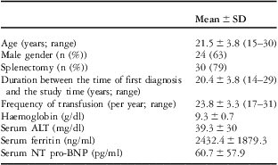
Clinical and biochemical characteristics of patients with β-thalassaemia major (n = 38) were summarised in Table 1. A total of 30 patients (79%) had splenectomy before the evaluation. The mean age at first diagnosis was 1.16 ± 0.4 (1–3 years). The duration from the first diagnosis was 20.4 ± 3.8 (14–29 years). There were no significant differences between the study population and control subjects with regard to age, gender, heart rate, and blood pressure.
Demographic, clinical, and biochemical characteristics of patients with β-thalassaemia major (TM) (n = 38).

ALT = alanine transaminase; NT pro-BNP = N-terminal pro–B-type natriuretic peptide
Values are expressed as mean ± SD unless otherwise defined
Correlation analysis revealed that the standard deviation of all NN intervals was not correlated with haemoglobin (R = −0.268, p = 0.104), patient's age (R = 0.158, p = 0.343), age at first diagnosis (R = −0.266, p = 0.106), frequency of transfusion (R = 0.015, p = 0.927), serum levels of ferritin (R = −0.199, p = 0.232), aspartate transaminase (R = −0.252 p = 126), and alanine transaminase (R = −0.258 p = 0.118).
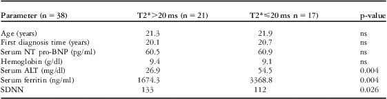
The mean cardiac T2* was 22.9 ± 13.3 ms in our study. Although left ventricle systolic functions were preserved in both groups, magnetic resonance imaging T2* was decreased in 17 patients, indicating cardiac iron involvement. We compared the heart rate variability analysis of patients with both reduced (T2* <20 ms) and preserved (T2* >20 ms) myocardial T2* values with control subjects. The standard deviation of all NN intervals was calculated to be significantly lower (112.0 ± 20.5 versus 133.0 ± 32.2 versus162.8 ± 32.9, p = 0.001, respectively) in β-thalassaemia major patients despite normal T2* magnetic resonance imaging values (Tables 2 and 3). The standard deviation of all NN intervals was not correlated with haemoglobin levels in this β-thalassaemia major population (p > 0.05).
Comparison of patients with β-thalassaemia major according to MRI T2* values.

ALT = alanine transaminase; ns = non-significant; NT pro-BNP = N-terminal pro–B-type natriuretic peptide; SDNN = standard deviation of NN interval
Comparison of B-TM patients with normal T2* values (>20 ms) with control group in terms of time domain heart rate analysis.

ns = non-significant; RMSSD = root of the mean squared difference of successive NN interval; SDNN = standard deviation of NN interval
In patients with β-thalassaemia major, ventricular extrasystoles were observed in only one patient (3%) and atrial extrasystoles were observed in two patients (5%) by 24-hour Holter monitorisation. In the control group, ventricular extrasystoles were observed in six patients (13%) and atrial extrasystoles were observed in 13 patients (29%) by 24-hour Holter monitorisation.
Discussion
Cardiovascular involvement is the leading cause of mortality and morbidity in patients with β-thalassaemia major.Reference Modell, Khan and Darlison 3 , Reference Borgna-Pignatti, Rugolotto and De Stefano 4 Iron-induced heart failure and arrhythmias are the main reasons of cardiovascular mortality.Reference Borgna-Pignatti, Rugolotto and De Stefano 14 When the capacity of the intracellular lysosomes is filled due to excessive iron loading; toxic non-transferrin bounded iron leads to cardiac failure.
Recent advances in magnetic resonance imaging (T2*) have increased its use for monitoring patients with thalassaemia. However, when we observe evident cardiac deterioration by transthoracic echocardiography and/or magnetic resonance imaging (T2* <20 ms), it is already the advanced stage of the disease.
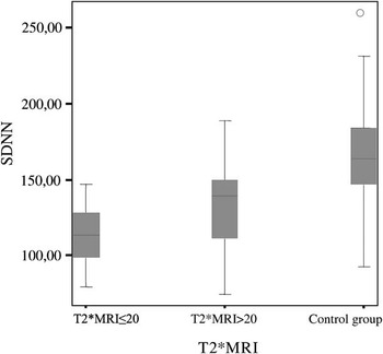
In our study, of the 38 patients with normal ejection fractions, 17 patients had decreased T2* values, indicating the deleterious effect of myocardial iron overload. At first sight, the remaining 21 patients who had normal T2* values (>20 ms) could be interpreted as off the cardiac disease. However, when we compared these patients with the normal subjects, the decreased time domain heart rate variability (including the standard deviation of all NN intervals, root mean square of successive differences) documented in these patients suggested an ongoing process before evident cardiac siderosis was demonstrated by T2* magnetic resonance imaging. Figure 1 shows the comparison of the standard deviation of all NN intervals values in different stages of β-thalassaemia major and the control group.
Comparison of SDNN values of B-TM subgroups (according to T2*MRI values) and control group. SDNN = standard deviation of NN interval, T2*MRI = T2* magnetic resonance imaging.

Heart rate variability analysis is an easy, non-invasive method that measures the beat-to-beat variability in R–R intervals. It reflects altering autonomic activity and its impact on cardiovascular function.Reference Hamaad, Lip and MacFadyen 15 Reduced heart rate variability is associated with a higher risk of arrhythmias after myocardial infarction and heart failure.Reference Odemuyiwa, Malik, Farrell, Bashir, Poloniecki and Camm 16 Chronic anaemia decreases the vagal tone, leading to a higher heart rate. However, decreased heart rate variability is not a normal physiological response or an adaptation to chronic anaemia. Therefore, decreased heart rate variability is always considered as a pathological parameter. Smilde et alReference Smilde, van Veldhuisen and van den Berg 9 reported a 13-year follow-up study with mild to moderate heart failure and showed that heart rate variability might be important in predicting the outcomes. Fetsch et alReference Fetsch, Reinhardt and Mäkijärvi 17 reported in their review that decreased heart rate variability is considered as a marker of impaired vagal activity of the heart and is strongly associated with the degree of myocardial damage.
Analysis of heart rate variability might be helpful in β-thalassaemia major patients by detecting an underlying substrate predisposing to arrhythmias.Reference Franzoni, Galetta, Di Muro, Buti, Pentimone and Santoro 18 Rutjanaprom et alReference Rutjanaprom, Kanlop and Charoenkwan 10 reported that heart rate variability is depressed in β-thalassaemia major patients. However, on the contrary they found that heart rate variability was significantly correlated with haemoglobin levels and suggested that anaemia influences the cardiac autonomic balance. In a study with 20 patients with β-thalassaemia major, De Chiara et alReference De Chiara, Crivellaro and Sara 19 specified abnormal heart rate variability as an early feature of cardiac disease with no evidence of ventricular dysfunction by radionuclide angiography.
Our study has some limitations. The standard deviation of all NN intervals was not correlated to serum levels of ferritin or haemoglobin. Yet the ferritin levels were significantly higher and the standard deviation of all NN intervals was significantly lower in the T2* ⩽ 20 ms group. We believe that the relatively small sample size might have influenced this result. The chelation regimens were not uniform in this β-thalassaemia major population. These variable therapeutic regimens could alter the heart rate variability measurements. Owing to the fact that the time from the first diagnosis and serum ferritin levels were different in every patient, it was reasonable to be on different therapies. On the other hand, as we did not evaluate patients serially, we could not discuss the longitudinal changes in myocardial involvement and heart rate variability in β-thalassaemia major. Prospective studies with long follow-up periods could specify the utility of heart rate variability analysis in predicting morbidity and mortality in patients with β-thalassaemia major. Arrhythmias seemed to be more frequent by 24-hour Holter monitorisation in the control group – 3% of TM patients had ventricular extrasystoles, 5% had atrial extrasystoles, whereas 13% of control group had ventricular extrasystoles, and 29% had atrial extrsystoles. The reduced heart rate variability in the patients did not seem to translate to increased risk of arrhythmias. The reduced heart rate variability might be explained by the chronic anaemia that characterises TM, which may lead to a persistent sinus tachycardia and a sustained decrease in autonomic fluctuations. Therefore, a control group with other forms of anaemia might have answered this problem.
In conclusion, heart rate variability parameters were significantly reduced in β-thalassaemia major patients with or without evident cardiac siderosis. The inhomogeneity of electrical behaviour of iron-loaded myocardial areas in β-thalassaemia major could be represented by reduced heart rate variability even before iron loading is demonstrated by T2* magnetic resonance imaging.
Acknowledgement
The authors have no conflict of interest to declare.We thank the staff of Tepecik Training and Research Hospital, Department of Pediatric Hematology.