Introduction
Reduced (left) hippocampal volume has consistently been observed in patients with post-traumatic stress disorder (PTSD) compared with trauma-exposed controls and trauma-unexposed healthy controls (Kitayama et al. Reference Kitayama, Vaccarino, Kutner, Weiss and Bremner2005; Smith, Reference Schneider2005; Karl et al. Reference Karl, Schaefer, Malta, Dorfel, Rohleder and Werner2006; Kuhn & Gallinat, Reference Kuhn and Gallinat2013). However, it is unclear whether a smaller hippocampus is a vulnerability factor for the development of PTSD or whether it is a consequence of stress/trauma exposure. The most compelling evidence for the former is provided by a twin study showing reduced hippocampal volume in the trauma-unexposed twin without PTSD (Gilbertson et al. Reference Gilbertson, Shenton, Ciszewski, Kasai, Lasko, Orr and Pitman2002). The latter is, in turn, suggested by primate studies (Sapolsky et al. Reference Sapolsky, Uno, Rebert and Finch1990; Gould et al. Reference Gould, Tanapat, McEwen, Flugge and Fuchs1998), as well as a human study reporting reduced hippocampal volume in trauma-exposed controls compared with trauma-unexposed healthy controls (Winter & Irle, Reference Winter and Irle2004). However, this was not found by another study comparing trauma-exposed and healthy controls (Bremner et al. Reference Bremner, Vythilingam, Vermetten, Southwick, McGlashan, Nazeer, Khan, Vaccarino, Soufer, Garg, Ng, Staib, Duncan and Charney2003).
Were stress/trauma exposure to be the cause of a smaller hippocampus in PTSD, it would be possible that successful treatment leads to hippocampal volume increase. Indeed, the hippocampus is known to be a highly plastic brain region that is both positively and negatively affected by experiences and hormones (Gould et al. Reference Gould, Tanapat, Rydel and Hastings2000). It is well known that hippocampal growth can be induced by treatment, such as electroconvulsive therapy in depressed patients (Nordanskog et al. Reference Nordanskog, Dahlstrand, Larsson, Larsson, Knutsson and Johanson2010; Tendolkar et al. Reference Teicher, Andersen, Polcari, Anderson, Navalta and Kim2013), and by lithium use in bipolar disorder (Yucel et al. Reference Yucel, McKinnon, Taylor, Macdonald, Alda, Young and MacQueen2007, Reference Yucel, Taylor, McKinnon, Macdonald, Alda, Young and MacQueen2008). A few longitudinal studies have investigated change in hippocampal volume following treatment in PTSD patients, but results are inconsistent. Three studies showed hippocampal growth after treatment with selective serotonin reuptake inhibitors (SSRIs) (Vermetten et al. Reference Tischler, Brand, Stavitsky, Labinsky, Newmark, Grossman, Buchsbaum and Yehuda2003; Bossini et al. Reference Bossini, Tavanti, Lombardelli, Calossi, Polizzotto, Galli, Vatti, Pieraccini and Castrogiovanni2007) or cognitive–behavioral therapy (CBT) (Levy-Gigi et al. Reference Levy-Gigi, Szabo, Kelemen and Keri2013), whereas others did not observe any change in hippocampal volume after treatment with phenytoin (Bremner et al. Reference Bremner, Mletzko, Welter, Quinn, Williams, Brummer, Siddiq, Reed, Heim and Nemeroff2005) or CBT (Lindauer et al. Reference Lindauer, Vlieger, Jalink, Olff, Carlier, Majoie, Den Heeten and Gersons2005). Of these studies, three did not include a control group (Vermetten et al. Reference Tischler, Brand, Stavitsky, Labinsky, Newmark, Grossman, Buchsbaum and Yehuda2003; Bremner et al. Reference Bremner, Mletzko, Welter, Quinn, Williams, Brummer, Siddiq, Reed, Heim and Nemeroff2005; Bossini et al. Reference Bossini, Tavanti, Lombardelli, Calossi, Polizzotto, Galli, Vatti, Pieraccini and Castrogiovanni2007), and could therefore not disentangle the effect of treatment from the effect of time. Moreover, all studies examined the PTSD group as a whole, whereas it is known that about 30 to 50% of patients do not respond to treatment (Bradley et al. Reference Bradley, Greene, Russ, Dutra and Westen2005). It is important to make this distinction and to compare patients who are in remission after treatment (remitted patients) with patients in whom symptoms persist (persistent patients), and with a control group. One study demonstrated reduced hippocampal volume in veterans with current PTSD, but not in (remitted) veterans with lifetime PTSD (Apfel et al. Reference Apfel, Ross, Hlavin, Meyerhoff, Metzler, Marmar, Weiner, Schuff and Neylan2011). This finding suggests that either hippocampal volume was increased in the remitted group, or raises a third hypothesis, i.e. that a smaller hippocampus is only observed in persistent patients and therefore constitutes a risk for the persistence of PTSD. The essential role of the hippocampus in learning and memory processes (Malenka & Nicoll, Reference Malenka and Nicoll1999) could potentially explain its importance in treatment response. However, as veterans in that study were not scanned pre-treatment, this differentiation could not be made. Answering this question is highly relevant for understanding vulnerability factors and treatment effects for hippocampal volume in PTSD.
In the current study, we collected magnetic resonance imaging (MRI) scans from 47 male war veterans with PTSD, 25 healthy male war veterans (combat controls) and 25 healthy non-military men. We measured hippocampal volumes to investigate whether trauma/stress exposure and PTSD status is related to reduced hippocampal volume. Since reduced volume of the left hippocampus was predominantly associated with PTSD, we analysed the left and right hippocampus separately. Our first aim was to test the hypotheses that hippocampal volumes of PTSD patients are smaller than hippocampal volumes of both control groups, and that combat controls have smaller hippocampal volumes than healthy controls. Furthermore, all veterans were scanned twice with a 6- to 8-month interval in which PTSD patients received trauma-focused therapy, the treatment for PTSD as recommended by international guidelines (Foa et al. Reference Foa, Keane, Friedman and Cohen2009). Trauma-focused therapy includes trauma-focused CBT or eye-movement desensitization and reprocessing (EMDR), which have been found to be equally effective in treating PTSD symptoms (Bisson et al. Reference Bisson, Ehlers, Matthews, Pilling, Richards and Turner2007). Based on post-treatment PTSD symptoms, patients were divided into a PTSD group who was in remission and a group in whom PTSD symptoms persisted. Pre- and post-treatment volume of the hippocampus was compared for the combat controls, remitted and persistent PTSD patients. With this longitudinal design our second aim was to investigate whether reduced hippocampal volume in PTSD patients normalizes after successful treatment.
Method
Participants
War veterans who were diagnosed with combat-related PTSD by a psychologist or psychiatrist at one of the four Military Mental Healthcare out-patient clinics were invited to participate in the current study. A total of 47 male war veterans with PTSD were included and 25 male veterans without a current psychiatric disorder were included as combat controls. Additionally, 25 non-military men without a current psychiatric disorder and who had not been exposed to enduring high levels of stress in their lives were included as healthy controls. All participants underwent 3-T MRI scanning. A second MRI scan was collected from all veterans with a 6- to 8-month interval in which PTSD patients received trauma-focused therapy (treatment as usual). To confirm and quantify the severity (or absence) of PTSD symptoms at both time points, the Clinician-Administered PTSD Scale (CAPS; Blake et al. Reference Blake, Weathers, Nagy, Kaloupek, Klauminzer and Charney1990) was applied by a trained researcher. PTSD patients were divided into remitted and persistent PTSD groups to investigate (the lack of) PTSD symptoms post-treatment. PTSD in remission was defined as a post-treatment CAPS score below 45 as this has previously been found to indicate the absence of clinically significant PTSD symptoms (Weathers et al. Reference Vythilingam, Luckenbaugh, Lam, Morgan, Lipschitz, Charney, Bremner and Southwick1999). A post-treatment CAPS score of 45 or above was taken as a measure for persistence of PTSD. To examine (co-morbid) psychiatric disorders at both time points the Structured Clinical Interview for DSM-IV Axis I disorders (SCID-I; First et al. Reference First, Spitzer, Gibbon and Williams2002) was administered. Subjects with current alcohol abuse and/or dependence or with a history of neurological illness were excluded. All participants gave written informed consent after having received complete written and verbal explanation of the study, in accordance with procedures approved by the University Medical Center Utrecht ethics committee and the declaration of Helsinki of 2008.
Image acquisition
A 3.0-T MRI scanner (Philips Medical System, The Netherlands) at the University Medical Center Utrecht was used to acquire a T1-weighed image (200 slices, repetition time = 10 ms, echo time = 3.8 ms, flip angle 8°, field of view = 240 × 240 × 160 mm, matrix of 304 × 299).
Image processing
The freely available and extensively validated Freesurfer software (version 5.1.0; http://surfer.nmr.mgh.harvard.edu) was used to estimate the volumes of the left and right hippocampus. Technical details on this analysis are described elsewhere (e.g. Dale et al. Reference Dale, Fischl and Sereno1999; Fischl et al. Reference Fischl, Sereno and Dale1999). For these volumes, neuroanatomical labels were automatically assigned. This automatic labeling was based on probabilistic information from a manually labeled set, and was previously shown to give similar results as when manually labeled (Fischl et al. Reference Fischl, Salat, Busa, Albert, Dieterich, Haselgrove, Van Der Kouwe, Killiany, Kennedy, Klaveness, Montillo, Makris, Rosen and Dale2002).
Before the group analyses on our regions of interest could be performed, all output was visually inspected to confirm that the hippocampus was properly segmented. Subcortical labeling was inspected following the standardized ENIGMA protocol (http://enigma.ini.usc.edu/protocols/imaging-protocols/quality-checking-subcortical-structures/).
Statistical analyses
Group differences for participant characteristics were analysed with analyses of variance for means and χ 2 tests for proportions.
For the MRI data, univariate analyses were performed to compare left and right hippocampal volume (pre-treatment) for PTSD patients, combat controls and healthy controls. Second, repeated-measures analyses with pre- and post-treatment as a within-subject factor and group (remitted patients, persistent patients, and combat controls) as the between-subjects factor were performed for the left and right hippocampus. Age and intracranial volume (determined from a T1-weighted image using Freesurfer) at the first scan were included as covariates in all MRI analyses.
Several post-hoc analyses were performed. First, the pre-treatment analyses were repeated with four groups, i.e. the two control groups and the remitted and persistent patients, to investigate if the findings could be explained by one of the PTSD groups in particular. Second, the pre- and post-treatment measurements were re-analysed with only medication-naive patients to investigate the effect of medication use, because medication is thought to influence hippocampal volume (Duman et al. Reference Duman, Nakagawa and Malberg2001). Furthermore, pre- and post-treatment hippocampal volumes were compared between medication-naive PTSD patients and patients using SSRIs pre-treatment. Third, as alcoholism was found to contribute to hippocampal volume deficits associated with PTSD (Hedges & Woon, Reference Hedges and Woon2010), analyses were repeated after excluding patients with lifetime (but not current) alcohol abuse and/or dependence. Finally, it was investigated whether there was a correlation between hippocampal volume and PTSD severity, and change in hippocampal volume and clinical improvement in the PTSD group.
Ethical Standards
All procedures contributing to this work comply with the ethical standards of the relevant national and institutional committees on human experimentation and with the Helsinki Declaration of 1975, as revised in 2008.
Results
Participants
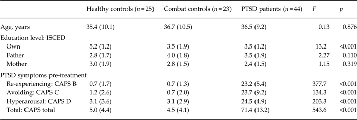
Participant characteristics are presented in Tables 1 and 2. Freesurfer analyses were not performed on MRI scans of two combat controls and one PTSD patient, due to poor scan quality (movement). One PTSD patient had severe temporal lobe atrophy and was therefore excluded from further analyses. Furthermore, one patient had not received treatment in between the two MRI scans and was also excluded. This yielded 44 PTSD patients, 23 combat controls and 25 healthy controls for the analyses. The three groups did not differ in age. The healthy control group had a higher education level compared with the veteran groups, but parental education level did not differ between the groups (Table 1).
Age, education level and PTSD symptoms pre-treatment of healthy controls, combat controls and PTSD patients

Data are given as mean (standard deviation).
PTSD, Post-traumatic stress disorder; ISCED, International Standard Classification of Education (Schneider, Reference Schneider2013); CAPS, Clinician Administered PTSD Scale (Blake et al. Reference Blake, Weathers, Nagy, Kaloupek, Klauminzer and Charney1990).
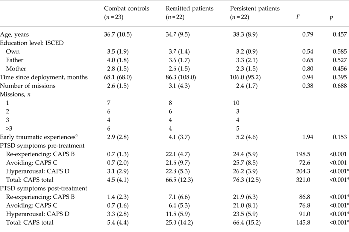
Age, education level, time since deployment, number of missions, number of early traumatic experiences and PTSD symptoms pre- and post-treatment of combat controls, remitted PTSD patients and persistent PTSD patients

Data are given as mean (standard deviation) unless otherwise indicated.
PTSD, Post-traumatic stress disorder; ISCED, International Standard Classification of Education (Schneider, Reference Schneider2013); CAPS, Clinician Administered PTSD Scale (Blake et al. Reference Blake, Weathers, Nagy, Kaloupek, Klauminzer and Charney1990).
a Early traumatic experiences were investigated with the Early Trauma Inventory – Self Report (Bremner et al. Reference Bremner, Bolus and Mayer2007).
* Mean values for remitted and persistent patients were significantly different.
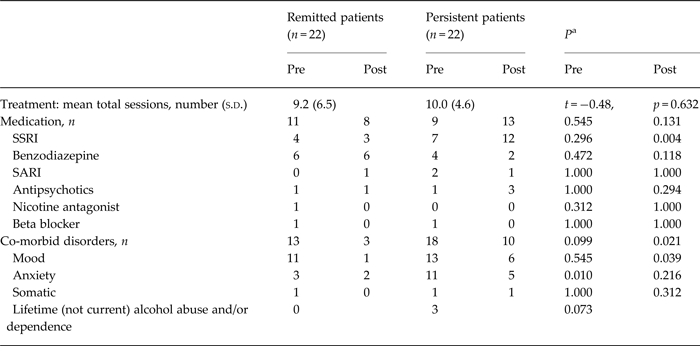
Based on post-treatment PTSD severity, 22 PTSD patients were classified as remitted and 22 as persistent patients. The three veteran groups did not differ in age, education level, months since deployment, number of missions, and early traumatic experiences (investigated with the Early Trauma Inventory – Self Report; Bremner et al. Reference Bremner, Bolus and Mayer2007) (Table 2). Pre-treatment total CAPS score was larger in patients than in controls, and larger in persistent patients than remitted patients. This variance between persistent and remitted patients was explained by a group difference in hyperarousal symptoms. Re-experiencing and avoiding and numbing symptoms did not significantly differ between remitted and persistent patients. All patients included in the analyses had received trauma-focused therapy, and number of treatment sessions did not differ between the two patient groups (Table 3). Remitted and persistent patients were comparable on pre-treatment medication use; however, persistent patients had more SSRI use post-treatment. Most patients had the same medication status or used the same type of medication throughout their treatment, and some patients changed medication. However, there were no group differences in the number of patients that changed medication status (remitted, n = 5; persistent, n = 6; p = 0.728) or type of medication (remitted, n = 3; persistent, n = 5; p = 0.434). Persistent patients had more co-morbid anxiety disorders pre-treatment and co-morbid depression post-treatment. The two patient groups did not significantly differ with respect to lifetime alcohol abuse and/or dependence (Table 3).
Number of treatment sessions, use of medication and presence of co-morbid disorders in remitted and persistent PTSD patients

PTSD, Post-traumatic stress disorder; pre, pre-treatment; post, post-treatment; s.d., standard deviation; SSRI, selective serotonin reuptake inhibitor; SARI, serotonin antagonist and reuptake inhibitor.
a p Values of medication and co-morbid disorder analyses are based on χ 2 analyses.
Relation to stress/trauma exposure
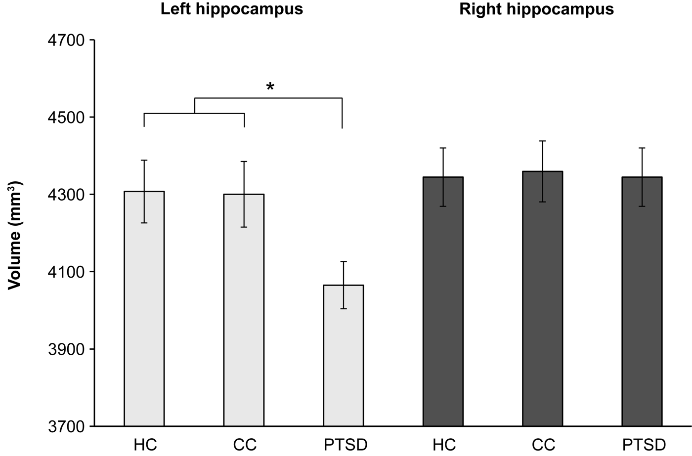
Results of the comparison of PTSD patients, combat controls and healthy controls are displayed in Fig. 1. The univariate analyses revealed a group difference in the left hippocampus (F 2,87 = 3.98, p = 0.022). Post-hoc t tests showed that PTSD patients differed significantly from the healthy controls (p = 0.019) and the combat controls (p = 0.027). The combat control and healthy control groups did not differ from each other (p = 0.952). No significant group difference was observed in the right hippocampus (F 2,87 = 2.27, p = 0.109).
Left and right hippocampal volumes of healthy non-military controls (HC), healthy war veterans (combat controls; CC) and post-traumatic stress disorder (PTSD) patients. Values are means, with standard errors represented by vertical bars. *Significant group difference (p < 0.05).

Effect of treatment
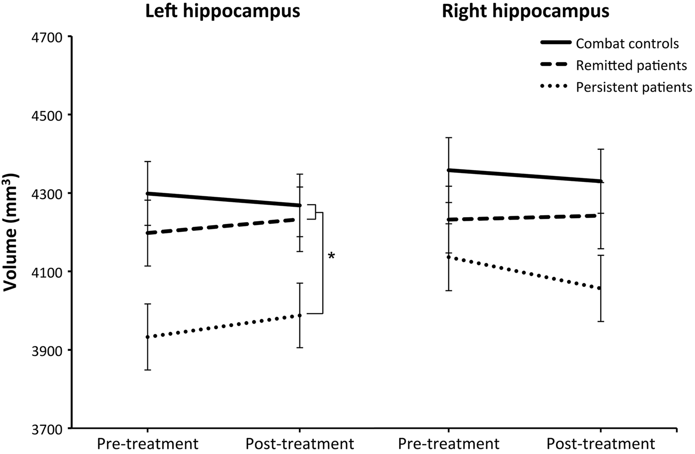
Results of the pre- and post-treatment analyses for the remitted and persistent patients and combat controls are displayed in Fig. 2. No significant interaction between group and time was observed. Instead, in the left hippocampus a significant main effect of group was found (F 2,62 = 4.72, p = 0.012). Persistent PTSD patients had a significantly smaller left hippocampus across both time points than both combat controls (p = 0.005) and remitted PTSD patients (p = 0.027). In the right hippocampus only a marginally significant group difference was found (F 2,62 = 2.86, p = 0.069). Post-hoc tests revealed a smaller right hippocampus in the persistent PTSD patients compared with combat controls (p = 0.037), whereas remitted patients did not differ from combat controls or persistent patients.
Effect of treatment on hippocampal volumes. Pre- and post-treatment hippocampal volumes (in mm3) of healthy war veterans (combat controls), post-traumatic stress disorder (PTSD) patients in remission (remitted patients) and PTSD patients in whom symptoms persisted after treatment (persistent patients) are displayed separately for the left and right hippocampus. Values are means, with standard errors represented by vertical bars. * Significant group difference (p < 0.05).

Post-hoc analyses
As the persistent patients probably explain the group difference for the comparison of PTSD patients, combat controls and healthy controls, the data were re-analysed with four groups. Again, a group difference was observed in the left hippocampus (F 3,86 = 4.08, p = 0.009). Indeed, only the persistent PTSD group differed from the healthy controls (p = 0.003), combat controls (p = 0.004) and from the remitted PTSD group (p = 0.049). There was no significant group difference in the right hippocampus.
Second, to investigate the potential effect of medication on our results, pre- and post-treatment measures of hippocampal volume were compared for medication-naive remitted patients (n = 10), medication-naive persistent patients (n = 8) and combat controls (n = 23). Again a group difference in the left hippocampus (F 2,36 = 3.74, p = 0.036) was observed, showing that this finding cannot be explained by medication use. In the right hippocampus, again no significant difference was observed (F 2,36 = 1.27, p = 0.289). The potential effect of SSRIs was analysed by comparing hippocampal volume, pre- and post-treatment, between patients using SSRIs pre-treatment and medication-naive patients in the whole PTSD sample (SSRI, n = 11; medication naive, n = 25), and in the persistent PTSD group only (SSRI, n = 7; medication naive, n = 14). These analyses revealed no significant hippocampal volume differences in the whole PTSD sample (group × time interaction, F 1,34 = 1.363, p = 0.251; main effect group F 1,34 = 0.002, p = 0.996) or in the persistent patients alone (group × time interaction, F 1,19 = 0.151, p = 0.702; main effect group F 1,19 = 0.138, p = 0.715).
Third, although the χ 2 test did not reveal a significant group difference for lifetime alcohol abuse/dependence, only a few patients (n = 3) in the persistent group had a history of alcohol dependence and/or abuse. Therefore, the original group analyses were repeated after excluding these three patients. Again, a significant main effect of group was found in the left hippocampus (F 2,58 = 6.109, p = 0.004). Furthermore, the marginally significant effect of group in the right hippocampus reached significance after excluding patients with a history of alcoholism (F 2,58 = 3.621, p = 0.033).
Finally, no significant correlations were observed in the PTSD group between PTSD severity (total CAPS score) and hippocampal volume, either pre-treatment (left, r = −0.089, p = 0.566; right, r = −0.044, p = 0.777) or post-treatment (left, r = −0.062, p = 0.689; right, r = −0.052, p = 0.736). Also, no correlations were observed in the PTSD group between clinical improvement (Δ total CAPS score) and change in hippocampal volume in the PTSD group as a whole (left, r = 0.011, p = 0.942; right, r = −0.175, p = 0.256) or when analysed separately for the remitted (left, r = 0.236, p = 0.290; right, r = 0.091, p = 0.688) and persistent patients (left, r = −0.117, p = 0.603; right, r = −0.125, p = 0.578).
Discussion
In addition to the replication of earlier findings that PTSD patients have smaller left hippocampal volumes than controls, we report that hippocampal volumes of combat controls and healthy non-military controls did not differ. This suggests that trauma or stress exposure during deployment does not result in decreased hippocampal volumes. Second, we found that hippocampal volume of PTSD patients did not normalize with successful treatment. Instead, we showed that (left) hippocampal volumes of PTSD patients in whom PTSD symptoms persisted after trauma-focused therapy were smaller than hippocampal volumes of combat controls and remitted patients both prior to treatment and after treatment. Previous studies on hippocampal volume and treatment in PTSD did not collect pre- and post-treatment MRI scans or did not differentiate between remitted and persistent patients. Therefore, we are the first to conclude that a smaller hippocampus represents a pre-treatment vulnerability factor for the persistence of PTSD.
Hippocampal volumes did not differ between trauma-exposed controls and healthy controls, suggesting that reduced hippocampal volume is not the consequence of stress or trauma exposure during deployment. This conclusion is supported by the twin study of Gilbertson et al. (Reference Gilbertson, Shenton, Ciszewski, Kasai, Lasko, Orr and Pitman2002) who showed reduced hippocampal volume in the non-deployed twin without PTSD (Gilbertson et al. Reference Gilbertson, Shenton, Ciszewski, Kasai, Lasko, Orr and Pitman2002), and by a study that found smaller hippocampal volumes in war veterans with PTSD, and war veterans without PTSD, but also in non-deployed reservists compared with healthy controls (Vythilingam et al. Reference Vythilingam, Heim, Newport, Miller, Anderson, Bronen, Brummer, Staib, Vermetten, Charney, Nemeroff and Bremner2005). In two non-military studies opposing results were found. A study on childhood abuse observed comparable hippocampal volumes of women with and without early childhood abuse (Bremner et al. Reference Bremner, Vythilingam, Vermetten, Southwick, McGlashan, Nazeer, Khan, Vaccarino, Soufer, Garg, Ng, Staib, Duncan and Charney2003), whereas a study that compared burns victims with and without PTSD and healthy controls observed reduced hippocampal volume in all burns victims (Winter & Irle, Reference Weathers, Ruscio and Keane2004). Two meta-analyses (Karl et al. Reference Karl, Schaefer, Malta, Dorfel, Rohleder and Werner2006; Woon et al. Reference Woon, Sood and Hedges2010) concluded that trauma exposure regardless of PTSD was associated with reduced (left) hippocampal volume. This conclusion is not supported by our data; however, the discrepancy in findings can potentially be explained by differences in type of trauma and, in particular, the timing of traumatic events. Early traumatic experiences have often been associated with reduced hippocampal volume (Vythilingam et al. Reference Vermetten, Vythilingam, Southwick, Charney and Bremner2002; Teicher et al. Reference Smith2003). It can therefore be postulated that negative environmental factors may influence hippocampal growth (in particular) during sensitive periods, putting these individuals at risk for PTSD. Building upon this hypothesis, trauma exposure in adulthood does not necessarily result in hippocampal volume reduction, which was indeed observed in the current study.
With this study we also investigated whether reduced hippocampal volume in PTSD patients normalizes after successful treatment, or if reduced hippocampal volume constitutes a vulnerability factor for the persistence of PTSD. We observed a smaller left hippocampus before and after treatment in persistent patients compared with remitted patients and combat controls. With this finding we support and extend the conclusion of Gilbertson et al. (Reference Gilbertson, Shenton, Ciszewski, Kasai, Lasko, Orr and Pitman2002) that a smaller hippocampus is a vulnerability factor for the persistence of PTSD. The present study also showed that a smaller hippocampal volume is not a necessary condition for developing PTSD, because mean hippocampal volume of remitted patients was comparable with that of combat controls and healthy controls, both pre- and post-treatment. Moreover, the current finding clarifies why not all previous MRI studies in PTSD have observed reduced hippocampal volume in patients, e.g. in a study with recently traumatized patients, PTSD was not associated with smaller hippocampal volumes (Bonne et al. Reference Bonne, Brandes, Gilboa, Gomori, Shenton, Pitman and Shalev2001). In line with earlier meta-analyses (e.g. Smith, Reference Schneider2005; Karl et al. Reference Karl, Schaefer, Malta, Dorfel, Rohleder and Werner2006), we also showed that reduced hippocampal volume in persistent patients was most pronounced in the left hippocampus. Group differences in the right hippocampus were marginally significant, though this difference became significant when excluding patients with a lifetime history of alcohol abuse and/or dependence.
The finding that reduced hippocampal volume constitutes a risk factor for the persistence of PTSD has potential implications for the prognosis and prediction of treatment success. PTSD is effectively treated with trauma-focused therapy (Bisson et al. Reference Bisson, Ehlers, Matthews, Pilling, Richards and Turner2007), which is based on extinction learning and aims at updating the traumatic memory by focusing on contextual (safety) information (Izquierdo et al. Reference Izquierdo, Cammarota, Vianna and Bevilaqua2004). The hippocampus is involved in learning, formation of contextual memories and consolidation of new memories (Fendt & Fanselow, Reference Fendt and Fanselow1999). Furthermore, hippocampal volume has been correlated with memory performance (Tischler et al. Reference Tendolkar, Van Beek, Van Oostrom, Mulder, Janzing, Voshaar and Van Eijndhoven2006; Pohlack et al. Reference Pohlack, Meyer, Cacciaglia, Liebscher, Ridder and Flor2014) and improvement after memory training (Engvig et al. Reference Engvig, Fjell, Westlye, Skaane, Sundseth and Walhovd2012). Therefore, it seems plausible to conclude that a larger hippocampus is associated with an enhanced ability to learn and store newly formed contextual memories, resulting in a better prospect to recover from PTSD. On the other hand, individuals with a smaller hippocampus might benefit from a different treatment approach. This is relevant because trauma-focused therapy is often experienced as strenuous and might initially worsen symptoms (Bisson et al. Reference Bisson, Ehlers, Matthews, Pilling, Richards and Turner2007). Therefore, more research on alternative treatment for this group is required.
Treatment response in our sample was not associated with change in hippocampal volume. Previous findings of treatment effects on hippocampal volume in PTSD patients were conflicting. Two studies observed hippocampal growth after treatment with SSRIs (Vermetten et al. Reference Tischler, Brand, Stavitsky, Labinsky, Newmark, Grossman, Buchsbaum and Yehuda2003; Bossini et al. Reference Bossini, Tavanti, Lombardelli, Calossi, Polizzotto, Galli, Vatti, Pieraccini and Castrogiovanni2007), whereas another study did not observe increase in hippocampal volume (Bremner et al. Reference Bremner, Mletzko, Welter, Quinn, Williams, Brummer, Siddiq, Reed, Heim and Nemeroff2005). These studies did not include a control group, which hampers the interpretation of the findings, because the effect of treatment cannot be separated from the potential effect of time. A study on the effect of CBT on hippocampal volume demonstrated a mean increase of 100 mm3 in PTSD patients after 12 weeks of CBT, while controls showed a hippocampal decrease of about 70 mm3 (Levy-Gigi et al. Reference Levy-Gigi, Szabo, Kelemen and Keri2013). Furthermore, a correlation between change in hippocampal volume and change in symptoms in the PTSD group was observed. We could neither replicate this pre- to post-treatment difference, nor this correlation. Remarkably, in that study the change in hippocampal volume in some patients was about 25%, which is very large and might be caused by movement or measurement error at one of two time points, thereby influencing hippocampal volume (change). Our findings support results from another CBT treatment study (Lindauer et al. Reference Lindauer, Vlieger, Jalink, Olff, Carlier, Majoie, Den Heeten and Gersons2005), in which patients who received CBT were compared with patients on a waiting list and a group of controls. In that study also no change in hippocampal volume was observed.
Limitations and future directions
The distinction between remitted and persistent patients resulted in some group differences probably related to post-treatment symptom severity, such as pre-treatment severity, co-morbidity and post-treatment medication use. As symptom severity did not correlate with left hippocampal volume within the groups, it is unlikely that group differences can be attributed to pre-treatment PTSD severity. Second, analyses were repeated with medication-naive patients and similar results were obtained. Third, it is also not likely that co-morbidity explains the findings, because the same pattern of results was observed when patients without co-morbidity were compared. Yet, based on these findings we cannot ascertain that medication and co-morbidity have not affected hippocampal volume, and future studies should confirm our results.
Education level differed between the veterans and non-military healthy controls. This is probably due to the fact that most military men joined the armed forces after high school, whereas the non-military men continued education. Parental education did not differ between groups.
We conclude that a smaller hippocampal volume is a vulnerability factor for treatment non-response in PTSD. This finding is of potential clinical significance in predicting persistence of PTSD; however, its exact predictive value should be further established. Furthermore, investigating (contextual) memory consolidation and retrieval in a group of remitted and persistent PTSD patients would inform us whether structural brain characteristics observed in this study indeed translate to behavioral differences.
Conclusion
Our findings suggest that a smaller (left) hippocampus is not the result of stress/trauma exposure. Furthermore, hippocampal volume does not increase with successful treatment. Instead, we demonstrate for the first time that a smaller (left) hippocampus constitutes a risk factor for the persistence of PTSD.
Acknowledgements
This study was financially supported by the Dutch Ministry of Defence.
Declaration of Interest
None.