INTRODUCTION
The setae which clothe the limbs and mouthparts of cirripedes have a number of important functions in feeding, sensory perception and reducing the fouling of the body surface and mantle lining by epizoites (Anderson & Anderson, Reference Anderson and Anderson1985), but very little is known about their detailed morphology. Thomas (Reference Thomas1970) defined decapod setae as ‘hair-like cuticular outgrowths articulating with the cuticle by a well-defined basal socket’ (see also Pohle & Telford, Reference Anderson1981). Garm (Reference Garm2004) refined this definition to ‘an elongate projection with a more or less circular base and a continuous lumen; the lumen has a semicircular arrangement of sheath cells basally’. The details of setal morphology are at or beyond the limits of light microscopy and few studies of cirripede structure have used scanning electron microscopy (SEM). Most studies on cirripede structure, functional morphology and classification have concentrated on shell morphology, structure of the cirri and mouthparts and the distribution of the setae on the appendages (Darwin, Reference Darwin1851, Reference Darwin1854; Pilsbry, Reference Pilsbry1916; Newman & Ross, Reference Newman and Ross1976; Foster, Reference Foster1978 and many other authors). Setal morphology has been used to a limited extent in cirripede taxonomy (Henry & McLaughlin, Reference Henry and Mclaughlin1975; Southward, Reference Southward1976; Foster, Reference Foster1978; Chan, Reference Chan2001; Pitombo, Reference Pitombo2004; Chan et al., Reference Chan, Tsang, Ma, Hsu and Chu2007, Reference Chan, Garm and Høeg2008, Reference Chan, Hsu and Tsai2009). Several studies on ecology and feeding have also figured some of the setae on the cirri (Anderson & Anderson, Reference Anderson and Anderson1985; Hunt & Alexander, Reference Hunt and Alexander1991; Chan et al., Reference Chan, Garm and Høeg2008).
Høeg et al. (Reference Høeg, Karnick and Frølander1994) were the first to use SEM to describe and classify the setae on the mouthparts of six barnacle species: three pedunculates and three balanomorphs. They recognized that there were inconsistencies in the terms used by previous authors to describe setae, the same terms being used by different authors to describe setae that are not the same. This problem was also highlighted by Chan et al. (Reference Chan, Garm and Høeg2008). Høeg et al. (Reference Høeg, Karnick and Frølander1994) suggested that setal morphology, as revealed by SEM, could make a useful contribution to elucidating cirripede phylogeny.
Similar inconsistencies in the terminology used to describe setae, particularly in the Decapoda, were noted by Watling (Reference Watling, Felgenhauer, Watling and Thistle1989). Garm (Reference Garm2004) addressed this problem in his study of the structure and function of mouthpart setae in seven species of decapods. He described seven categories of setae. He concluded that his classification reflected mechanical function rather than evolutionary history. Using Garm's setal classification Chan et al. (Reference Chan, Garm and Høeg2008) compared setal morphology and cirral setation in six species of pedunculate and sessile cirripedes and related their observations to differing feeding strategies. They concluded that the changes in feeding behaviour seen during cirripede evolution were accompanied by an increase in the variety of the setae and changes in the setation patterns.
The present paper extends the work of Høeg et al. (Reference Høeg, Karnick and Frølander1994). The range of setal types seen in a wider variety of cirripedes is described. A three level classification is suggested that might be applied to setae throughout the Cirripedia, bringing consistency to setal descriptions, thus enabling setal characteristics to be used as part of the character sets used in future studies of cirripede phylogeny and taxonomy. The problems of reconciling the proposed setal categories with those defined by Garm (Reference Garm2004) for the Decapoda are discussed.
MATERIALS AND METHODS
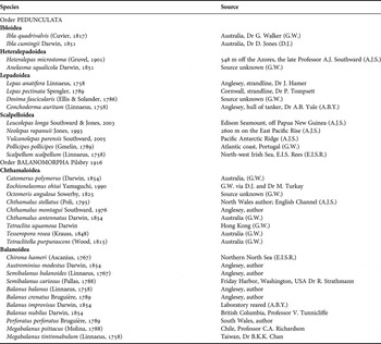
The pedunculate and sessile cirripedes examined in this study included species collected from intertidal, sub-littoral and deep-sea vent locations. The 33 species studied are listed in Table 1.
Species examined with sources of specimens. Nomenclature based mainly on Southward (Reference Southward, Crothers and Hayward2008).

For fixation of those species where living specimens were available, bodies were removed still attached to the opercular plates by cutting the opercular membrane with a fine scalpel. They were placed in filtered seawater and relaxed by the addition of MS 222 (3-amino benzoic acid ethyl ester methane sulphonate salt, SIGMA A 5040) powder to the seawater. Once relaxed, with the cirri unfurled, they were rinsed in three changes of filtered seawater before fixation in 2.5% glutaraldehyde in filtered seawater for 4 hours. Fixed material was rinsed in filtered seawater followed by three changes of tap water. Bodies were then separated from the opercular plates, before soaking in a dilute detergent (Decon) solution for 15 minutes followed by 15 seconds ultrasonication (Felgenhauer, Reference Felgenhauer1987). Cleaned material was then rinsed with three changes of distilled water and, either dehydrated through a graded series of ethanol and finally into acetone before critical-point drying from liquid carbon dioxide, or stored in 70% ethanol. Dissection to yield individual limbs and mouthparts was usually carried out on material in 70% ethanol. Mouthparts for most but not all the species, cirri, the oral cone and in some cases whole bodies were then mounted on aluminium stubs using double-sided Sellotape before being gold-coated and examined in a Cambridge Stereoscan 120 scanning electron microscope (SEM) operated at 10 kV. Specimens already preserved in alcohol or formalin, were thoroughly rinsed in tap water before dissection, cleaning with detergent, ultrasonication, rinsing and dehydration as above.
To avoid losing very small items such as dissected mouthparts or very small limbs, they were dehydrated and dried inside small, custom-made, polythene tubes closed at both ends with fine plankton netting. To dissect very small specimens, fine stainless steel entomological pins, mounted on the end of cocktail sticks with Araldite, were used. To transfer and mount these smaller specimens, eyelashes also fixed to cocktail sticks and moistened with saliva were used. Eyelashes are flexible and therefore do not damage dried delicate structures.
RESULTS
General setal morphology
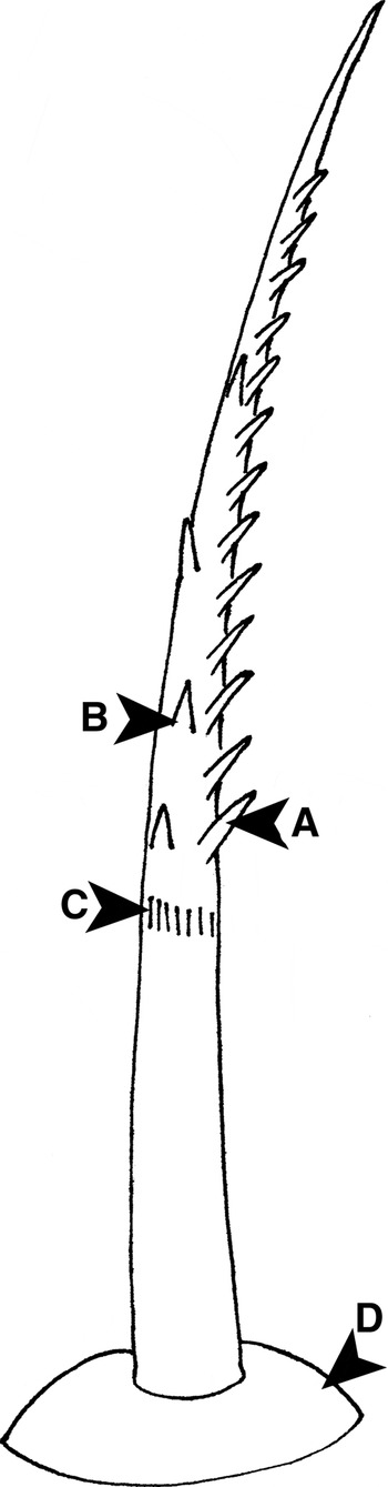
Cirripedes conform to the basic crustacean setal morphology already described in numerous studies and Figure 1 illustrates the basic features of a denticulate seta.
Diagram illustrating the basic morphology of a denticulate seta. Only one row of denticles is shown: (A) denticle; (B) supplementary denticle; (C) annulus; (D) setal socket.

The setal socket and setal shaft
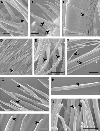
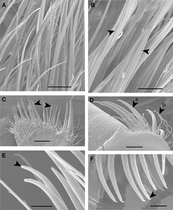
Where the seta articulates with the cuticle surface, there is usually a raised circular area of thin, flexible cuticle from the centre of which the setal shaft arises (Figures 1 & 2A). This socket in most cases allows the seta to move relative to the surface from which it arises. This supra-cuticular articulation is the normal situation in the Cirripedia. However, in particularly robust and rigid setae, such as many of those on the rami of Conchoderma auritum it is difficult to see a socket (Figure 2B) and these setae might be thought to be large denticles. Their true nature may only be confirmed by observing the mode of formation during the moulting cycle (see below), or whether a lumen is present. In several species, setae on the anterior face of the rami of those cirri comprising the cirral net were found to have hinged sockets allowing them to fold flat against the face of the ramus facilitating withdrawal of the cirri into the mantle cavity (Figure 2C). Setae found on the lateral surfaces of the mandibles and some setae on the maxillae of all the cirripedes examined, have the base of the setal shaft sunk into a pit in the cuticle (Figure 2D). This arrangement is described as infra-cuticular articulation and is the normal situation in the Decapoda that have been studied so far (Watling, Reference Watling, Felgenhauer, Watling and Thistle1989; Garm, Reference Garm2004).
The setal socket and the setal shaft. (A) Balanus crenatus, supra-cuticular articulation of setae on the exopodite of cirrus 2. Arrow indicates setal socket; (B) Conchoderma auritum, rigid seta, Type 1b1, on the distal region of a ramus of cirrus 1: (a) setal socket and (b) annulus; (C) Ibla quadrivalvis, hinged setae lying along the face of a ramus of cirrus 4. Arrow indicates the hinged socket; (D) Heteralepas microstoma, Type 2b1 setae on the lateral face of a mandible. Arrow indicates infra-cuticular socket; (E) Balanus balanus, shafts of setae on the exopodite of cirrus 1. Arrows indicate clearly marked annuli; (F) Lepas anatifera, part of the shaft of a stiff seta. Arrows indicate one of the surface ridges (a) and a needle-shaped denticle (b); (G) Leucolepas longa, Type 1b3 setae on the distal edge of maxilla 1. Arrows indicate the angular twisted setal shafts; (H) Neolepas rapanui, distal region of a Type 1a2 seta from the anterior faces of the rami of cirri 4–6. Arrow indicates the angular nature of the shaft; (I) Tesseropora rosea, tips of Type 1b2 setae on the distal edge of maxilla 1. Arrows indicate sub-terminal pores; (J) Semibalanus balanoides, tips of Type 1a4 setae on proximal region of the endopodite of cirrus 1. Arrows indicate the terminal pores. Scale bars: (A) 10 µm; (B) 50 µm; (C) 50 µm; (D) 10 µm; (E) 10 µm; (F) 10 µm; (G) 20 µm; (H) 5 µm; (I) 10 µm; (J) 5 µm.

The setal shaft often exhibits a discontinuity approximately one-third to one-half of the distance along its length (Figures 1 & 2E), which is the point at which the characteristic pattern of denticulation begins, or, is the point of transition between two differing patterns of cuticular ornamentation. This feature is called the annulus and marks the position of the base of the epidermal invagination in which the new seta forms during the moulting cycle (Nilsson-Cantell, Reference Nilsson-Cantell1921; Drach, Reference Drach1939; Reaka, Reference Reaka1975; Watling, Reference Watling, Felgenhauer, Watling and Thistle1989).
In fresh and fixed specimens setal shafts may be straight or more frequently slightly curved. They may be flexible or stiff. Stiff shafts are often strongly ridged (Figure 2F) or may be relatively massive (Figure 2B). Most setal shafts are circular in section but some on the mouthparts appear to be flattened (Figure 2D). An unusual feature of the eolepadines were setae with an angular cross-section and a spiral twist, most clearly seen in the robust setae on the distal edge of the first maxillae (Figure 2G) and in the distal regions of the long collecting setae of cirri 3–6 (Figure 2H).
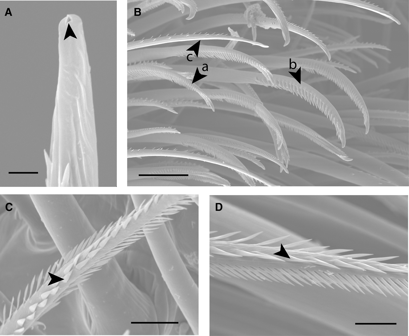
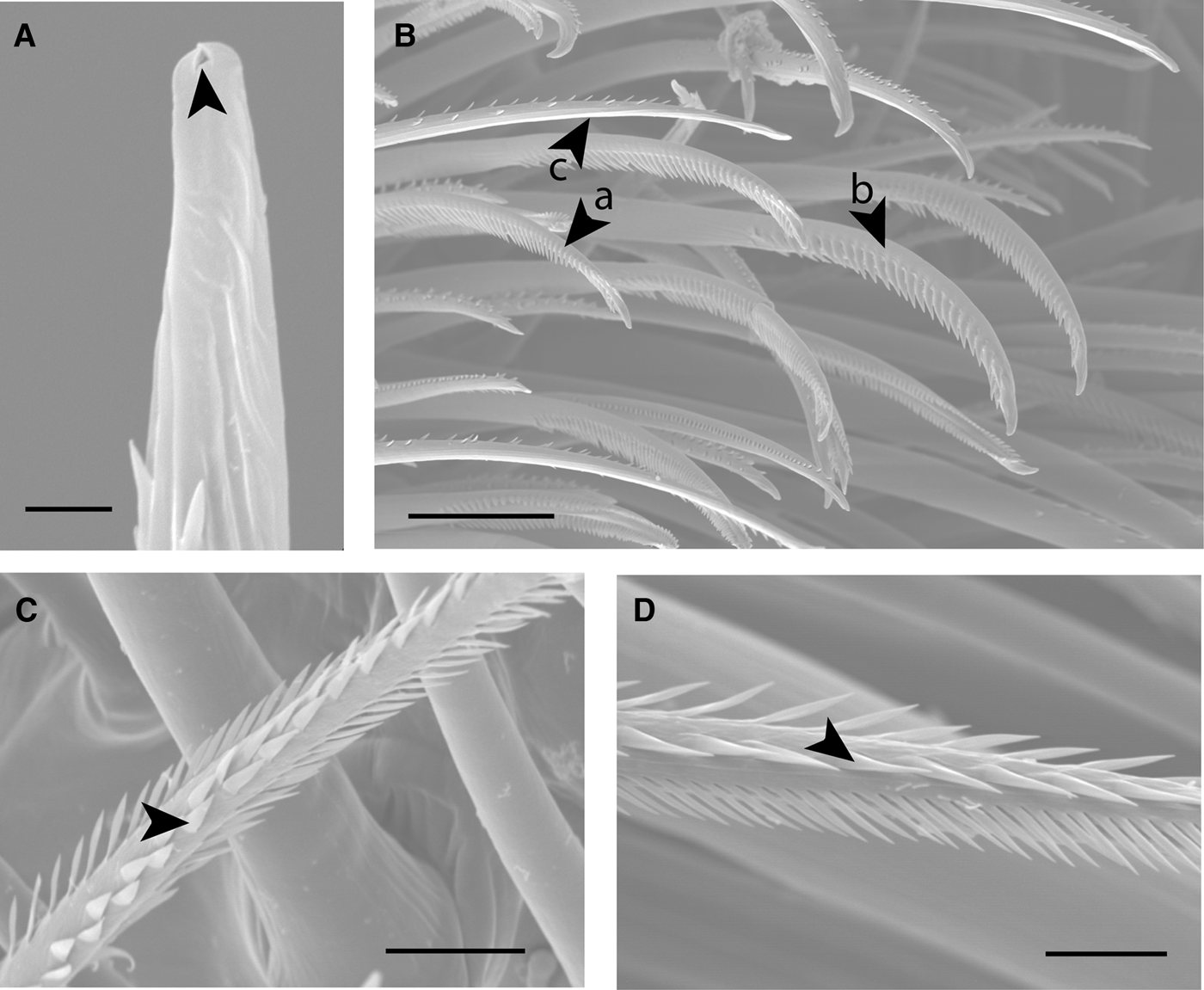
Sub-terminal or terminal pores were found in several different types of setae in a number of different locations (Figure 2I, J). Pollicipes pollicipes, Catomerus polymerus, Octomeris angulosa, Tetraclita squamosa, Tesseropora rosea, Tetraclitella purpurascens and Megabalanus tintinnabulum, were found to have some setae with bifid tips and a very small terminal pore (Figure 4A).
Outgrowths from the setal shaft
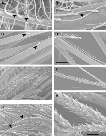
Cuticular outgrowths from the setal shaft can broadly be divided into setules and denticles. In this study the term setule is applied to very long (up to 20 µm), slender, flexible, usually flattened outgrowths from the setal shaft. Setules do not taper: they are the same width throughout their length. They usually arise seamlessly from the shaft (Figure 3A) but may, in some cases arise from pits (Figure 3B).
The variety of setules and denticules arising from the setal shaft. (A) Tesseropora rosea, part of the shaft of a Type 3b, setulate seta on the posterior border of the coxopodite of cirrus 1. The arrows indicate the setules arising seamlessly from the shaft; (B) Heteralepas microstoma, part of the shaft of a Type 3a, setulate, seta found on the exopodite of cirrus 1. The arrow indicates a setule arising from a pit in the setal shaft; (C) Pollicipes pollicipes, part of the shaft of a seta from the ramus of cirrus 2. Arrows indicate the seamless origin of the denticles; (D) Ibla quadrivalvis, part of the shaft of a Type 2a3 seta on a ramus of cirrus 4, with rigid needle-shaped denticles; (E) Balanus perforatus, shafts of setae, on the exopodite of cirrus 1, with flexible denticles; (F) Balanus balanus, part of the shaft of a seta on the anterior face of a ramus of cirrus 4, with long, slender needle-shaped denticles; (G) Chthamalus antennatus, bidentate Type 2d3 setae (arrows), with large conical denticles, on the tip of the endopodite of cirrus 2; (H) Balanus nubilus, part of the shaft of a Type 2e3 seta from the median face of the mandibular palp, with broad, flattened (foliate) denticles. Scale bars: (A) 2 µm; (B) 5 µm; (C) 10 µm; (D) 10 µm; (E) 10 µm; (F) 10 µm; (G) 20 µm; (H) 5 µm.

Cuticular outgrowths that cannot be described as setules are here termed denticles. Denticles arise seamlessly from the surface of the setal shaft (Figure 3C) and may be rigid (Figure 3D, F) or flexible (Figure 3E), varying in length from 2–15 µm. Some are needle-shaped (Figures 2F, 3D & 3F) others taper to a point from a broader base (Figure 3C). Some are stout but blunt while others are conical (Figure 3G). Some are flattened and leaf-shaped (foliate; Figure 3H).
Distribution of setules and denticles on the setal shaft
In setulate setae the setules are usually distributed along the whole length of the setal shaft but sometimes they are confined to the distal region beyond the annulus. In the species studied they were found to be arranged either in two irregular rows on opposite sides of the shaft or irregularly over half the circumference of the shaft. In setulate setae the annulus is often not easy to see.
In most denticulate setae the denticles arise distally, beyond the annulus, and are usually arranged in two rows. The denticular layout may be irregular immediately distal to the annulus, but further along the shaft the denticles take up the pattern characteristic of a particular setal type (see below). Most setae taper distally and the denticular layout may alter towards the tip of the setal shaft: in some denticles are present right to the tip, but in others the tip is free of ornamentation.
In denticulate setae with curved setal shafts (Figure 4B) the rows of denticles tend to be on the inner face of the curve. However, in P. pollicipes, C. polymerus, O. angulosa, T. squamosa, T. rosea and T. purpurascens, setae were found with denticles on the outer face of the curve (Figure 4B) while the inner face of the curve was without denticles.
(A) Pollicipes pollicipes, distalmost region of a Type 2f2 seta from the anterior face of a ramus of cirrus 4 showing the bifid tip and pore; (B) Pollicipes pollicipes, setae on the endopodite of cirrus 1: (a) Type 2d1 seta with denticles on the inner face of the curved shaft; (b) Type 2d2 seta; (c) Type 2a7 with denticles on the outer face of the curved shaft; (C) Scalpellum scalpellum, part of the shaft of a Type 2c1 seta on the exopodite of cirrus 1. Arrow indicates supplementary denticles; (D) Lepas anatifera, part of the shaft of a bipectinate seta, Type 2c1, on the exopodite of cirrus 2. Arrow indicates supplementary denticles. Scale bars: (A) 2 µm; (B) 50 µm; (C) 10 µm; (D) 10 µm.
In denticulate setae of a given type, some of the setae may have extra denticles on the opposite side of the setal shaft to the rows of denticles which define the type. These denticles can be scattered apparently at random (Figure 1) or arranged in highly ordered rows (Figure 4C, D). These ‘supplementary’ denticles can be recognized because the spacing and/or characteristics of the denticles differ from those in the rows which characterize the setal type. They are also seen on setulate setae.
Where setae are not crowded together it is relatively easy to work out the disposition of denticles or setules and hence assign a seta to a category or type. However, in dense, complex setal fields, such as those found on the inner/anterior faces of the maxillipeds in various balanomorph species it is often extremely difficult to work out the setal structure because the view of a particular seta may be obscured by other setae. It would be possible to isolate individual setae for study on the rami of the captorial cirri of the larger cirripedes, but it would be difficult in the complex setal fields on the maxillipeds. In such fields, setae are often found which are intermediate between defined setal types, as one setal type will grade into the adjacent type without a sharp boundary.
Description of setal types
In the 33 barnacle species examined 43 setal types have been described. These have been allocated to five Groups:
Group 1, simple: shaft without setules or denticles. A pore may be present at or close to the setal tip.
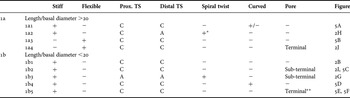
Table 2 summarizes the characteristics of the setal types comprising Group 1;
Characteristics of setal types in Group 1: simple Setae.

A, angular; C, circular; Prox., proximal; TS, transverse section; *, spiral twist distal to annulus; **, in Chthamalus spp. setal shaft projects beyond pore on one side.
Group 2, denticulate: shaft with denticles usually in a row or rows distal to the annulus. A pore may be present at the setal tip.
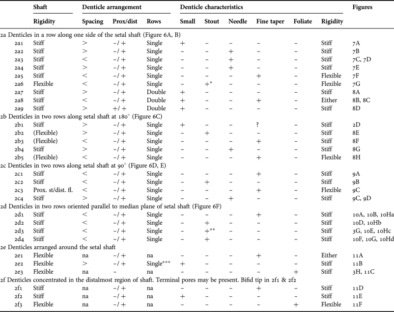
Table 3 summarizes the characteristics of the setal types comprising Group 2.
Characteristics of setal types in Group 2: denticulate setae.
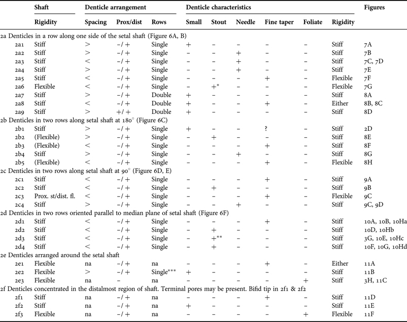
+, feature present; –, absent; (Flexible), flexibility uncertain; >spacing greater than denticle length; <spacing less than denticle length; na, not applicable; prox/dist, proximal or distal; +*, +**, single, ***, see text.
In considering the features used to characterize the Group 2 setal types in Table 3 some qualifications must be made.
It is usually easy to determine from an SEM image whether or not a setal shaft is stiff or flexible but sometimes there may be an element of doubt. In most rows of denticles along the setal shaft the denticles are aligned in a single line but in Types 2a8 and 2a9 the denticles are in pairs. In Type 2a7 there is an irregular double row of denticles which may change to a single line towards the setal tip. The use of the term ‘small’ to describe a denticle is a relative not an absolute term: a small denticle has a length considerably less than the diameter of the setal shaft. Denticles defined as ‘stout’ would have a round or oval section at the base. In Type 2a6 (+* in Table 3) the denticles are relatively long and taper to a fine point. In Type 2b2 and Type 2c2 the denticle is much shorter and blunt. The Types 2d2, 2d3 and 2d4 setae all have quite distinct denticular shapes illustrated in Figure 10H. The morphology of Type 2d3 setae is an important characteristic in Chthamalus taxonomy and there are several sub-types. In C. stellatus (Figure 10E; +** in Table 3) the distal region of the Type 2d3 setae resembles the Type 2d1 setae of C. montagui (Figure 10B). Type 2e2 (single*** in Table 3) has multiple single rows of small denticles around the setal shaft (Figure 11B).
Group 3, Setulate: shaft with setules.
Type 3a: setules arising from pits or sockets in the shaft (Figure 3B).
3a1: setules distributed over 180° of the shaft circumference.
3a2: setules arising in 2 irregular rows on opposite sides of the shaft (Figure 12A).
Type 3b: setules arise from the shaft without discontinuity (Figure 3A).
3b1: setules distributed over 180° of the shaft circumference (Figure 12B).
3b2: setules arising in 2 zones on opposite sides of the setal shaft.
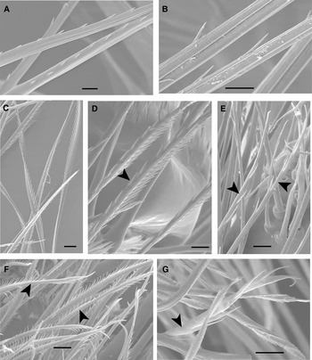
The arrangement of the setules often changes as the setal shaft tapers towards the tip. Proximally the setules may be distributed over half the circumference or in two zones on opposite sides of the setal shaft (Figure 12A). Distally, as the shaft tapers, the setules are often arranged in single rows along opposite sides of the shaft. The annulus is rarely observed. Some setulate setae have setules confined to the distal region of the shaft (Figure 12B), but usually setules extend proximally almost to the socket (Figure 12A).
Group 4, plumodenticulate: shaft with setules proximal to the annulus and denticles distal to the annulus.
Type 4: only one type was seen (Figure 12C).
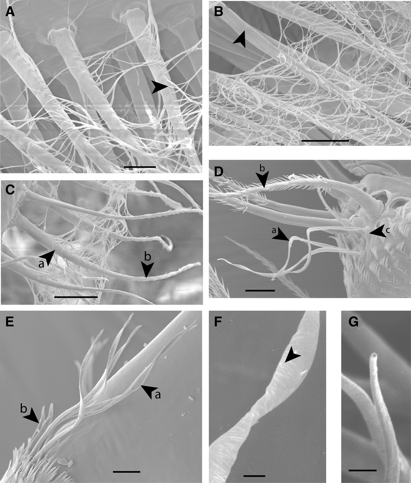
Group 5, aesthetascs: slender shaft usually twisted distal to the annulus (Figures 12D & 12E).
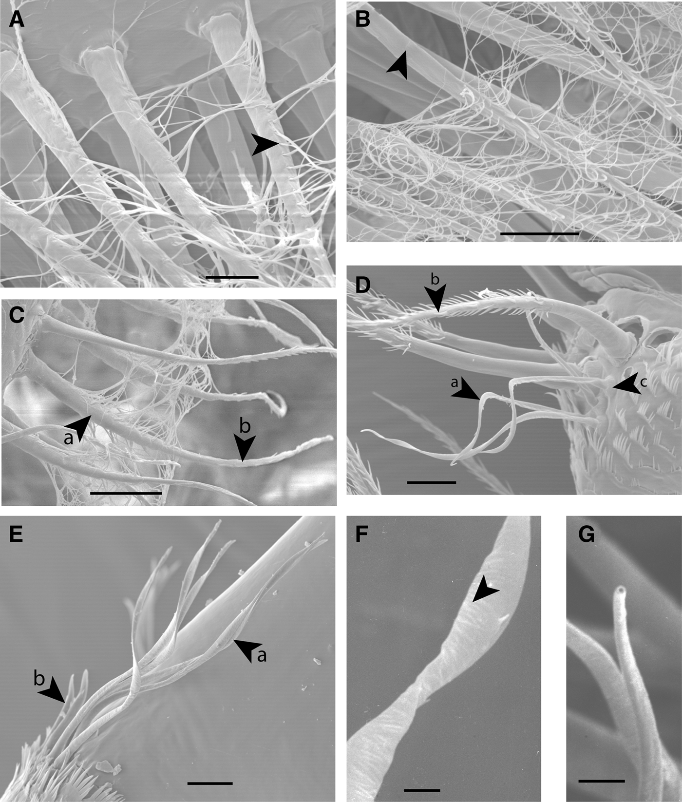
Type 5: these slender setae appear flattened in cross-section distal to the annulus, having a thin shaft wall (Figure 12F) and often a terminal pore (Figure 12G). The twisted appearance is not a fixation artefact as the twisted shaft is clearly visible in fresh material.
Distribution of setae on the limbs and prosoma in the barnacles studied
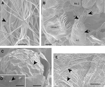
With the exception of the two Ibla species, setae were found only on the mouthparts, cirri and penis. In I. quadrivalvis and I. cumingii denticulate setae (Type 2b3) were also found on the prosoma (Figure 13A) and on the ventral cone situated between the bases of the cirri just posterior to the oral cone (Figure 13B). Darwin (Reference Darwin1851, p.187) described this structure as a ‘conical prominence clothed with bristles’ lying ‘over the infra-oesophageal ganglion’. There are many gland pores on the lateral faces of this structure that are probably the pores of the sub-oesophageal ‘salivary’ glands seen in many cirripedes (Bocquet-Védrine & Ovechko, Reference Bocquet-Védrine and Ovechko1960; Walley, Reference Walley1965). The eolepadines Leucolepas longa, Vulcanolepas parensis and Neolepas rapanuii also have gland pores on a protuberance in a similar position but without setae (Figures 13C & 13D). Darwin (Reference Darwin1854) reported setae on the prosoma of Chthamalus species. There are processes arising from the prosoma cuticle in C. montagui but they do not seem to have a basal socket and are here interpreted as long flexible denticles up to 40 µm in length (Figure 13E). Anelasma squalicola, an ectoparasite on sharks, was the only species examined which was found to have no setae, confirming the observations of Johnstone & Frost (Reference Johnstone and Frost1927).
Occurrence of setal types in the barnacle species studied
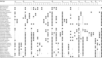
The presence or absence of the different setal types in the species examined are summarized in Table 4. Some setal types are widespread but others seem to be restricted to particular species or taxonomic groups. Types that are recorded as common to different species fall into several distinct sub-types varying in the spacing, length or characteristics of the denticles. These finer variations could prove useful in differentiating genera or species.
Distribution of setal types across the taxa examined.

■ setal type recorded; ?, setal type not seen but may be present; no symbol, setal type thought to be absent.
The most commonly occurring setal types were 1b2, 2b1, 2c1, 2e1 and 5. Type 1b2 (Figures 2I & 5C) is found only along the distal edge of the first maxilla. In some of the largest examples the basal socket and sub-terminal pore are difficult to see and in Lepas anatifera (Høeg et al., Reference Høeg, Karnick and Frølander1994) and P. pollicipes the dorsalmost projections on the edge of the first maxilla seem to be large denticles rather than setae. (The Type 1b3 setae of the Eolepadidae also found on the distal edge of the first maxilla can be regarded as homologous with Type 1b2.) The sub-terminal pores in these setae suggest a chemo-sensory function (Høeg et al., Reference Høeg, Karnick and Frølander1994).
Simple setae. (A) Semibalanus balanoides, Type 1a1 setae on the endopodite of cirrus 3; (B) Balanus crenatus, Type 1a3 setae on the distal face of maxilla 2. Arrows indicate annuli; (C) Tesseropora rosea, Type 1b2 setae on distal border of maxilla 1; (D) Dosima fascicularis, Type 1b4 setae (a) together with Type 5 setae (b) on the posterior distal border of a ramus segment of cirrus 4; (E) Balanus balanus, tips of Type 1b5 setae on the distal border of maxilla 1 showing terminal pores (arrow); (F) Chthamalus stellatus, tips of Type 1b5 setae on the distal border of maxilla 1 showing asymmetrical cuticular extension beyond terminal pores (arrow). Scale bars: (A) 20 µm; (B) 20 µm; (C) 100 µm; (D) 50 µm; (E) 5 µm; (F) 5 µm.

Type 2b1 setae are found on the lateral faces of the mandibles (Figure 2D) and sometimes on the first maxillae. These were the only cirripede setae that were found to have infra-cuticular articulation. The size and spacing of the lateral denticles and the characteristics of the setal shaft vary between species. In order to compare Type 2b1 setae from different species it is important to look at setae from a similar location in the setal field. Towards the edge of the setal field the denticulation may become sparser or even disappear altogether, particularly where they grade into adjacent fields of slender denticles.
Type 2c1 setae (Figure 9A) are found on the rami of cirri 1–3, often as part of the complex fields of setae on the median faces of the rami. They were not found on the anterior faces of cirri 4–6 forming part of the cirral net.
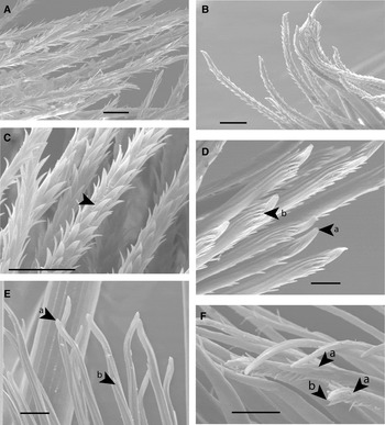
Type 2e1 setae are found on both limbs and mouthparts but very commonly on maxilla 2 (Figure 11A).
Type 5 setae are usually found on the posterior distal borders of the ramus segments of cirri 4–6, in close association with whatever setal type is characteristic of this position in a given species (Figures 5D & 12D–G). They are also found along the length of the antenniform rami of cirri 1–3 and on the penis. The precise morphology varies from species to species. The external morphology suggests that these may be sensory setae and their possible function is discussed below.
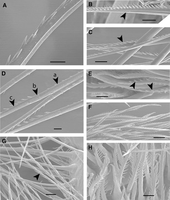
Certain setal types occur in particular species or taxonomic groups (see Table 4). The two Ibla spp. are characterized by Type 2a setae (Table 3; Figure 6A). Those setae with two rows of denticles that are present are Type 2b3 (Table 3; Figures 6C & 8F) in contrast to other cirripedes where Type 2c1 (Table 3; Figures 6D & 9A) is the most common type. Heteralepas microstoma is unique in having Type 2a9 setae (Figure 8D), and among the pedunculates examined in having setulate, Type 3a, and plumodenticulate, Type 4, setae on the exopodite of cirrus 1. The three members of the Eolepadidae examined, L. longa, N. rapanuii and V. parensis, are characterized by 5 setal types: Types 1a2, 1b3, 2a6, 2c2 and 2c3 (Figures 2H, 2G, 7G, 9B & 9C). The Type 2c2 and 2c3 setae comprise the prominent field of anteriorly directed setae on the proximal podomeres of cirrus 1 and cirrus 2. The Type 2c3 setae are up to 1 mm long. These setae are overlain on the median face of the ramus by the Type 2c2 setae.
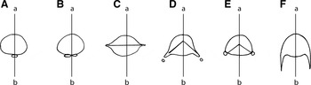
Diagram illustrating the alignment of the rows of denticles on the setal shaft in Types 2a, 2b, 2c and 2d. The line (a)–(b) represents the median plane of the setal shaft. (A) Types 2a1–2a6; (B) Types 2a7–2a9; (C) Types 2b1–2b5; (D) Types 2c1–2c3; (E) Type 2c4; (F) Types 2d1–2d4.

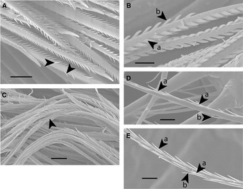
Examples of Types 2a1–2a6 setae. (A) Tetraclitella purpurascens, part of the shaft of a Type 2a1 seta from the anterior face of a ramus of cirrus 5. Note the small denticles in a single row; (B) Lepas anatifera, part of the shaft of Type 2a2 setae on the anterior face of a ramus of cirrus 4. Note the long slender needle shaped denticles in a single row; (C) Balanus nubilus, Type 2a3 setae on the anterior face of the rami of cirrus 4. Note the single row of regularly arranged needle shaped denticles; (D) Tetraclitella purpurascens, Type 2a3 setae with supplementary denticles (arrow) on the exopodite of cirrus 1; (E) Balanus crenatus, Type 2a4 setae (arrows) on the anterior face of a ramus of cirrus 3. Note the well-spaced distally directed needle-shaped denticles; (F) Ibla cumingii, Type 2a5 setae (arrows) on the proximal region of the endopodite of cirrus 1; (G) Neolepas rapanuii, Type 2a6 setae (arrow) on the exopodite of cirrus 2. Scale bars: (A) 10 µm; (B) 20 µm; (C) 20 µm; (D) 10 µm; (E) 10 µm; (F) 10 µm; (G) 10 µm.

Examples of Type 2a7, 2a8 and 2a9 setae, and Type 2b setae. (A) Tesseropora rosea, part of the shaft of a Type 2a7 seta on the anterior face of a ramus of cirrus 4. Note the irregular double row of denticles; (B) Chirona hameri, part of the shaft of a Type 2a8 seta (arrow) on the anterior face of a ramus of cirrus 4. Note the double row of slender denticles; (C) Scalpellum scalpellum, Type 2a8 setae (arrow) on the anterior face of a ramus of cirrus 4; (D) Heteralepas microstoma, Type 2a9 setae on the anterior face of a ramus of cirrus 5: (a) proximal region of seta, (b) annulus and (c) distal region of seta; (E) Ibla quadrivalvis, Type 2b2 setae on the exopodite of cirrus 1. Arrows indicate the stout denticles; (F) Ibla cumingii, Type 2b3 bipectinate setae on the exopodite of cirrus 1 with two rows of slender needle shaped denticles; (G) Balanus crenatus, Type 2b4 seta (arrow) on the anterior face of a ramus of cirrus 4. Note the long, slender, distally directed denticles; (H) Chthamalus antennatus, Type 2b5 setae on proximal segments of the exopodite of cirrus 1. Note the long flexible denticles. Scale bars: (A) 20 µm; (B) 10 µm; (C) 20 µm; (D) 20 µm; (E) 20 µm; (E) 10 µm; (F) 20 µm; (G) 20 µm; (H) 10 µm.

Examples of Type 2c setae. (A) Pollicipes pollicipes, Type 2c1 setae on the exopodite of cirrus 1. Arrows indicate randomly arranged supplementary denticles; (B) Leucolepas longa, part of the shaft of a Type 2c2 seta from a ramus of cirrus 2. Note the 2 rows of stout denticles (a) and the regularly arranged supplementary denticles (b); (C) Neolepas rapanuii, distal regions of Type 2c3 setae on proximal podomeres of the endopodite of cirrus 1. The arrow indicates the flexible shaft with flexible denticles; (D) Balanus crenatus, Type 2c4 seta on the anterior face of a ramus of cirrus 4: (a) long, slender, distally directed denticles on either side of the face of the seta and (b) supplementary denticle on the opposite face of the seta; (E) Perforatus perforatus, part of the shaft of a Type 2c4 seta on the anterior face of a ramus of cirrus 6: (a & b) as for (D). Scale bars: (A) 20 µm; (B) 5 µm; (C) 10 µm; (D) 10 µm; (E) 10 µm.

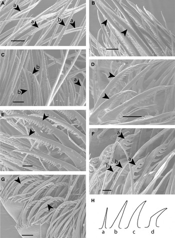
Pollicipes pollicipes, C. polymerus, O. angulosa, T. rosea, T. squamosa and T. purpurascens have a number of setal types in common. They all have Type 2a7 or 2a1 (Figures 8A & 7A) on cirri 3–6 comprising the cirral net (Type 2a1 may have arisen by simplification of Type 2a7). P. pollicipes, C. polymerus and O. angulosa all have Type 2f1 setae (Figure 11D) on the posterior distal borders of the podomeres of the rami of cirri 3–6. All six species have Type 2f2 setae (Figure 11E) comprising the setal tufts located on the anterior faces of the rami between the bases of the captorial setae. P. pollicipes, C. polymerus, O. angulosa and T. rosea all have the robust Type 2d4 setae (Figure 10F–H) in various locations on cirri 1–3. P. pollicipes also has Type 2d2 setae (Figure 10C, H) on the exopodite of cirrus 3. Type 2d2 setae are found on the endopodite of cirrus 3 in T. purpurascens (Figure 10D), T. squamosa and M. tintinnabulum.
Examples of Type 2d setae. (A) Ibla cumingii, (a) Type 2d1 setae on the exopodite of cirrus 2 and (b) Type 2a2 seta; (B) Chthamalus montagui, Type 2d1 setae on distal podomeres of the exopodite of cirrus 2; (C) Pollicipes pollicipes, setae on the exopodite of cirrus 3: (a) Type 2d2 and (b) Type 2d4; (D) Tetraclitella purpurascens, Type 2d2 setae (arrows) on the rami of cirrus 3; (E) Chthamalus stellatus, Type 2d3 setae on the terminal podomere of the exopodite of cirrus 2; (F) Octomeris angulosa, setae on the distal podomeres of the exopodite of cirrus 2: (a) Type 2d4 and (b) Type 2d1; (G) Tesseropora rosea, Type 2d4 setae (arrows) on the endopodite of cirrus 3; (H) diagram illustrating the morphology of the denticles of Type 2d setae: (a) Type 2d1, (b) Type 2d2, (c) Type 2d3 and (d) Type 2d4. Scale bars: (A) 20 µm; (B) 10 µm; (C) 50 µm; (D) 50 µm; (E) 10 µm; (F) 20 µm; (G) 50 µm.

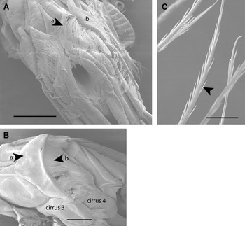
Several setal types are absent from pedunculates but are found in balanomorphs. With the exception of H. microstoma setulate setae were not found in pedunculates but were found in most balanomorphs, the exceptions being Eochionelasmus ohtai, C. polymerus, O. angulosa and T. purpurascens. However, whereas the tetraclitids and the balanids have Type 3b setae (Figure 3A), the Chthamalus spp. have Type 3a (Figures 3B & 12A). In those balanomorphs that have setulate setae they are universally found fringing the posterior border of the coxopodite of cirrus 1. In the following balanids, Balanus spp., Semibalanus spp., Perforatus perforatus and Austrominius modestus there is a field of very long, flexible setulate setae arising from the base of cirrus 3 (Figure 14A) which in the living animal would hang down into the mantle cavity. This setal field is not present in Chirona hameri. Megabalanus psittacus has a flange with setulate setae at the base of cirrus 3 but it is not such an elaborate shape as the crescentic structure seen in M. tintinnabulum (Figure 14B) (see Darwin Reference Darwin1854, plate XXV, figure 1). Type 1b5 setae (Figure 5E) were also absent from the pedunculates with the exception of P. pollicipes. They were found in all the balanomorphs examined with the exception of Balanus nubilus (but see Høeg et al., Reference Høeg, Karnick and Frølander1994), M. psittacus and M. tintinnabulum. Another setal type, Type 2e3 (Figures 3H & 11C), was found on the median face of each mandibular palp in the balanoids and coronuloids but observations on the pedunculates were not adequate to determine whether or not this setal type was present. However Høeg et al. (Reference Høeg, Karnick and Frølander1994) found this type in Pollicipes cornucopia (= P. pollicipes) but not in L. anatifera. In the same location in Chthamalus stellatus there is a field of specialized setae but of a different type, Type 2f3 (Figure 11F).
Examples of Type 2e and Type 2f setae. (A) Conchoderma auritum, Type 2e1 setae fringing maxilla 2; (B) Heteralepas microstoma, Type 2e2 setae on maxilla 2; (C) Semibalanus cariosus, part of the distal region of a Type 2e3 seta from the median face of the mandibular palp. Note the wrinkled setal shaft (arrow); (D) Pollicipes pollicipes, Type 2f1 setae on the posterior distal border of a ramus segment of cirrus 2: (a) bifid setal tip with pore and (b) ‘collar’ of denticles; (E) Tesseropora rosea, Type 2f2 setae between the bases of the captorial setae on the anterior face of the rami of cirrus 4: (a) bifid setal tip with pore and (b) sparse denticles on setal shaft; (F) Chthamalus stellatus, (a) Type 2f3 seta on the median border of the mandibular palp and (b) pore in setal tip. Scale bars: (A) 20 µm; (B) 20 µm; (C) 10 µm; (D)10 µm; (E) 10 µm; (F)10 µm.

Examples of Type 3 setulate setae (A & B), Type 4 plumodenticulate setae (C) and Type 5 setae, aesthetascs (D–G). (A) Chthamalus montagui, Type 3a2 setae (arrows) on posterior border of the coxopodite of cirrus 2; (B) Balanus nubilus, Type 3b1 setae arising from the posterior border of the coxopodite of cirrus 1, showing the origin of the setules distal to the annulus (arrow). The setules are located on one side of the shaft; (C) Chthamalus montagui, Type 4, setae along the posterior border of the coxopodite of cirrus I: (a) setulate section of shaft and (b) denticulate section of shaft; (D) Tetraclitella purpurascens, Type 5 setae (a) arising from the posterior surface of the endopodite of cirrus 2, together with Type 2c1 setae with supplementary denticles (b). (c) Socket of Type 5 seta; (E) Tesseropora rosea, Type 5 setae (a) arising from the posterior distal border of a podomere of a ramus of cirrus 4: (b) associated Type 1b1 setae; (F) Dosima fascicularis, part of distal setal shaft. Note the delicate wrinkled cuticle of the shaft wall (arrow); (G) D. fascicularis, tip of Type 5 seta showing the terminal pore. Scale bars: (A) 10 µm; (B) 20 µm; (C) 20 µm; (D) 20 µm; (E) 20 µm; (F) 2 µm; (G) 2 µm.
(A) Ibla quadrivalvis, Type 2b3 setae arising from the prosoma cuticle. Note the setal socket (arrow); (B) Ibla quadrivalvis, ventral cone (v.c.) between cirri 1 and 2 behind the oral cone: (a) setae on ventral cone, (b) location of gland pores on the lateral face of the ventral cone and (c) opening of the maxillary gland at the base of maxilla 2 (Mx 2); (C) Leucolepas longa, ventral cone with pores of sub-oesophageal glands; (D) Leucolepas longa, pores of the sub-oesophageal glands (arrow) on the ventral cone; (E) Chthamalus montagui, long cuticular processes (arrows) arising from the prosoma cuticle. Scale bars: (A) 20 µm; (B) 100 µm; (C) 100 µm; (D) 20 µm; (E) 10 µm.

(A) Balanus crenatus, posterior view of thorax. The arrows (a) indicate the very long setulate setae extending from the base of cirrus 3 (b); (B) Megabalanus tintinnabulum, dorso-lateral view of thorax showing extension (a) from base of cirrus 3 bearing a fringe of setulate setae (b); (C) Balanus balanus, unidenticulate distal region of a Type 2c4 seta on the anterior face of the ramus of cirrus 4. Arrow indicates supplementary denticle. Scale bars: (A) 500 µm; (B) 1 mm; (C) 20 µm.

DISCUSSION
The classification proposed here, provides a framework for describing setal structure in cirripedes. Assignment to a type, within a three level scheme, is based on the presence or absence of denticles and setules on the setal shaft, their disposition on the setal shaft and their characteristics. A difficulty with the scheme is in finding sufficient names to describe the great variety of setal types observed. For this reason code numbers and letters are used which, though not directly descriptive, can be related to photographic or diagrammatic images. The system put forward here can be expanded and modified to accommodate more types as revealed by the study of further species.
Comparison with previous descriptions of setal morphology in cirripedes
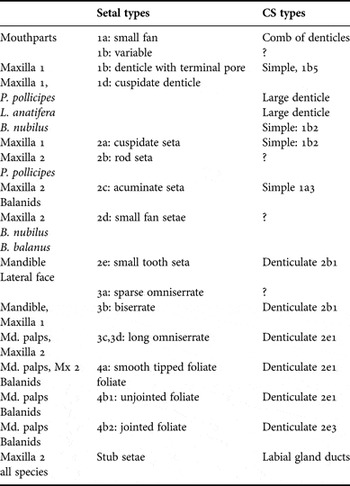
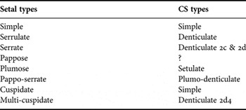
Most of the species studied by Anderson & Anderson (Reference Anderson and Anderson1985), Hunt & Alexander (Reference Hunt and Alexander1991) and Høeg et al. (Reference Høeg, Karnick and Frølander1994) were examined in the present study. Thus, it is possible to compare the setal types described by these authors with those described in the present study (see Tables, pp. 19, 20).
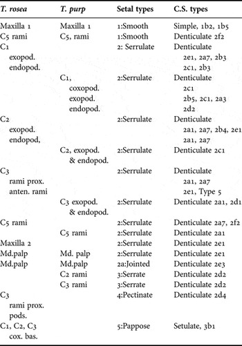
Anderson & Anderson (Reference Anderson and Anderson1985) compared the functional morphology of Tesseropora rosea and Tetraclitella purpurascens. Using optical microscopy they described five types of setae on the limbs and mouthparts. They divided their Type 2 serrulate setae into five sub-types but 9 setal types were seen in those setal fields described as serrulate. Table 5 equates the setal types described in various locations with the setal types seen in the same locations in this study.
Setal types in Tesseropora rosea and Tetraclitella purpurascens described by Anderson & Anderson (1985) compared with the current study

anten., antenniform; bas., basipodite; C1, cirrus 1; cox., coxopodite; C.S., current study; Md., mandibular; pods., podomeres; prox., proximal.
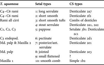
Hunt & Alexander (Reference Hunt and Alexander1991) studying feeding in Tetraclita squamosa described 10 setal categories based on those of Anderson & Anderson (Reference Anderson and Anderson1985). It was not possible to equate all their types to those described here but where comparisons were possible they are set out in Table 6. Høeg et al. (Reference Høeg, Karnick and Frølander1994) used SEM to describe the major denticles and setae on the mouthparts of three pedunculate species and three balanomorph species. They devised a classification scheme with six categories. Category 1, denticles, are not setae as defined here or by previous authors but some of their ‘denticles’ probably are setae. Category 6, stub setae, are not setae either but the ducts of labial glands opening on the second maxillae (Walley, unpublished data). The types and sub-types observed by Høeg et al. (Reference Høeg, Karnick and Frølander1994) are compared with those described here in Table 7.
Setal types in Tetraclita squamosa described by Hunt & Alexander (Reference Hunt and Alexander1991) compared with the current study.

ant., anterior; C., cirrus; CS, current study; Md., mandibular.
Setal types described by Høeg et al. (1994) compared with the current study.

CS, current study; Md., mandibular.
Chan et al. (Reference Chan, Garm and Høeg2008) followed Garm (Reference Garm2004) and used 8 broad categories to describe the setae that they observed in Ibla cumingii, Octolasmis warwickii, Capitulum mitella, Pollicipes polymerus, Tetraclita japonica japonica and Megabalanus volcano. Ibla cumingii was examined in the current study. With the exception of O. warwickii and C. mitella, species closely related to the remaining three species were also studied. Thus it is possible to suggest how their categories equate to those described here (see Table 8). They concluded that the development of the balanomorph form was accompanied by a greater variety of setae and more complex setation patterns, associated with changes in feeding strategy. The observations made in the current study broadly support this conclusion. They assumed that setal functions seen in decapods hold true for cirripedes, for example ‘Pappose setae can be used for water current generation and filter-feeding purposes’ and they speculated that the plumose setae on the coxopodite of cirrus 1 and cirrus 2 in Tetraclita and Megabalanus could serve in microfiltration. It seems unlikely that either of these suggestions is correct. The setulate (= plumose) setae are usually positioned where they could not be reached by the maxillipeds to transfer food particles to the mouthparts. The generation of water currents seems to result from movements of the whole body and limbs (Crisp & Southward, Reference Crisp and Southward1961). It is much more likely that the plumose/pappose (=setulate) setae in the balanomorphs are involved in the direction of the currents within the mantle cavity.
Comparison of setal types described by Chan et al. (Reference Chan, Garm and Høeg2008) with the current study.

CS, current study.
Comparison of cirripede and decapod setal classifications
Garm (Reference Garm2004) is the most recent of a series of authors (Thomas, Reference Thomas1970; Drach & Jacques, Reference Drach and Jacques1977; Factor, Reference Factor1978; Pohle & Telford, Reference Pohle and Telford1981; Watling, Reference Watling, Felgenhauer, Watling and Thistle1989; Lavalli & Factor, Reference Lavalli and Factor1992) to tackle the problem of devising a system for the classification of crustacean and more particularly decapod setae. From SEM studies of setae on the mouthparts of seven decapod species, covering a range of families, Garm (Reference Garm2004) defined seven types which he considered to provide an adequate framework for describing the setae that he observed.
There are many points of agreement between the setal groups defined by Garm (Reference Garm2004) and those described in the present study. In both cases the groups are defined with reference to the nature and arrangement of the outgrowths from the setal shaft. The main point of disagreement is in defining when an outgrowth is a denticle and not a setule. Thomas (Reference Thomas1970) restricted the use of the term ‘setule’ to the very long slender outgrowths seen in plumose, pappose and plumodenticulate setae. Thomas described setules as arising from indentations in the setal shaft but does not use this feature—basal articulation—as defining a setule. He uses the term ‘denticle’ for the other types of cuticular outgrowths referring to setae with such structures as denticulate. Pohle & Telford (Reference Pohle and Telford1981) used the terms setule and denticule to describe the two most common cuticular outgrowths from the setal shaft. They clearly differentiated between the long, slender, setules and the shorter tooth or needle-shaped denticules. They state ‘While denticule-like structures are direct and continuous extensions from the shaft wall, setules are articulated at their bases’. Examining their images it appears that the ‘articulation’ takes the form of an indentation in the shaft surface from which the setule arises. As successive authors described setal morphology in decapods, basal articulation became the defining characteristic of a setule rather than its long, slender shape. Watling (Reference Watling, Felgenhauer, Watling and Thistle1989, figure 1b) shows setules as slender outgrowths from the setal shaft ‘of uniform width from base to tip, and forming an articulated or flexible junction with the shaft’. However, he did not figure or describe what he understood by the term ‘flexible junction’. Garm (Reference Garm2004) accepted Watling's definition and his figure 4G shows a seta bearing two rows of needle-shaped processes which is said to show a transition from setules to denticles between annulus and setal tip although the structures and their origin from the setal shaft appear the same throughout. Figure 8B in Garm (Reference Garm2004) shows part of a setal shaft with tapering denticles on one side and ‘setules’ with serrated borders on the opposite side. But the ‘setules’ show no evidence of articulation—either a socket (as illustrated in Figure 4E) or a ‘flexible junction’. Chan et al. (Reference Chan, Garm and Høeg2008) adopted Garm's definition of a setule and used his setal categories in their study of setal morphology and cirral setation in cirripedes. They used the term ‘setule’ for ‘any ornament on a seta that has an articulated base irrespective of its morphology’. However, the current study has shown that very few setae in cirripedes have cuticular outgrowths that are articulated at the base. The only setae that were found to have articulated outgrowths were setulate setae in the Chthamalus species and Heteralepas microstoma. There seems no logical reason for using basal articulation as an over-riding defining feature and so the original definition of the term setule as a very long and very slender outgrowth from the setal shaft is used here.
The ‘serrulate’ category defined by Chan et al. (Reference Chan, Garm and Høeg2008), is described as ‘slim with a naked proximal part but with small setules distally which can be arranged in rows (normally three) or scattered randomly along the shaft’. Observations in the current study do not support this interpretation. Most of the cuticular outgrowths arising from the setal shafts in those setal types encompassed by the ‘serrulate’ category arise seamlessly from the setal shaft without any discontinuity and are described as denticles in this paper. Furthermore, in cirripedes two rows of denticles rather than three are the norm.
The seven setal categories described by Garm (Reference Garm2004) were: pappose, plumose, serrulate, serrate, papposerrate, simple and cuspidate. With regard to basic setal morphology Garm's study confirms that setal articulation in the Decapoda is usually infra-cuticular, only the plumose setae have supra-cuticular articulation. In the Cirripedia, however, setal articulation is usually supra-cuticular. In both groups there are robust, rigid, setae notably on the first maxilla, where the basal attachment does not allow movement relative to the surrounding cuticle. A clearly marked annulus was observed on setal shafts in both groups. Although decapod setae tend to be much more elaborate than cirripede setae, presumably because of the more complex mechanical functions involved in feeding and grooming, it is possible to compare Garm's setal types with the groups described in the present study. The pappose and plumose setae equate to the setulate Group 3. The serrulate setae are said to have three rows of small setules distal to the annulus but Garm (Reference Garm2004) also states ‘the articulation with the shaft is often difficult to detect’ so perhaps the serrulate setae should be regarded as denticulate in which case the serrulate and serrate setae equate to the denticulate Group 2. The papposerrate equate to the plumodenticulate Group 4. The term ‘plumodenticulate’ is used in the current study because it is a long established and well understood term as shown in Table 2 of Garm (Reference Garm2004). Both the simple and cuspidate setae equate to the Group 1 simple setae. There is no equivalent to the cirripede Group 5. In cirripedes there are no functional antennae or antennules in the adults which in other Crustacea carry the aesthetascs. However, it seems highly likely that sensory setae fulfilling the same functions as aesthetascs will be found. The setae comprising Group 5 have features in common with those described for aesthetascs in other crustaceans (Derby, Reference Derby, Felgenhauer, Watling and Thistle1989), for example, a thin wall to the setal shaft and in some cases a terminal pore. The Group 5/Type 5 setae are located in positions on the limbs where they could pick up chemical signals during feeding (see Results above) or those on the penis could detect pheromones during the search for receptive individuals prior to insemination. It is suggested that these unusual setae are the functional equivalent of aesthetascs.
Some of the serrulate and serrate setae described by Garm (Reference Garm2004) as having three rows of ‘setules’ or as having ‘setules’ on the opposite side of the setal shaft from the two rows of denticles (see figure 8B in his paper) could be interpreted as being equivalent to the setae with supplementary denticles seen in cirripedes. It would be possible to use the scheme put forward here to describe decapod setae, for example, the serrate seta (figure 8A in his paper) could be placed in Type 2c1 in the current study. Garm also makes the point that some types, such as the simple setae, are based on shared morphological characteristics that have been convergently acquired through the loss of setules/denticles which also seems to be the case in cirripedes.
Setal morphology, cirripede phylogeny and taxonomy
One of the most important developments in cirripede phylogeny was the adoption of the balanomorph form. There are several aspects of setal morphology that are associated with this change.
RETENTION OF SETAL TYPES
Darwin (Reference Darwin1854) suggested that the change from the pedunculate to the sessile barnacles could be envisaged by comparing the anatomy, shell structure, and mode of shell growth of Pollicipes and Catophragmus (=Catomerus). This was further explored by Anderson (Reference Anderson1983) who compared the anatomy and functional morphology of Catomerus polymerus and the pollicipoid scalpellid Calantica villosa. Anderson (Reference Anderson1983) stated ‘Functional studies of C. polymerus and C. villosa have now shown that an evolution of catophragmid balanomorphs from calanticine scalpellids is functionally feasible, upholding Darwin's proposal. C. polymerus retains the scalpellid mode of planktivorous extension feeding……The basic adaptive significance of the foreshortened form and flattened operculum of C. polymerus lies in allowing a species with this mode of feeding to inhabit a high energy intertidal environment.’ Both authors emphasized the essential similarities between the soft body anatomy of Catomerus and Pollicipes/Calantica. They both drew attention to the characteristic setation of the posterior four pairs of cirri in Catomerus where each podomere carries several pairs of long antero-lateral setae and has a bunch of short forwardly projecting setae between them on the anterior face of the podomere, a feature also seen in Calantica (and Pollicipes). Darwin (Reference Darwin1854) also pointed out that the anatomy and morphology of Octomeris could be derived from that of Catophragmus which also exhibits this setal arrangement on cirri 3–6. The current study has shown that the similarity extends beyond the disposition of the setae to the setal types (see Table 5): the long antero-lateral setae on the anterior face of the podomeres of cirri 4–6 in Pollicipes pollicipes, C. polymerus and also Octomeris angulosa are Type 2a7 and the setae comprising the dense bunch of shorter setae between the bases of the Type 2a7 setae are Type 2f2. All three species have type 2f1 setae (Figure 11D) on the posterior distal borders of the podomeres of the rami of cirri 3–6. Both setal types have bifid tips (Figures 4A & 11D, E) which are also seen on some of the other setal types found in these species and which were observed in T. japonica and M. volcano by Chan et al. (Reference Chan, Garm and Høeg2008). There is further setal similarity between these three species in that Type 2d4, bidentate setae are found on the rami of the first three cirri. So in the transition from the pedunculate to the sessile form, setal type, as well as setal disposition on the cirri, is retained.
THE DEVELOPMENT OF SETULATE SETAE IN BALANOMORPH CIRRIPEDES
The development of setulate setae appears to be correlated with the adoption of the balanomorph form. In the balanids, chthamalids and tetraclitids (except for Tetraclitella) examined in this study the posterior border of the coxopodite of cirrus 1 is fringed with setulate setae: Type 3a in the chthamalids and Type 3b in the tetraclitids and balanids (Table 4). It has been suggested that setulate setae are involved in microfeeding; however, they are usually (but not always) positioned where they could not be reached by the maxillipeds to transfer food particles to the mouthparts. It seems much more probable that these linear fields of flexible setulate setae are involved in keeping the body surface free of epizoites and in the direction of water currents through the mantle cavity for respiration and the oxygenation of developing embryos. Such currents could then be exploited for micro-feeding by those species showing a ‘strong pumping beat’ (Anderson, Reference Anderson1981; Anderson & Southward, Reference Anderson, Southward and Southward1987).
SETAL TYPES ON THE MOUTHPARTS OF BALANOMORPH CIRRIPEDES
If we consider the mouthparts there are several morphological and setal characteristics that seem to be associated with the evolution of the balanomorph form. Type 2e3 setae were found on the mandibular palps of all the balanoids and tetraclitids but were absent from the pedunculates with the exception of P. pollicipes. In Chthamalus stellatus these setae are replaced by a field of another specialized setal type, Type 2f3. Type 1b5 setae were found on maxilla 1 in all the balanomorphs examined, except for Balanus nubilus, Megabalanus psittacus and Megabalanus tintinnabulum. However Høeg et al. (Reference Høeg, Karnick and Frølander1994) reported, what appear to be setae of this type from B. nubilus, arranged in small groups on the distal margin of the first maxilla. Usually they are simple setae with a terminal pore (Figure 5E) but the Type 1b5 setae in the Chthamalus species have an asymmetrical cuticular extension beyond the terminal pore (Figure 5F). These setae are probably chemosensory.
The second maxillae of most lepadomorphs are simple structures bearing setae and the pores of the labial glands. Pollicipes has a dorsal notch and a second maxilla of almost identical structure is seen in Catomerus. This dorsal notch is probably the forerunner of the development of a distal mobile maxillary palp as described in Tetraclita squamosa by Hunt & Alexander (Reference Hunt and Alexander1991) and which is particularly well developed in the balanids, P. perforatus (Anderson, Reference Anderson1981), B. nubilus and M. psittacus. In the balanids examined in this study the distal surface of the maxillary palps were clothed with simple, often flexible, type 1a3 setae. The two exceptions were Chirona hameri and M. tintinnabulum, both of which have denticulate, Type 2e1, setae in this position as seen in the tetraclitids.
SETAL MORPHOLOGY AND CIRRIPEDE TAXONOMY
Classical cirripede taxonomy was based on shell morphology and to a lesser extent body morphology including mouthparts and the characteristic distribution patterns of setae on the limbs, but not much attention was paid to setal morphology. The emphasis on shell morphology enables comparisons to be made with palaeontological evidence allowing deductions to be made about the evolutionary history of modern species and groups. However, Darwin (Reference Darwin1854) expressed the opinion that there were dangers in relying too much on shell structure in working out relationships and said that more attention should be paid to soft body morphology. The setal morphologies revealed in this study in most cases reflect the accepted taxonomic groups but there are anomalies in the Balanoidea (see below).
Cases of convergent evolution are also apparent for example in the Group 1 simple setae (see above) and in Group 2, denticulate setae where robust, Type 2d3 and Type 2d4, setae are found in a number of cirripede species inhabiting locations exposed to severe wave action but the morphology of the denticles in different genera is distinct. Another example of convergent or perhaps parallel evolution is provided by the setulate setae fringing the posterior border of the coxopodite of cirrus 1 in balanomorphs: in the chthamalids these fringing setae are Type 3a whereas in the balanids and tetraclitids they are Type 3b. This structural difference suggests the possibility of two independent lines of evolution for these setae.
If we consider the balanoid spp. examined in this study and compare the setae comprising the cirral net, on the anterior face of the rami of cirri 4–6, C. hameri with Type 2a8 setae and M. tintinnabulum with Type 2a1 setae (also seen in M. volcano Chan et al., Reference Chan, Garm and Høeg2008) are clearly distinct from the other balanoid species. Megabalanus psittacus, however, has Type 2a3 setae as seen in B. nubilus (Figure 7C). The Balanus species (with the exception of B. nubilus), the Semibalanus species and Perforatus perforatus, all have Type 2c4 with characteristic long, slender, distally directed denticles. Austrominius (Elminius) modestus has Type 2a4 setae with a single row of the same long slender denticles as Type 2c4 setae, which could be interpreted as being derived from the Type 2c4 by simplification. Indeed in A. modestus further simplification is observed in that some of the setae of the cirral net have no denticles and are Type 1a1. Balanus balanus is particularly interesting in that the distalmost region of the longest setae on the rami of cirri 4–6 has a single row of closely set denticles along the anterior face of the shaft with more widely spaced supplementary denticles on the posterior face of the shaft (Figure 14C), suggesting how the denticular layout seen in the Type 2a3 setae of B. nubilus (Figure 7C) and M. psittacus might have arisen. The marked difference between the setae of the captorial cirri in B. nubilus and the other Balanus spp. and their resemblance to those of M. psittacus suggests that perhaps B. nubilus should not be included in the genus Balanus. However M. tintinnabulum has setae on cirri 4–6 which clearly resemble those seen in Tetraclita squamosa and are of the same type as those of M. volcano (Chan et al., Reference Chan, Garm and Høeg2008). The morphology of cirri 1–3 and the oral cone in M. tintinnabulum also bear a striking resemblance to T. squamosa. The setae on the distal region of the maxillary palps are denticulate (see above)—not a balanid feature but typical of the tetraclitids. Perhaps it might be more appropriate to include M. tintinnabulum and M. volcano with the tetraclitids?
Recent studies have tested the accepted orthodoxies of cirripede classical taxonomy by applying the techniques of molecular biology. Pérez-Losada et al. (Reference Perez-Losada, Harp, Høeg, Achituv, Jones, Watanabe and Crandall2008) reviewed previous attempts at elucidating barnacle phylogeny and then applied cladistic techniques to both molecular and morphological data from a very wide range of cirripedes. They used DNA sequences from three nuclear genes and 44 morphological characters collected from 76 thoracican and 15 rhizocephalan species, but setal morphology was not used in the range of morphological characters subjected to cladistic analysis. Of the 76 thoracican species studied 24 were examined in the current study. Analysis of the data led them to conclude that the Iblomorpha were a monophyletic group which diverged very early in cirripede evolution. The current study has also revealed a number of characteristics that set the two Ibla spp. apart from the rest of the cirripedes studied, for example the preponderance of unidenticulate setal types (2a1, 2a2 and 2a3) and the presence of setae on the prosoma and thorax. Those bidenticulate setae that are present are Type 2b3 in contrast to the other cirripedes examined where Type 2c1 is predominant.
Pérez-Losada et al. (2008) concluded that the plateless condition of the Heteralepadomorpha was not a primitive feature but had probably arisen at least twice within the Lepadomorpha. In the current study H. microstoma was shown to differ from all the other species examined in having Type 2a9 denticulate setae and was unique among the pedunculates in having setulate setae, Type 3a1, and plumodenticulate setae, Type 4. It would be interesting to see whether it shares these characteristics with other Heteralepas species and with Paralepas.
Analyses using the DNA data, the morphological characters and a combination of the two data sets, all resulted in the deep-sea vent scalpellomorphs Neolepas, Leucolepas and Vulcanolepas being clustered together. This is certainly reflected in their setal morphology. Neolepas rapanuii, Leucolepas longa and Vulcanolepas parensis are characterized by 5 setal types: 1a2, 1b3, 2a6, 2c2 and 2c3 not seen in any other species examined.
On the basis of molecular evidence Pérez-Losada et al. (2008) separated Calantica from Pollicipes. Representatives of both genera have been used as models when considering the transition from the pedunculate to the sessile form (Darwin, Reference Darwin1854; Anderson, Reference Anderson1983). There can be little doubt that the ancestor of the balanomorph cirripedes was closely related to a pedunculate scalpellid ancestor closely resembling these genera. Both Calantica and Pollicipes have a similar arrangement of setae on the anterior faces of the rami of cirri 4–6: a dense tuft of slender setae between paired long, lateral setae. This arrangement is also seen in Catomerus. However the paired long lateral setae, in Calantica spinosa (plate 1C in Foster, Reference Foster1978) appear to be Type 2a1 with a single row of well-spaced small denticules whereas P. pollicipes has Type 2a7 with a more closely spaced irregular double row of small denticules, a characteristic it shares with C. polymerus. Both P. pollicipes and C. polymerus have slender Type 2f2 setae comprising the anterior setal tuft. The nature of the setae in the setal tuft in Calantica is not known. If we consider the stout dentate setae seen on cirri 1, 2 and 3 in Pollicipes, and on cirrus 2 in Calantica and Catomerus, P. pollicipes has both Type 2d2 and 2d4 on the rami of cirrus 3. Catomerus has Type 2d4 on the terminal podomeres of the rami of cirrus 2. It is not clear from the images in Foster (Reference Foster1978) to which Type the stout dentate setae seen in C. spinosa belong. Thus in its setal morphology C. polymerus may have a greater affinity with Pollicipes than with Calantica.
Within the Balanomorpha the data presented by Pérez-Losada et al. (2008) seems to suggest a split, grouping Catomerus with the chthamalids and grouping the tetraclitids together with the balanids. This split is mirrored in the current study by some but not all features of setal morphology. For example, the chthamalids have Type 3a setulate setae, whereas the tetraclitids and the balanids have Type 3b setulate setae. Similarly all the balanomorphs studied have specialized setae along the median face of the mandibular palps, but while the tetraclitids and the balanids have Type 2e3 (as does Catomerus), C. stellatus has Type 2f3. When we consider the setae of cirri 4–6 comprising the ‘cirral net’ the situation is different. Catomerus, the chthamalids and the tetraclitids all have setae with relatively small denticles along one side of the setal shaft (Type 2a) (a characteristic also possessed by M. tintinnabulum and M. volcano see above), whereas the majority of the balanids (as currently defined) examined have setae with very characteristic long slender needle-shaped denticules directed distally along the setal shaft. The separation of A. modestus and Elminius kingii from the rest of the balanids is surprising: both species exhibit typical balanid features in particular the field of very long setulate setae arising from the base of cirrus 3 and the morphology of the setae on cirri 4–6.
To conclude, the five Groups of setal types described here are possibly functional groupings as suggested by Garm (Reference Garm2004). Study of the more detailed setal morphology reveals characteristics that are useful in elucidating relationships within crustacean groups such as the Decapoda or the Cirripedia rather than between groups. The demonstration in the current study that setal type can be correlated to some extent with taxonomic position within the Cirripedia supports the conclusion of Høeg et al. (Reference Høeg, Karnick and Frølander1994) that setal structure and distribution of different setal types ‘offer a new character set with likely phylogenetic significance at both lower and higher taxonomic levels’. However, the setal morphology described here would be of limited use in field taxonomy as most of the features described are beyond the limits of good resolution for optical microscopy.
ACKNOWLEDGEMENTS
This study was carried out in the School of Ocean Sciences, Bangor University. I am most grateful for permission to use equipment and materials over a number of years and particularly for the support of Dr Graham Walker and Dr Ian Lucas who introduced me to the techniques necessary for the use of the scanning electron microscope and the preparation of the specimens. I would also like to thank all those people who very kindly sent me specimens. David Roberts was most helpful in preparing the figures. I would like to thank my husband Ivor Rees for his endless patience during the long gestation of this paper and the referees for their critical and valuable comments.