INTRODUCTION
The Andaman and Nicobar Islands are endowed with invaluable diversity of marine fauna. Geographically, the islands are the summits of a submarine range extending from the Arakan Yomas of Burma in the north to the Sumatra in the south (Tikadar & Das, Reference Tikadar and Das1985). The groups include 572 islands, islets, reefs and isolated rocks and are situated within the latitude 6°45′N to 14°N and longitude 92°E to 94°E.
Sea slugs are the least studied fauna from Indian waters (Apte, Reference Apte2009) and this is also the case in the Andaman and Nicobar Islands. However, the taxonomy and ecology of opisthobranchs from these islands have recently received some attention (Ramakrishna et al., Reference Ramakrishna, Sreeraj, Sivaperuman and Kumar2010; Sreeraj et al., Reference Sreeraj, Rajan, Raguraman, Raghunathan, Rajkumar, Immanuel, Ramakrishna, Ramakrishna, Raghunathan and Sivaperuman2010, Reference Sreeraj, Sivaperuman and Raghunathan2012a, Reference Sreeraj, Sivaperuman and Raghunathanb). To date, a total of 162 species was reported from the Andaman Islands (Sreeraj et al., Reference Sreeraj, Sivaperuman and Raghunathan2011, Reference Sreeraj, Sivaperuman and Raghunathan2012a); 52 opisthobranch species were recorded from the Nicobar group, of which 11 were new records for Indian waters (Sreeraj et al., Reference Sreeraj, Sivaperuman and Raghunathan2012b). Current studies on nudibranchs reveal that the Andaman and Nicobar Islands manifest biodiversity of extraordinary range within a limited geographical area. These islands are a virtual bioreserve and represent a rich repository, unique both in terms of biodiversity and abundance.
The following characteristics are diagnostic for the genus Herviella: a single row of cerata in the right liver; anterior of foot expanded and rounded; rhinophores simple; jaws high anteriorly and narrow posteriorly, masticatory edge with a single row of denticles; penial stylet present; and female ducts serial with the spermatheca (i.e. it is formed by a swelling of the oviduct or vagina) (Baba, Reference Baba1966b).
MATERIALS AND METHODS
Field observation of an intertidal rock pool was carried out on 21 October 2012. A pair of specimens was collected from the shallow rock pool of Burmanallah, located at 11°33′29″N and 92°43′53″E on Port Blair, South Andaman (Figure 1).
Black round with spot showing the species collected from the Andaman Sea.

The specimens were photographed live with a Sony cyber shot 14 m pixel camera. A stereo zoom microscope (Model-ZEISS Axiovert 40 cfl) was utilized for preparation and photographs of the radula, jaw plates. The radula was extracted in 10% hyperchlorite and it was subjected to microscopic characterization. The specimens were preserved in 4% formalin after studying the morphological characters in live material. One specimen was submitted to the Zoological Survey of India for reference (Reg. No. ZSI-8077).
The immediate habitat of Herviella mietta was sea anemone and the area was encrusted with many types of coral, seaweeds, gastropods (chitons, limpets, cowries, etc.), cnidarians (anemone, hydroids, corals and soft coral), echinoderms (sea cucumbers, urchins and brittle stars), sponges, and other associated motile invertebrates, such as polychaetes.
RESULTS
SYSTEMATICS
Subclass OPISTHOBRANCHIA Milne-Edwards, 1848
Order NUDIBRANCHIA Blainville, 1814
Family GLAUCIDAE Gray, 1827
Genus Herviella Baba, Reference Baba1949
Herviella mietta Marcus & Burch, Reference Marcus and Burch1965
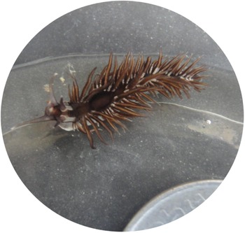
(Figure 2)
Herviella mietta.

SYNONYMY
Herviella mietta Marcus & Burch, Reference Marcus and Burch1965: 252, 253, figures 31–35 (western Pacific, Eniwetok Atoll).
DESCRIPTION
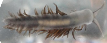
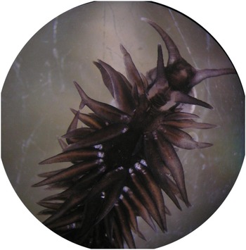
The morphological characters were as follows: body length 15 to 18 mm, body colour white ventrally (Figure 3) and black above, the pigmentation extending onto the cerata except at the tips, and on the head, oral tentacles (dorso-medially), and rhinophores, middle third of the rhinophores were black with a short pigmentation line below, carrot-shaped cerata that are predominantly black in colour and black pigment on the body, head with a black pattern, the rhinophores with a black band and the cerata clear with yellow digestive glands (Figure 4).
Herviella mietta (ventral side).

Anterior cerata and rhinophore (400×).

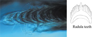
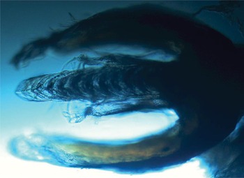
The uniseriate radula with 18 teeth, the central cusp short and pointed with 8–9 thin denticles on each side of the central cusp, the outermost denticles as long as the central cusp (Figures 5, 6 & 7).
Radula teeth (400×).

Radula covered with jaw plate (400×).

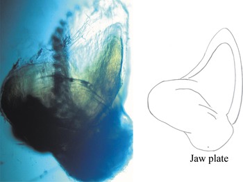
Jaw plate (400×).

DISCUSSION
The present finding is the first report of Herviella mietta from the Andaman and Nicobar Islands and a new biodiversity record for the Indian subcontinent. Herviella mietta was originally reported from Eniwetok Atoll, western Pacific (Marcus & Burch, Reference Marcus and Burch1965) and it is now known to be prevalent to the Pacific region (Young, Reference Young1967; Gosliner, Reference Gosliner1980; Johnson & Boucher, Reference Johnson and Boucher1983; Nakano, Reference Nakano2004; Coleman, Reference Coleman2008; Gosliner et al., Reference Gosliner, Behrens and Valdés2008). This is the most distinctive species of the genus; however, H. mietta and H. albida Baba, Reference Baba1966a have a similar pattern of black pigment on the head.
The distribution is restricted to the Indo-West Pacific Ocean (Marcus & Burch, Reference Marcus and Burch1965) and reported from Japan (Nakano, Reference Nakano2004). Ten species of Herviella are known from the western Pacific Ocean, namely H. exigua (Risbec, Reference Risbec1928), H. yatsui (Baba, Reference Baba1930), H. affinis Baba, Reference Baba1960, H. claror Burn, 1963, H. evelinae (Er. Marcus, Reference Marcus1965), H. mietta Marcus & Burch, Reference Marcus and Burch1965, H. albida Baba, Reference Baba1966a, H. burchi Burn, Reference Burn1967, Herviella africana Edmunds, 1970, and Herviella cloaca Rudman, 1980—these are summarized by Burn (Reference Burn1967) and Rosenberg (Reference Rosenberg2012).
The frequent large scale climatic oscillations and local disturbance on the reefs of this archipelago as well as other parts of the Indian subcontinent add pressures on sustaining biodiversity in this region. During the study of Lakshadweep Archipelago (a similar group of islands) a total of 60 species from 25 families were found; 52 species were new records to Lakshadweep and 40 were new records to the Indian coast (Apte, Reference Apte2009). A study of intertidal reefs of the Gulf of Kutch revealed 21 new local records and 13 records new to the Indian subcontinent. This report of the rare species Herviella mietta as new from this archipelago as well as new to India is further evidence of the Andaman and Nicobar Islands being a region of high opisthobranch diversity and this species needs to be explored further.
ACKNOWLEDGEMENTS
We thank Pondicherry University for providing the necessary facilities and Mr C.R. Sreeraj, Research Scholar, Zoological Survey of India, Andaman, for his help during this study.