Introduction
When an animal is presented with movement of the entire visual world (i.e., optic flow), an optomotor response is initiated in the direction of the flowfield, thereby minimizing any motion on the retina. This is known as the optokinetic response (OKR) and is one of several behaviors that help to stabilize the retinal image (Waespe & Henn, Reference Waespe and Henn1987). Without retinal image stabilization, both visual acuity and relative velocity discrimination suffer (Westheimer & McKee, Reference Westheimer and McKee1975; Nakayama, Reference Nakayama1985).
In all vertebrate classes, retinal-recipient nuclei in the pretectum and accessory optic system (AOS) are involved in the analysis of optic flow and the generation of OKR. In birds, these structures are the nucleus lentiformis mesencephali (LM) of the pretectum and the nucleus of the basal optic root (nBOR) of the AOS. The mammalian homologs of the LM and nBOR are, respectively, the nucleus of the optic tract (NOT) of the pretectum and the medial terminal nucleus (MTN) and lateral terminal nucleus of the AOS (Simpson, Reference Simpson1984; Fite, Reference Fite1985; McKenna & Wallman, Reference McKenna and Wallman1985a; Weber, Reference Weber1985; Simpson et al., Reference Simpson, Giolli and Blanks1988; Gamlin, Reference Gamlin2006; Giolli et al., Reference Giolli, Blanks and Lui2006). In several reports, the LM and nBOR have been erroneously grouped together as the AOS. Despite their similarity in function, Giolli et al. (Reference Giolli, Blanks and Lui2006) have emphasized that the optokinetic nuclei in the pretectum and AOS differ with respect to their connectivity and neurochemistry.
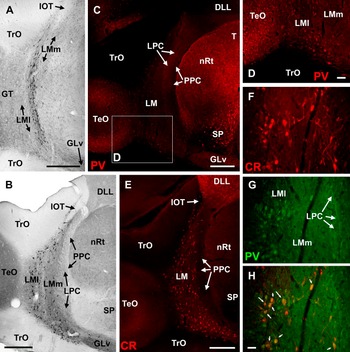
The nBOR and LM have similar projection pathways to the cerebellum (Cb). From both nuclei, there are direct mossy fiber projections to the posterior Cb (folia VI–IXcd) as well as indirect projections to the vestibulocerebellum (folia IXcd and X) via the medial column of the inferior olive (IO). Thus, there is a convergence of information from these two pathways in folium IXcd (Clarke, Reference Clarke1977; Brecha et al., Reference Brecha, Karten and Hunt1980; Gamlin & Cohen, Reference Gamlin and Cohen1988b; Arends & Voogd, Reference Arends and Voogd1989; Lau et al., Reference Lau, Glover, Linkenhoker and Wylie1998; Wylie et al., Reference Wylie, Winship and Glover1999; Crowder et al., Reference Crowder, Winship and Wylie2000; Wylie, Reference Wylie2001; Pakan & Wylie, Reference Pakan and Wylie2006). As shown in Fig. 1A, the IO-projecting LM cells are found caudally in the LM, localized in a thin strip along the border of the medial and lateral subnuclei (LMm and LMl, respectively; Gamlin & Cohen, Reference Gamlin and Cohen1988b; Wylie, Reference Wylie2001). These IO-projecting cells are fusiform neurons that are oriented dorsoventrally (Pakan et al., Reference Pakan, Krueger, Kelcher, Cooper, Todd and Wylie2006). The cerebellar-projecting neurons are large (Fig. 2) multipolar neurons found throughout LMl and LMm (Fig. 1B; Gamlin & Cohen, Reference Gamlin and Cohen1988b; Pakan et al., Reference Pakan, Krueger, Kelcher, Cooper, Todd and Wylie2006). Likewise, in nBOR, the cerebellar-projecting neurons are large multipolar cells, whereas the IO-projecting neurons tend to be small fusiform neurons localized to the dorsal nBOR (Brecha et al., Reference Brecha, Karten and Hunt1980; Wylie, Reference Wylie2001; Wylie et al., Reference Wylie, Pakan, Elliott, Graham and Iwaniuk2007).
Photomicrographs show cells in the retrogradely labeled cells in the nucleus LM after injections of cholera toxin subunit B in the IO (A) or vestibulocerebellum (VbC; B) (adapted from Pakan et al., Reference Pakan, Krueger, Kelcher, Cooper, Todd and Wylie2006). The cells projecting to the VbC are large multipolar cells found throughout the medial and lateral subnuclei (LMm and LMl), whereas those projecting to the IO are localized to a strip located caudally, along the border of LMm and LMl. C–H show the expression of PV and CR in the LM and surrounding regions. PV was strongly expressed in nRt, triangularis (T), subpretectalis (SP), and the optic tectum (TeO) (C). Expression in the LM was not as intense; however, numerous PV-immunopositive (PV+ve) cell bodies were observed in LMl and the LPC (D and G). PV expression was apparent in the neuropil of LMm, but fewer neurons were PV+ve (D and G). CR was strongly expressed in the anterior dorsolateral thalamus, pars lateralis (DLL) and large multipolar cells throughout LM (E and F). F–H show a section reacted for both CR (F, red) and PV (G, green). The overlay (H) shows that many cells express both PV and CR, some of which are indicated by the white arrows. For all panels, left is lateral. GLv, lateral geniculate nucleus, pars ventralis; IOT, isthmo-optic nucleus; PPC, nucleus principalis precommissuralis; TrO, tractus opticus. Scale bars: 500 μm in A–C and E, 100 μm in D, and 50 μm in H.

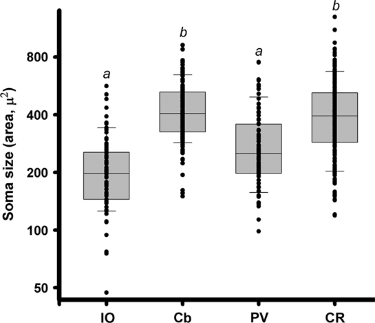
A comparison of the sizes of neurons in nucleus LM. Box plots show the cross-sectional areas of PV- and CR-immunopositive neurons in LM, as well as LM neurons retrogradely labeled from injections into the vestibulocerebellum and IO. The measurements were subject to a log transformation to normalized the data sets. The boundaries of the box indicate the 25th and 75th percentiles, and the horizontal line marks the median. Bars mark the 90th and 10th percentiles. “a” indicates a significant difference from all other categories (Tukey Honestly Significant Difference (HSD)). “b” indicates significant difference from PV and IO groups (Tukey HSD).

In a recent report, we showed that a substantial proportion of the cerebellar-projecting nBOR neurons express the calcium-binding proteins parvalbumin (PV) and calretinin (CR), whereas very few of the IO-projecting neurons express either of these (Wylie et al., Reference Wylie, Pakan, Gutierrez-Ibanez and Iwaniuk2008). In this report, we investigate whether the equivalent pathways originating in LM also differ in their expression of calcium-binding proteins. Calcium-binding proteins are expressed in LM; CR immunoreactivity is intense in the perikarya and neuropil of LM, whereas PV immunoreactivity is moderate in the perikarya and neuropil (Pfeiffer & Britto, Reference Pfeiffer and Britto1997; De Castro et al., Reference De Castro, Cobos, Puelles and Martinez1998). In contrast, calbindin is not expressed in perikarya, but immunoreactivity is moderate in the neuropil (Pfeiffer & Britto, Reference Pfeiffer and Britto1997). De Castro et al. (Reference De Castro, Cobos, Puelles and Martinez1998) reported that many of the cerebellar-projecting neurons in LM (which they refer to as the external pretectal nucleus) express CR, but other calcium-binding proteins and the IO-projecting neurons have not been investigated.
Materials and methods
The methods reported herein conformed to the guidelines established by the Canadian Council on Animal Care and were approved by the Biosciences Animal Care and Policy Committee at the University of Alberta. We used the similar animals as those of our previous paper that examined the expression of calcium-binding proteins in nBOR neurons projecting to the IO and Cb (Wylie et al., Reference Wylie, Pakan, Gutierrez-Ibanez and Iwaniuk2008). The details of the methods can be found in this paper. Briefly, Silver King and homing pigeons were anesthetized with an intramuscular injection of ketamine (65 mg/kg)/xylazine (8 mg/kg), and animals were placed in a stereotaxic device with pigeon ear bars and beak adapter so that the orientation of the skull conformed to the atlas of Karten and Hodos (Reference Karten and Hodos1967). Sufficient skull and dura were removed to expose the brain surface and allow access to either the IO or the Cb. For the IO injections, we targeted the medial column, which is the region that responds to optokinetic stimuli and receives input from LM and nBOR (Clarke, Reference Clarke1977; Gamlin & Cohen, Reference Gamlin and Cohen1988b; Wylie et al., Reference Wylie, Linkenhoker and Lau1997; Winship & Wylie, Reference Winship and Wylie2001, Reference Winship and Wylie2003; Wylie, Reference Wylie2001). For the cerebellar injections, we targeted folium IXcd of the vestibulocerebellum. Previous studies using anterograde techniques have shown that the LM projection to the Cb is a bilateral mossy fiber projection, which terminates in the granule layer as mossy fiber rosettes. This projection is primarily directed to IXcd but also includes folia VI–IXab (Clarke, Reference Clarke1977; Gamlin & Cohen, Reference Gamlin and Cohen1988b). The projection to IXcd originates mainly in LMl, whereas the projection to folia VI–IXab is mainly from LMm (Pakan & Wylie, Reference Pakan and Wylie2006). All target sites were localized using stereotaxic coordinates (Karten & Hodos, Reference Karten and Hodos1967), and single-unit recordings made with glass micropipettes (tip diameters 4–5 μm) confirmed that neurons were responsive to optokinetic stimuli. Once responsive neurons were located, we replaced the recording electrode with a micropipette (tip diameter 20 μm) containing a fluorescent retrograde tracer (red Lumafluor, fluorescent latex microspheres; LumaFluor Corporation, Naples, FL), 1% cholera toxin subunit B–AlexaFluor 594 conjugate (Molecular Probes, Eugene, OR), or 10% biotinylated dextran amine (BDA) (mini-ruby D-3312; Invitrogen, Carlsbad, CA). These were pressure injected using a Picospritzer II (General Valve Corporation, Fairfield, NJ). For the cerebellar injections, there is no issue of potential leakage into other structures that receive input from LM: As long as the injection is confined to the Cb, only the large multipolar LM neurons will be labeled (Gamlin & Cohen, Reference Gamlin and Cohen1988b). These neurons project only to the Cb (Pakan et al., Reference Pakan, Krueger, Kelcher, Cooper, Todd and Wylie2006). The LM does not project to any other brain stem structure in the vicinity of, or caudal to, the IO. Thus, for the injections targeting IO, we are confident that the retrogradely labeled cells would only represent those projecting to the IO.
Postsurgically, the birds were given an intramuscular injection of buprenorphine (0.012 mg/kg) as an analgesic. After a recovery period of 2–5 days, the animals were deeply anesthetized with sodium pentobarbital (100 mg/kg) and immediately perfused with heparinized phosphate-buffered saline (0.9% NaCl, 1 ml/100 ml heparin, and 0.1 M phosphate buffer). As outlined in Wylie et al. (Reference Wylie, Pakan, Gutierrez-Ibanez and Iwaniuk2008), serial sections in the coronal plane (40 μm thick) were collected throughout the rostrocaudal extent of the Cb, pretectum, and IO and were immunohistochemically stained for PV and CR. Sections were viewed with a fluorescence microscope (Leica DMRE, Richmond Hill, ON, Canada), and images were acquired using a Retiga EXi FAST Cooled mono 12-bit camera (Qimaging, Burnaby, BC, Canada) and analyzed with Openlab imaging software (Improvision, Lexington, MA).
Nomenclature of LM
For the nomenclature of LM, we relied on Gamlin and Cohen (Reference Gamlin and Cohen1988a,Reference Gamlin and Cohenb) who divided the LM into medial and lateral subdivisions (LMm and LMl), both of which contain large and small cells (Gottlieb & McKenna, Reference Gottlieb and McKenna1986; Zayats et al., Reference Zayats, Eyre, Nemeh and Tombol2003). Continuous with the LMl at its lateral and caudal aspects is the tectal gray (GT), which contains mainly small cells. The LMm, LMl, and the rostral part of GT all receive retinal input (Gamlin & Cohen, Reference Gamlin and Cohen1988a). Previously, the LMm was known as the LM magnocellularis, and the LMl and GT were included as the LM parvocellularis (e.g., Karten & Hodos, Reference Karten and Hodos1967). The LMm is bordered medially by the nucleus laminaris precommissuralis (LPC), a thin strip of cells that does not receive input from the retina. Medial to LPC is the nucleus principalis precommissuralis, which is lateral to the nucleus rotundus (nRt). In Nissl-stained sections, the layers of the pretectum are relatively easy to distinguish, although the border between GT and LMl can be difficult to localize.
Results
The results presented are based on experiments performed on 12 pigeons. Five pigeons received injection of retrograde tracer in the Cb and five received injection in the medial column of the IO. The remaining two did not receive injections, but sections through the pretectum were processed for PV and CR immunoreactivity to examine their respective distributions. Examples of injection sites in the IO and Cb can be found in Wylie et al. (Reference Wylie, Pakan, Gutierrez-Ibanez and Iwaniuk2008).
Fig. 1C shows a section through the LM immunoreacted for PV. Consistent with previous reports (Pfeiffer & Britto, Reference Pfeiffer and Britto1997; Heyers et al., Reference Heyers, Manns, Luksch, Gunturkun and Mouritsen2008), PV was strongly expressed in neurons in nRt, triangularis (T), subpretectalis, and the optic tectum. Expression in the LM was not as intense, but numerous PV+ve cell bodies were present in LMl, as shown in the inset (Fig. 1D). PV expression was apparent in the neuropil of LMm, but fewer neurons were PV+ve (Fig. 1G).
Fig. 1E shows a section through the LM immunoreacted for CR. Consistent with previous studies (Pfeiffer & Britto, Reference Pfeiffer and Britto1997; Heyers et al., Reference Heyers, Manns, Luksch, Gunturkun and Mouritsen2008), CR was strongly expressed in the anterior dorsolateral thalamus, pars lateralis. In LM, large multipolar cells throughout LM were strongly CR+ve (Fig. 1E and 1F). These cells clearly resembled those labeled from injections of retrograde tracer in the Cb, with respect to size, morphology, and distribution (Fig. 1B). Fig. 1F–1H shows a section reacted for both CR (panel F, red) and PV (panel G, green). The overlay (panel H) shows that many cells express both PV and CR and are therefore double labeled (white arrows). Overall, CR+ve cells were more abundant in LM compared to PV+ve cells. In a survey through the rostrocaudal extent of LM in two cases, we counted 1291 CR+ve and 558 PV+ve LM neurons, 173 of which were double labeled. That is, 13.4% of CR+ve neurons were also PV+ve and 31.0% of PV+ve neurons were also CR+ve.
Fig. 2 shows a plot of the sizes of CR+ve and PV+ve LM neurons, as well as the sizes of LM neurons that project to the Cb and IO and other structures. The cells that project to the Cb are larger than those that project to the IO (mean cross-sectional area, Cb = 416 μ 2 and IO = 216 μ 2), although there is overlap in their distributions. The CR+ve cells were large (422 μ 2), with a distribution very similar to that of the cerebellar-projecting LM cells. The PV+ve neurons were slightly smaller (291 μ 2), with the distribution overlapping both the cerebellar- and the IO-projecting LM neurons. An analysis of variance of cell size revealed a significant difference among the four categories in Fig. 2 (F = 689.87, df = 4, 868, P < 0.01). The CR+ve and cerebellar-projecting cells were not significantly different from each other but were significantly larger than all other groups (post hoc Tukey Honestly Significant Difference (HSD), P < 0.05). The PV+ve neurons were significantly different from all other groups.
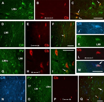
Fig. 3 shows several examples of retrogradely labeled LM neurons, from injections of red LumaFluor in the Cb, in sections immunoreacted for CR (panels A–C and D–F), PV (panels G–I), and both PV and CR (panels J–M and N–Q). Many of the cerebellar-projecting neurons were CR+ve (Fig. 3C, 3F, 3M, and 3Q). Across all five cases, of 588 retrogradely labeled cells, 306 (52%) were CR+ve. In contrast, far fewer cells were PV+ve (Fig. 3I, 3M, and 3Q). Across all five cases, of 897 retrogradely labeled cells, 135 (15%) were PV+ve. In sections that were immunoreacted for both PV and CR, it was clear that almost all those retrogradely labeled cells that were PV+ve were also CR+ve (Fig. 3M and 3Q). From these sections, of 49 retrogradely labeled cells that were PV+ve, 48 were also CR+ve.
Expression of calcium-binding proteins in cerebellar-projecting neurons in the nucleus LM. Cells were retrogradely labeled with red LumaFluor from injections into the vestibulocerebellum (B, E, L, and P), and the sections were reacted for CR (A and D), PV (G), or both PV and CR (J–K and N–O). The overlays are also shown (C, F, I, M, and Q). The arrows indicate retrogradely labeled cells that were also immunopositive for CR and/or PV. In M and Q, the larger stylized arrow indicates a retrogradely labeled cell that was PV+ve and CR+ve. In Q, the small arrows indicate three retrogradely labeled cells that were CR+ve but not PV+ve. All scale bars = 100 μm.

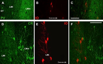
Fig. 4 shows examples of retrogradely labeled LM neurons from injections of BDA (red) in the IO in sections immunoreacted for PV (panels A–C) and CR (panels D–F). Although the retrogradely labeled cells were often surrounded by CR+ve and PV+ve neurons, very few of the IO-projecting neurons were immunopositive. Only 4.5% of the IO-projecting neurons were CR+ve (9 of 202 retrogradely labeled cells) and 5.8% were PV+ve (12 of 207 retrogradely labeled cells). In sections that were reacted for both PV and CR, none of the retrogradely labeled cells were positive for both CR and PV.
Expression of calcium-binding proteins in IO-projecting neurons in the nucleus LM. Cells were retrogradely labeled with red BDA from injections into the IO (B and E), and the sections were reacted for either CR (D) or PV (A). The overlays are also shown (C and F). None of the retrogradely labeled cells was immunopositive for the calcium-binding proteins. All scale bars = 100 μm.

Discussion
The restricted expression of the calcium-binding proteins in neuronal populations in the central nervous system has been described in several species (e.g., Celio, Reference Celio1990; Van Brederode et al., Reference Van Brederode, Mulligan and Hendrickson1990; Resibois & Rogers, Reference Resibois and Rogers1992; Pfeiffer & Britto, Reference Pfeiffer and Britto1997; Pritz & Siadati, Reference Pritz and Siadati1999). Calcium-binding proteins have numerous cellular functions (e.g., Kohr et al., Reference Kohr, Lambert and Mody1991; Yamaguchi et al., Reference Yamaguchi, Winsky and Jacobowitz1991; Baimbridge et al., Reference Baimbridge, Celio and Rogers1992; Schwaller et al., Reference Schwaller, Meyer and Schiffmann2002; Schwaller, Reference Schwaller, Lajtha and Banik2007, Reference Schwaller2009), although their role from a systems perspective is not well understood. Nonetheless, an analysis of the differential expression of calcium-binding proteins has been very useful in elucidating neural circuitry, particularly in sensory systems (Blümcke et al., Reference Blümcke, Hof, Morrison and Celio1990; Van Brederode et al., Reference Van Brederode, Mulligan and Hendrickson1990; Wild et al., Reference Wild, Williams, Howie and Mooney2005). For example, Heyers et al. (Reference Heyers, Manns, Luksch, Gunturkun and Mouritsen2008) examined the expression of calcium-binding proteins in the tectofugal and thalamofugal visual pathways of the zebra finch (Taeniopygia guttata). In both the diencephalon and the telencephalon, PV was primarily expressed in structures of the thalamofugal pathway, whereas calbindin was expressed in the tectofugal pathway.
The extent to which this differential expression applied to other nuclei of the visual system is not, however, well understood. The present study and our study of the nBOR (Wylie et al., Reference Wylie, Pakan, Gutierrez-Ibanez and Iwaniuk2008) show that calcium-binding proteins are differentially expressed within a visual nucleus. For both nBOR and LM, few of the IO-projecting neurons expressed either CR or PV (nBOR <2%; LM <5%). In contrast, almost half of the cerebellar-projecting neurons in LM and nBOR express CR (52% for both LM and nBOR), and a smaller proportion express PV (15% in LM; 34% in nBOR), although most of these PV+ve neurons also expressed CR (90% in nBOR; 95% in LM). Why calcium-binding proteins are expressed differently in LM and nBOR is unknown.
As LM and nBOR neurons detect optic flow that results from self-motion (e.g., Burns & Wallman, Reference Burns and Wallman1981; Morgan & Frost, Reference Morgan and Frost1981; Winterson & Brauth, Reference Winterson and Brauth1985; Wylie & Frost, Reference Wylie and Frost1990), one could imagine that they are active almost continuously and require substantial calcium buffering to prevent excitotoxicity. A recent report has suggested that CR, but not PV, protects neurons against glutamate excitotoxicity (D’Orlando et al., Reference D’Orlando, Celio and Schwaller2002). As retinal ganglion cell axons are glutamatergic (Cardozo et al., Reference Cardozo, Buijs and Van der Want1991), perhaps, the large multipolar neurons in LM and nBOR that project directly to the Cb receive direct retinal input, whereas the IO-projecting neurons do not receive glutamatergic input. If this is the case, enhanced CR expression might help prevent overexcitation of the cerebellar-projecting pathway. Related, CR−/− mice show an increased excitability in cerebellar granular cells (Gall et al., Reference Gall, Roussel, Susa, D’Angelo, Rossi, Bearzatto, Galas, Blum, Schurmans and Schiffmann2003).
On classifying the LM as an accessory optic nucleus
The nBOR is a nucleus of the AOS and, strictly speaking, homologous to the mammalian MTN (Simpson, Reference Simpson1984). The LM, homologous to the mammalian NOT, resides within the pretectum (Simpson et al., Reference Simpson, Giolli and Blanks1988) but is often considered, erroneously, part of the AOS (Giolli et al., Reference Giolli, Blanks and Lui2006). This is due to the fact that their response properties are quite similar: Neurons in LM and nBOR (and their mammalian homologs) show direction selectivity in response to large-field visual stimuli and are critical for mediating the OKR (e.g., Fite et al., Reference Fite, Reiner and Hunt1979; Burns & Wallman, Reference Burns and Wallman1981; Morgan & Frost, Reference Morgan and Frost1981; Gioanni et al., Reference Gioanni, Rey, Villalobos, Richard and Dalbera1983a,Reference Gioanni, Villalobos, Rey and Dalberab; Simpson, Reference Simpson1984; McKenna & Wallman, Reference McKenna and Wallman1985b; Winterson & Brauth, Reference Winterson and Brauth1985; Simpson et al., Reference Simpson, Giolli and Blanks1988). Giolli et al. (Reference Giolli, Blanks and Lui2006) have noted that connections and neurochemistry of the pretectum and AOS are quite similar in mammals, although there are a few differences. The same can be said of the nBOR and LM. The LM and nBOR have reciprocal projections, and both project to the Cb, IO, pontine nuclei, ventral tegmental area, and dorsal thalamus (Clarke, Reference Clarke1977; Brecha et al., Reference Brecha, Karten and Hunt1980; Gamlin & Cohen, Reference Gamlin and Cohen1988b; Wylie et al., Reference Wylie, Linkenhoker and Lau1997, Reference Wylie, Glover and Lau1998, Reference Wylie, Winship and Glover1999). Both nBOR and LM receive input from the visual Wulst, although from different regions (Wylie et al., Reference Wylie, Ogilvie, Crowder, Barkley and Winship2005). The nBOR, unlike the LM, projects heavily to the interstitial nucleus of Cajal and other accessory oculomotor nuclei and the oculomotor complex (Brecha et al., Reference Brecha, Karten and Hunt1980). The present study speaks to more similarities with respect to the connectivity and neurochemistry of the pretectum and AOS in birds. For both LM and nBOR, the projection to the IO is from neurons that generally do not express CR or PV and the projection to the Cb is from large multipolar neurons, many of which express CR (Pakan et al., Reference Pakan, Krueger, Kelcher, Cooper, Todd and Wylie2006; Wylie et al., Reference Wylie, Pakan, Elliott, Graham and Iwaniuk2007, Reference Wylie, Pakan, Gutierrez-Ibanez and Iwaniuk2008). However, the projection from the LM to the Cb targets folia VI–IXcd, whereas that from the nBOR is almost exclusive to IXcd (Clarke, Reference Clarke1977; Brecha et al., Reference Brecha, Karten and Hunt1980; Gamlin & Cohen, Reference Gamlin and Cohen1988b; Pakan & Wylie, Reference Pakan and Wylie2006).
Acknowledgments
This research was supported by funding from the Natural Sciences and Engineering Research Council of Canada (NSERC) to D.R.W. J.M.P.P. was supported by graduate fellowships from NSERC, the Alberta Ingenuity Fund, and a Killam Scholarship. C.G.-I. was supported by a graduate fellowship from Ministerio de Planificación of the Chilean government. D.R.W. was supported by funding from the Canada Research Chairs Program.