INTRODUCTION
Microsporidia are obligate intracellular parasites that are ubiquitous among organisms ranging from insects to mammals, including humans (Sprague et al. Reference Sprague, Becnel and Hazard1992). Nosema bombycis, a pathogen of the economically important silkworm Bombyx mori, was the first identified microsporidia (Nägeli, Reference Nägeli1857). Nosema bombycis can cause pebrine, which can lead to a large amount of destruction in the silkworm industry due to horizontal and vertical transmission. In 1985, humans were found to be infected by Enterocytozoon bieneusi (Desportes et al. Reference Desportes, Charpentier, Galian, Bernard, Cochand-Priollet, Lavergne, Ravisse and Modigliani1985); thereafter, 14 other species in five microsporidia genera were discovered and associated with human illnesses (Weber and Bryan, Reference Weber and Bryan1994; Didier et al. Reference Didier, Snowden and Shadduck1998). Microsporidiosis has been recognized in various groups of people including AIDS patients, cancer patients, organ transplant recipients, diabetics, travellers, children and the elderly (Didier et al. Reference Didier, Snowden and Shadduck1998).
Some microbes, such as bacillaceae, yeast, microsporidia and other species, produce spores or endospores to ensure survival through periods of environmental stress. When conditions change and are favourable, a spore can reactivate itself and enter a vegetative state. Understanding the construction and protein composition of the spore wall is important for pathogen research. The model organism Saccharomyces cerevisiae can form spores to resist a wide variety of environmental conditions (Coluccio et al. Reference Coluccio, Rodriguez, Kernan and Neiman2008). The spore wall of S. cerevisiae contains four different layers. The two innermost layers consist mainly of mannans and glucan and are similar in composition to the vegetative cell wall (Manners et al. Reference Manners, Masson and Patterson1973). The outer two layers are unique to the spore. The outermost shell primarily consists of dityrosine cross-linked macromolecules (Briza et al. Reference Briza, Winkler, Kalchhauser and Breitenbach1986). The next layer is composed of chitosan and appears to contribute to resistance against enzymatic degradation in addition to acting as the substrate of the dityrosine layer (Briza et al. Reference Briza, Ellinger, Winkler and Breitenbach1988). It has been further shown that the spore wall of S. cerevisiae contains at least 20 different proteins whose functions include catalytic activity, water retention, virulence, adhesion, chitin degradation, remodelling of the glucan network and other activities (Orlean, Reference Orlean, Pringle, Broach and Jones1997; De Groot et al. Reference De Groot, Ram and Klis2005; Lesage and Bussey, Reference Lesage and Bussey2006).
Compared with S. cerevisiae, little information is known about the microsporidial spore wall. It appears that the spore wall of microsporidia is comprised of a proteinaceous electron-dense exospore, a chitinous electron-transparent endospore and a plasma membrane (Bigliaedi et al. Reference Bigliaedi, Selmi, Lupetti, Corona, Gatti, Scaglia and Sacchi1996; Vavra and Larsson, Reference Vavra, Larsson, Wittner and Weiss1999). This unique structure provides protection against environmental stress and allows the spore to survive before it infects the host cell (Shadduck and Polley, Reference Shadduck and Polley1978). Additionally, the spore wall can maintain a relatively high hydrostatic pressure and water permeability to control polar tube ejection (Frixione et al. Reference Frixione, Ruiz, Santillán, de Vargas, Tejero and Undeen1992). However, little has been known about the formation of the spore wall layers and their component proteins. Only 13 spore wall proteins have been reported, and their functions have not been fully identified. Endospore Protein (EnP) 1 of Encephalitozoon cuniculi and Spore Wall Protein (SWP) 26 of N. bombycis are thought to be associated with host cell adhesion via a heparin binding motif (Southern et al. Reference Southern, Jolly, Lester and Hayman2007; Li et al. Reference Li, Wu, Pan, He, Zhang, Hu and Zhou2009). SWP5 of N. bombycis interacts with polar tube proteins and may function in modulating N. bombycis infection (Li et al. Reference Li, Pan, Li, Huang, Chen, Geng, Yang, Wang and Zhou2012) and host cell invasion (Cai et al. Reference Cai, Lu, Qiu, Li and Feng2011).
Chitin has been confirmed to be a component of the fibrils that form bridges across the endospore and part of the fibrillar system of the exospore in Encephalitozoon intestinalis (Vavra and Larsson, Reference Vavra, Larsson, Wittner and Weiss1999). However, only one chitin-associated protein has been previously reported: EcCDA of E. cuniculi was demonstrated to function in chitin deacetylation during all sporogonic stages (Brosson et al. Reference Brosson, Kuhn, Prensier, Vivarès and Texier2005). In this study, we used 2D gel electrophoresis (2D-PAGE) to identify the spore wall proteins of N. bombycis. Via MALDI-TOF/TOF MS analysis, three spots on the gel were identified as the same protein, NbHSWP12 (GenBank accession No. EF683112), which has been described as a hypothetical spore wall protein (Wu et al. Reference Wu, Li, Pan, Tan, Hu, Zhou and Xiang2008). A comparative genomic analysis indicated that Nbhswp12 homologue genes are widely present in different species of microsporidia. Immunoblotting and immunofluorescence indicated that NbHSWP12 localizes to the N. bombycis spore wall. Interestingly, NbSWP12 could bind the chitin coat of N. bombycis.
MATERIALS AND METHODS
Microsporidia
Nosema bombycis isolate CQ1 is a characterized strain maintained in our laboratory (Xu et al. Reference Xu, Pan, Fang, Li, Tian, Li, Zhou and Xiang2006a). It was isolated from infected silkworms in Chongqing and conserved in the China Veterinary Culture Collection Center (CVCC No. 102059). Spores were produced and harvested as previously described (Wu et al. Reference Wu, Li, Pan, Tan, Hu, Zhou and Xiang2008).
Protein extraction and 2D gel electrophoresis
Spores were disrupted in lysis buffer (109 cells mL−1) using acid-washed glass beads (Sigma, diameter: 425–600 μm) in a vortex oscillator at 4 °C overnight. Proteins were isolated by centrifugation at 12 000 g for 10 min and used for immunoblotting. Spore germination was induced by 0·1 mol L−1 K2CO3 at 28 °C for 30 min. The sediment was dispersed with PBS and loaded onto a discontinuous Percoll gradient (30, 45, 60, 75 and 90% v/v). Centrifugation was performed at 14 000 g for 40 min in a CP100MX ultracentrifuge (Hitachi Koki Co., Ltd). The material in the 60% Percoll phase was collected and again subjected to Percoll gradient centrifugation. Then, spore coats were recovered from the 60% Percoll gradient layer, and spore wall proteins were extracted in a solution containing 7 m urea, 2 m thiourea, 4% CHAPS, 100 mm DTT and 0·2% SDS at room temperature overnight. Insoluble substances were removed by centrifugation. An Ettan IPGphor II unit (Amersham Biosciences) was used for 2D electrophoresis. 80 μg of spore wall proteins mixed with rehydration buffer were applied to 13 cm IPG gel strips with a non-linear separation range of pH 3–10 (Roche) and rehydrated at 50 V for 12 h. Then, proteins were isoelectrically focused at 15 °C under the following conditions: 100 V for 1 h, 200 V for 2 h, 500 V for 1 h, 1000 V for 0·5 h, 3000 V for 0·5 h, 5000 V for 0·5 h and 8000 V until the voltage reached 80 000 V h−1. After equilibration with a buffer containing 6 m urea, 2% SDS, 50 mm Tris–HCl (pH 8·8), 20% glycerol, 130 mm DTT, and 130 mm iodoacetamide, strips were placed on top of a 12% SDS–PAGE gel and transferred at 120 V for 1·5 h. The gel was stained with silver (Ameresco).
MALDI-TOF/TOF MS analysis
Protein spots were excised from the 2D-gel, destained, digested and analysed by MALDI-TOF/TOF MS (4800 Plus MALDI-TOF/TOFTM Analyzer, Applied Biosystems, USA) as described previously (Cai et al. Reference Cai, Lu, Qiu, Li and Feng2011). The data obtained were used to search against our local N. bombycis database (http://microbe.swu.edu.cn/silkpathdb/) using MASCOT version 2.1 (Matrix Science) with the following parameter settings: trypsin cleavage, carbamidomethyl-Cys set as fixed modification, peptide mass tolerance set to ±100 ppm, fragment mass tolerance set to ±0·8 Da, and maximum missed cleavages set to 1.
Analysis of gene transcription patterns
RT-PCR was used to analyse the transcription of Nbhswp12. Fourth instar silkworm larvae were challenged by feeding them mulberry leaves that had N. bombycis spores (107 spores/100 larvae) dispersed on their surface. The midguts of N. bombycis-infected silkworms at 1–8 days post-infection were collected and ground in Tripure (Roche) for RNA extraction. Prior to the synthesis of sscDNA by reverse transcription using M-MLV, DNaseI (Promega) treated RNA was tested for genomic DNA contamination by PCR using the following primers: 5′-TGGACGCCATTAGACAAG-3′ and 5′-GGCTCGAGTTCATAACATCTAACCAATA-3′, which are specific for the N. bombycis tubulin-β gene. The Nbhswp12 primers for RT-PCR were as follows: 5′-CGGGATCCATGAAAGATTTTAAAAAGAA-3′ and 5′-GCGTCGACCTTAGTCCTCTCTAATGCTT-3′. The tubulin-β gene was used as an internal control.
Sequence analysis and polyclonal antibody production
The Nbhswp12 gene was amplified from N. bombycis genomic DNA using the primers described above and cloned into the p-Cold I vector. The recombinant plasmid was transformed into Escherichia coli Rosetta cells, which expressed recombinant NbHSWP12 following induction for 20 h at 16 °C with 0·2 mm IPTG. The fused expression proteins were purified using the Ni-NTA Superflow Cartridge (QIAGEN). Five mice were intradermally immunized with recombinant NbHSWP12 (100 μg/mouse) homogenized with Freund's adjuvant (1 : 1; Sigma) four times (on days +1, +8, +16 and +24). One week after the fourth injection, antisera were collected and stored at −20 °C.
Immunoblotting
For immunoblot analysis, proteins were separated by SDS–PAGE and transferred to a PVDF membrane (Roche). After blocking for 1 h at 37 °C in TTBS (0·05% Tween 20 in TBS) with 5% (w/v) skim milk, membranes were incubated with 1 : 1500 dilutions of anti-NbHSWP12 or negative control sera in TTBS for 1 h at 37 °C. Following several washes, membranes were reacted with peroxidase-labelled goat anti-mouse IgG (Roche) at a dilution of 1 : 5000 for 1 h. After three washes, bands were detected by adding the substrate diamino benzidine tetrahydrochloride (DAB).
Indirect immunofluorescence assay
Spores were placed in a 1·5-mL centrifuge tube and treated with 0·5% TritonX-100 for 0·5 h. The spores were then washed three times by centrifugation at 3000 g for 1 min and fixed with 4% paraformaldehyde in PBS at 4 °C for 10 min. The fixed spores were washed, and experiments were carried out at 37 °C as follows: spores were blocked in PBS containing 10% (w/v) goat serum and 0·5% (v/v) BSA for 1 h and incubated with 1 : 100 dilutions of anti-NbHSWP12 or control serum in PBS for 1 h. After several washes, spores were incubated with a 1 : 64 dilution of FITC-conjugated goat IgG specific to mouse immunoglobulins (Sigma) for 1 h, and DNA was stained with 1 μg mL−1 DAPI (Sigma) for 5 min. After three washes, the spores were suspended using Fluoromount™ Aqueous Mounting Medium (Sigma) and mounted with a coverglass. Fluorescence was observed and imaged with a Nikon Diaphot microscope.
Preparation of deproteinized chitin spore coats
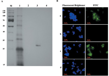
Deproteinized N. bombycis spore coats, which are composed of chitin, were isolated via hot alkaline treatment. The spores were treated with 1 m NaOH at 100 °C for 1 h. Insoluble spore material was recovered by centrifugation at 12 000 g for 1 min and boiled in a buffer containing 10% SDS, 20% 2-ME, and 0·3 m Tris–HCl (pH 6·8) for 10 min. The deproteinized chitin spore coats were recovered after five washes with 10% SDS and deionized water in turn. SDS–PAGE and indirect immunofluorescence assays were performed to detect the presence of proteins in deproteinized chitin coats. 5 μg mL−1 of Fluorescent Brightener 28 (Sigma) were used to stain chitin spore coats in the immunofluorescence assay.
Chitin combination assay
A total of 106 deproteinized chitin spore coats were incubated with 2 μg recombinant NbSWP12 or NbRicin-B-Lectin 21 (NbRBL 21) proteins in a 1·5 mL tube at room temperature for 2 h. PBST (0·05% Tween 20 in PBS) containing 50, 200 or 500 mm NaCl was used to wash the samples sequentially by centrifugation. The insoluble chitin coats were subjected to the indirect immunofluorescence assay or boiled in protein extraction buffer, after which proteins were separated by SDS–PAGE.
RESULTS
2D gel electrophoresis of spore wall proteins and identification of NbHSWP12
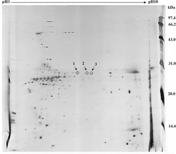
We observed more than 150 spots on our 2D map of the proteins found in germinated N. bombycis spore coats, and most of these spots distributed in the region, between 20–31 kDa and pI 5–7. Only a few spots were observed in the high molecular weight area, particularly in the basic segment. Proteins distributed in the acidic or basic terminals resulted in two areas of entrainment and formed few spots (Fig. 1).
Two-dimensional electrophoresis of Nosema bombycis spore wall proteins. The three circled spots were all identified as NbHSWP12 with MALDI-TOF/TOF MS.

Protein spots were excised and analysed by MALDI-TOF/TOF MS, and three spots were unambiguously identified as NbHSWP12, a protein predicted by N. bombycis genomic data (Tables S1, S2 and S3 – in Online version only). This protein was previously described as hypothetical spore wall protein (GeneBank accession No. EF683112) by Wu et al. (Reference Wu, Li, Pan, Tan, Hu, Zhou and Xiang2008).
Characterization of NbHSWP12
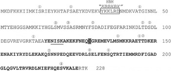
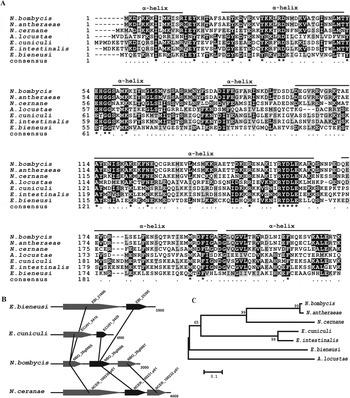
Nbhswp12 is 684 bp in length and encodes a 228-amino acid protein with a predicted pI of 6·49 and a molecular weight of 26·6 kDa, which was in accordance with its location on the 2D gel. As shown in Fig. 2, a BAR-2 domain (e-value: 1.35e-03) was predicted by conserved domain analysis using the BLAST program (http://www.ncbi.nlm.nih.gov) and the Pfam database (http://pfam.janelia.org/). Additionally, an N-terminal protein-heparin interaction motif HBM2 (‘XBBXBX’, amino acids 33–38) and 16 phosphorylation sites catalysed by protein kinase C, casein kinase II, cGMP-dependent protein kinase and calmodulin-dependent protein kinase II were predicted (http://www.cbs.dtu.dk). In particular, most phosphorylation sites were specifically catalysed by protein kinase C at a serine site. Based on NetNglyc and NetOglyc (http://www.cbs.dtu.dk) analysis, a potential N-glycosylation site (NISK) was predicted at the 117–120 position, but no O-glycosylation sites were predicted. Moreover, NbHSWP12 was found to be composed of seven α-helices (http://bioinf.cs.ucl.ac.uk/psipred/), which is consistent with the BAR family of proteins (Fig. 3A).
Sequence features of NbHSWP12. The heparin-binding motif (HBM) is indicated, and the potential N-glycosylation site is underlined. The shaded letter is Cys. The numbers indicate phosphorylation sites as follows: 1, protein kinase C phosphorylation sites; 2, casein kinase II phosphorylation sites; 3, cGMP-dependent protein kinase; 4, calmodulin-dependent protein kinase II. The predicted BAR-2 domain (e-value: 1.35e-03) sequence is in bold.

HSWP12 is relatively conserved in Microsporidia. (A) Amino acid sequence alignment of SWP12 from seven microsporidia. Identical and similar residues are highlighted in black and grey, respectively. The secondary structure of NbSWP12, which is mainly composed of alpha helices, was aligned. (B) Gene order conservation and divergence between Enterocytozoon bieneusi, E. cuniculi, Nosema bombycis and N. ceranae. Nbswp12 and its orthologues are coloured black. (C) The phylogenetic tree of hswp12 genes in microsporidia. The neighbour-joining tree was generated with MEGA5.0 software.

HSWP12 is conserved in microsporidia
To identify homologous genes, a Blast search (http://www.ncbi.nlm.nih.gov/) was performed with the NbHSWP12 amino acid sequence. Plausible Nbhswp12 gene homologues were identified in several microsporidia. When Nbhswp12 homologues within the Nosema genus were compared, only 6 amino acids differed between N. bombycis and Nosema antheraeae (GenBank accession No. KC193258), and there was more than 50% identity between N. bombycis and Nosema ceranae (GenBank accession No. NW_003314251). Furthermore, the hswp12 sequence similarity between E. cuniculi (GenBank accession No. CAD24911) and E. intestinalis (GenBank accession No. NC_014415) was above 80%. Twelve conserved amino acid sites were identified in the N-terminal and C-terminal domains of the proteins. Two conserved motifs, ‘YEHGG’ and ‘RYDLE’, were found in all hswp12 homologous genes, indicating that they are functionally necessary. All Nbhswp12 homologue genes exhibited similar sequence features. NbHSWP12 and its homologues were also conserved in terms of secondary structure, as they all contain seven alpha-helices, although an additional β-strand was present in E. cuniculi at position 25–26 and E. intestinalis at position 20–29 (Fig. 3A). Furthermore, conservation and divergence analysis of hswp12 homologues suggested that the orientation of hswp12 was conserved in N. bombycis, N. ceranae, E. cuniculi and E. bieneusi. All genes exhibited a strong degree of synteny (Fig. 3B). A phylogenetic tree generated with microsporidial hswp12 genes demonstrates that hswp12 was conserved in the evolutionary process (Fig. 3C).
Transcription and expression of Nbhswp12
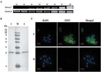
To examine the transcriptional pattern of Nbhswp12, RT-PCR was performed using cDNA generated from the midguts of silkworms at 1–8 days post-infection. As shown in Fig. 4A, transcription of Nbhswp12 began at 3 days post-infection, and transcriptional levels gradually rose from 3–8 days post-infection. Immunoblotting indicated that a single 27 kDa protein was recognized by NbSWP12 antiserum in both the total spore proteins and the spore coat proteins (Fig. 4B). These data suggest that NbHSWP12 localized to the mature spore wall.
Expression analysis of NbSWP12. (A) RT-PCR analysis neighbour P12 expression patterns in the midguts of infected silkworms at 1–8 days post-infection. The tubulin-β gene was used as an internal control. (B) Immunoblotting with NbSWP12 antiserum revealed a single 27 kDa protein that was detected both from total proteins (lane 1) and spore wall proteins (lane 2). (C) Indirect immunofluorescence assay with mouse antisera to NbSWP12 (a) and negative control (b). The permeabilized N. bombycis spores displayed a bright green fluorescent signal at the spore wall compared with the negative control, revealing localization to the spore wall. The scale bar represents 5 μm.

NbHSWP12 localizes at the spore wall
To determine its cellular location, NbHSWP12 was analysed by IFA. Nosema bombycis spores were incubated with the anti-NbHSWP12 polyclonal antibody and exhibited a bright green fluorescent signal at the spore wall, while no green fluorescent signal was observed in the negative control (Fig. 4C). These data suggest that NbHSWP12 is a spore wall protein, which is consistent with the prediction, and therefore we renamed it spore wall protein 12 (NbSWP12).
NbSWP12 proteins bind to deproteinized spore wall chitin
Microsporidia are protected by a thick wall that contains proteins and chitin (Vavra and Larsson, Reference Vavra, Larsson, Wittner and Weiss1999). Deproteinized N. bombycis spore coats composed of chitin were extracted with hot NaOH buffer. SDS–PAGE and IFA results indicated that the hot alkaline-treated spore coats did not contain protein (Fig. 5). After incubation and several washes, we determined that the recombinant NbSWP12 protein could adhere to the isolated spore wall chitin. As expected, deproteinized chitin-rich spore coats that were incubated with the recombinant NbSWP12 protein displayed a bright green fluorescent signal, while no green fluorescent signal was observed in the negative control (Fig. 5B). Therefore, recombinant NbSWP12 protein has the ability to directly bind deproteinized chitin coat.
NbSWP12 binding to deproteinized Nosema bombycis spore wall chitin. Silver staining (A) and IFA assays (B) showed that spores treated with NaOH (lane 2 in A and b in B) contained no detectable protein compared with natural spores (lane 1 in A and a in B). Heterologously expressed NbSWP12 (lane 3 in A and c in B) bound well to deproteinized spore coat chitin. In contrast, heterologously expressed NbRBL21 could not bind to deproteinized chitin coats (lane 4 in A and d in B). Chitin was stained with fluorescent brightener. The scale bar represents 5 μm.

DISCUSSION
Despite extensive characterization of the spore wall structure of microsporidia, very little has been known about its component proteins and deposition mechanism. Up to now, only 13 proteins that participate in the formation of the spore wall had been identified in different microsporidia. Four were found in E. cuniculi. SWP1 was found to be expressed on the surface of early sporonts and then target to the exospore in mature spores (Bohne et al. Reference Bohne, Ferguson, Kohler and Gross2000). The E. cuniculi chitin deacetylase enzyme (EcCDA), which is located at the plasma membrane-endospore interface, was the only reported spore wall protein to potentially function in the deacetylation of chitin into chitosan in the spore wall (Brosson et al. Reference Brosson, Kuhn, Prensier, Vivarès and Texier2005). EnP1 and EnP2 (or SWP3) were found to associate with the chitin-rich layer of the spore wall (Peuvel-Fanget et al. Reference Peuvel-Fanget, Polonais, Brosson, Texier, Kuhn, Peyret, Vivarès and Delbac2006; Xu et al. Reference Xu, Takvorian, Cali, Wang, Zhang, Orr and Weiss2006b). Additionally, EnP1 is also expressed on the anchoring disk complex and the exospore surface, and this protein was reported to be involved in modulating host cell adherence and infection (Southern et al. Reference Southern, Jolly, Lester and Hayman2007). SWP1 and SWP2 of E. intestinalis, which localize to the exospores of developing and mature spores, are differentially expressed and appear to complex on the surface of mature spores (Hayman et al. Reference Hayman, Hayes, Amon and Nash2001). EhSWP1a and EhSWP1b which localize to the exospore, are polymorphic and of interest for epidemiological studies (Polonais et al. Reference Polonais, Mazet, Wawrzyniak, Texier, Blot, El Alaoui and Delbac2010). Within the Nosema genus, 14 hypothetical spore wall proteins have been predicted in N. bombycis, while three exospore proteins (SWP32, SWP26 and SWP5) and two endospore proteins (SWP30 and SWP25) have been identified (Wu et al. Reference Wu, Li, Pan, Tan, Hu, Zhou and Xiang2008, Reference Wu, Li, Pan, Zhou and Xiang2009; Cai et al. Reference Cai, Lu, Qiu, Li and Feng2011; Li et al. Reference Li, Pan, Li, Huang, Chen, Geng, Yang, Wang and Zhou2012). SWP26 was suggested to function in spore adherence and host infection processes (Li et al. Reference Li, Wu, Pan, He, Zhang, Hu and Zhou2009; Zhu et al. Reference Zhu, Shen, Hou, Zhang, Geng, Tang, Xu and Guo2013). SWP5 was observed to interact with polar tube proteins, which may be involved in modulating the course of infection (Li et al. Reference Li, Pan, Li, Huang, Chen, Geng, Yang, Wang and Zhou2012) and protect spores from phagocytic uptake (Cai et al. Reference Cai, Lu, Qiu, Li and Feng2011). Despite these data, more information about spore wall proteins is necessary to elucidate the composition of the spore wall and its role in resistance and infection.
In our study, NbSWP12 was first detected from isolated spore wall proteins by 2D gel. Immunoblotting and immunofluorescence verified that NbSWP12 is expressed in the spore coat of N. bombycis. Thus, NbSWP12 is a new spore wall protein identified in the Nosema genus. Furthermore, recombinant NbSWP12 protein was able to combine with commercial chitin (data not shown), which challenged us to detect the NbSWP12 in combination with natural spore wall chitin. We developed a new procedure to obtain deproteinized chitin from spore coats with hot alkaline buffer. This method is convenient and effective. Immunoblot and IFA results showed that the NbSWP12 protein can bind with the deproteinized chitin coat. These data indicate that NbSWP12 is a chitin-binding spore wall protein that may participate in the formation of the spore wall.
Recent reports have shown that SWP1, a 51 kDa spore wall protein, localizes to the exospore in E. cuniculi and E. intestinalis (Bohne et al. Reference Bohne, Ferguson, Kohler and Gross2000; Hayman et al. Reference Hayman, Hayes, Amon and Nash2001). The corresponding gene, swp1, has also been identified in Encephalitozoon hellem (Polonais et al. Reference Polonais, Mazet, Wawrzyniak, Texier, Blot, El Alaoui and Delbac2010). Similar to these results, we determined that the swp12 gene is widely present among microsporidia. Two conserved motifs, ‘YEHGG’ and ‘RYDLE’, may be key residues in terms of SWP12 function. Our research provides a reference for other microsporidial species. Furthermore, a BAR (Bin-Amphiphysin-Rvs) domain, which has not been reported in other microsporidia, is predicted in NbSWP12 and its homologues. BAR domains are curvature-sensing modules found in many different proteins with diverse functions including endocytosis, exocytosis, intracellular transport, organelle biogenesis, signal transmission, cell division and migration (Balguerie et al. Reference Balguerie, Sivadon, Bonneu and Aigle1999; Takei et al. Reference Takei, Slepnev, Haucke and De Camilli1999; Harada et al. Reference Harada, Furuta, Takeuchi, Itakura, Takahashi and Umeda2000; Lee et al. Reference Lee, Marcucci, Daniell, Pypaert, Weisz, Ochoa, Farsad, Wenk and De Camilli2002; Peter et al. Reference Peter, Kent, Mills, Vallis, Butler, Evans and McMahon2004). With 2D gel electrophoresis, three different spots were identified as NbSWP12, suggesting that the protein exhibits varying degrees of post-translational modification. NbSWP12 may be phosphorylated and is only composed of α-helices, which is similar to other BAR-containing proteins. Our data show that NbSWP12 has the characteristics of a BAR protein, and, combined with its ability to bind chitin, this suggests that the protein may play a role in spore maintenance and signal transmission. The specific functions of NbSWP12 will be further explored.
In conclusion, this is the first report to characterize spore wall protein 12 of N. bombycis. Recognition of the adherence of NbSWP12 to deproteinized N. bombycis spore coat chitin provides important data regarding the localization and function of NbSWP12. These data will be helpful in understanding the protection and invasion mechanisms of microsporidia.
ACKNOWLEDGEMENTS
Thanks to all who provided us with the software utilized in this study free of charge.
FINANCIAL SUPPORT
This work was supported by the National Basic Research Programme of China (973 Programme, No. 2012CB114604); the Key Project of the Natural Science Foundation of China (No. 30930067); the National High-tech R&D Programme (863 Programme, No. 2012AA101301); and the Doctoral Innovation Fund of Southwest University (No. KB2010004 for ZhM).