Implications
Docosahexaenoic acid (DHA; c22:6 n-3) and eicosapentaenoic acid (EPA; c22:5 n-3) are important n-3 fatty acids to improve human health. DHA is important in brain for cognitive development. This study has been carried out with growing pigs as model for humans. The n-3 fatty acid such as α-linolenic acid (ALA; c18:3 n-3) is a precursor for DHA and EPA via enzymatic steps. EPA can be increased in the liver by dietary ALA. Feeding of linoleic acid (LA; c18:2 n-6), a precursor for a competitive pathway, inhibits the synthesis of EPA. The potential for increasing brain DHA via an increase in dietary ALA is limited.
Introduction
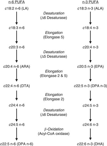
Arachidonic acid (ARA; c20:4 n-6), eicosapentaenoic acid (EPA; c20:5 n-3) and docosahexaenoic acid (DHA; c22:6 n-3) are long-chain polyunsaturated fatty acids (LC-PUFA) and well known to play important regulatory functions in the immune, nervous and cardiovascular system (Innis, Reference Innis2007; Calder, Reference Calder2009; Calder and Yaqoob, Reference Calder and Yaqoob2009; Russo, Reference Russo2009). The n-6 LC-PUFA can be synthesized from linoleic acid (LA; c18:2 n-6) and those of the n-3 family from α-linolenic acid (ALA; c18:3 n-3). Synthesis of EPA and DHA from ALA is achieved by a sequence of desaturation and chain elongation steps, controlled by the enzymes Δ5- (FADS1) and Δ6 desaturase (FADS2) and presumably elongase 2 (ELOVL2) and -5 (ELOVL5) (see Figure 1). DHA can be generated from EPA that requires an additional chain-shortening step (i.e. β-oxidation) in the peroxisome (Sprecher, Reference Sprecher2000; Igarashi et al., Reference Igarashi, Ma, Chang, Bell and Rapoport2007a). The conversion of n-6 LC-PUFA from LA (resulting in the production of ARA) sharing the same enzymes resulting in enzymatic competition between the biosynthetic pathways of n-3 and n-6 LC-PUFA.
General metabolic pathway of n-6 and n-3 fatty acids (adapted from Igarashi et al., Reference Igarashi, Ma, Chang, Bell and Rapoport2007a); LA = linoleic acid; ALA = α-linolenic acid; ARA = arachidonic acid; EPA = eicosapentaenoic acid; DTA = docosatetranoic acid; DPA = docosapentaenoic acid; DHA = docosahexaenoic acid.

The requirement of LC-PUFA for brain growth and development is of current interest. LC-PUFA, in particular DHA and ARA, accumulate rapidly during the brain growth spurt, the most critical stage of brain development that takes place during the later part of gestation and early postnatal life. ARA is essential for normal brain growth, playing a role in synaptic transmission (Bazan, Reference Bazan2003; Sang and Chen, Reference Sang and Chen2006). Changes in brain DHA concentrations have been demonstrated to be positively correlated with cognitive development (Innis, Reference Innis2007). In addition, the dietary supplementation of DHA could improve the cognitive dysfunction due to aging (McCann and Ames, Reference McCann and Ames2005; Cole and Frautschy, Reference Cole and Frautschy2006). The regulatory mechanisms of the conversion of LC-PUFA into ARA, EPA and DHA, and the consequences of LA- and ALA-rich diets consumed during childhood remain to be elucidated. In adult humans, it has been estimated that the β-oxidation of ALA is ± 20% to 30%, which is much lower than that of LA, which was estimated to be 12% to 19% (DeLany et al., Reference DeLany, Windhauser, Champagne and Bray2000). The oxidative losses may partly explain why the conversion of ALA to EPA and DHA in adults is low (Burdge and Calder, Reference Burdge and Calder2005). In addition, human food sources such as cereals, sunflower and soybean oil are rich in LA (>50% of the fat) and have been dominant in the Western diet over the last decades. Consumption of LA-rich diets could also result in inadequate conversion of ALA to DHA and reduced concentrations of DHA in the developing brain (Russo, Reference Russo2009). Dietary studies about the effect of LA are mostly carried out at an isocaloric basis. LA is then replaced by other energy sources such as oleic acid (18:1 n-9). However, it has been reported recently that oleic acid itself can reduce Δ6-desaturase activity (Portolesi et al., Reference Portolesi, Powell and Gibson2008).
This study focuses on the interactions between the n-3 and n-6 LC-PUFA biosynthetic pathways in liver and brain using young, growing pigs as a model for human infancy. This animal model has three major advantages over rodents when studying lipid metabolism. First, brain anatomy and morphology and the timing of the brain growth spurt in pigs and humans are similar (Pond et al., Reference Pond, Boleman, Fiorotto, Ho, Knabe, Merssmann, Savell and Su2000; Duhaime, Reference Duhaime2006). Second, the anatomy of the digestion system, including liver, stomach and intestine, and also many of the pathways of lipid metabolism in pigs are rather similar to human (Moughan et al., Reference Moughan, Cranwell, Darragh and Rowan1991; Innis, Reference Innis1993). Third, the risk for obesity in young, growing pigs is low, enabling large study contrasts in absolute intakes of LC-PUFA precursors, rather than exchanging for other nutrients to maintain isocaloric intake. Here, we have fed young, growing pigs either low or high amounts of LA and ALA, with equal difference between low and high in the intake within the respective fatty acid, and investigated the effect on the fatty acid composition in liver and brain. In the same tissues, we have determined the mRNA levels of Δ6- and Δ5 desaturase and ELOVL2 and ELOVL5 by quantitative PCR.
Material and methods
Animals and housing
In all, 32 female pigs were selected from eight litters. From each litter, four gilts were allotted to one of the four dietary treatments. On arrival, pigs had a body weight (BW) of 16 kg (s.d. = 1.8). The pigs were housed individually during an experimental period of 4 weeks. They were weekly weighed and feed intake was recorded daily. The experimental protocol was approved by the Animal Experimental Committee of Wageningen University, The Netherlands.
Diets
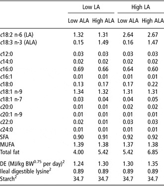
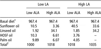
This study was carried out as a 2 × 2 factorial design with daily intakes of LA and ALA as independent variables. Differences between low and high intakes were designed to be identical for LA and ALA: low ALA and LA intakes were 0.16 and 1.32, and high ALA and LA intakes were 1.48 and 2.65 g/kg BW0.75 per day, respectively. The dietary energy percentage from LA was 3.4, 3.3, 6.6 and 6.4 and those of ALA was 0.4, 3.7, 0.4 and 3.5 for the diets low LA/low ALA, low LA/high ALA, high LA/low ALA and high LA/high ALA, respectively. The size of the experimental contrasts was, in part, based on recent studies with humans showing an effect of dietary LA on EPA in plasma phospholipids (Goyens et al., Reference Goyens, Spilker, Zock, Katan and Mensink2006). In addition, the low LA and ALA intakes were slightly above minimal requirement figures for pigs (National Research Council, 1998; Schellingerhout, Reference Schellingerhout2002). The addition of LA and ALA was on top of a basal diet. The intakes of fatty acids are presented in Table 1. With the exception of LA and ALA, the intakes of SFA (saturated fatty acids) and MUFA (monounsaturated fatty acids) were kept constant. This was done by optimizing the dosages of analyzed sunflower oil, linseed oil, high-oleic acid sunflower oil and palm oil. The diets did not contain n-3 or n-6 LC-PUFA other than LA and ALA. The composition of the basal diet and the inclusion rates of the oils in the treatments are presented in Table 2. The low LA/low ALA diet was formulated to contain sufficient ileal digestible amino acids in relation to energy (CVB Table Pigs, 2007).
Experimental design: average intakes of DE, starch, ileal digestible lysine, fat and fatty acidsFootnote 1 in g/kg BW0.75 per day

DE = digestible energy; LA = linoleic acid; ALA = α-linolenic acid; SFA = saturated fatty acid; MUFA = monounsaturated fatty acid.
1 Based on analyzed values.
2 Based on calculated values (CVB Table Pigs, 2007).
Dietary ingredients (in g/kg, relative to the low LA/low ALA diet) of the dietary treatments with differences in LA and ALA intakes

LA = linoleic acid; ALA = α-linolenic acid; HOSF = high-oleic sunflower oil.
1The basal diet contained (g/kg, as fed) corn starch, 160.1; wheat, 160.5; barley, 321.0; wheat bran, 53.5; molasses, 32.1; soybean meal, 64.2; peas, 53.5; potato protein, 53.5; wheat gluten, 53.5; limestone, 14.1; monocalcium phosphate, 13.7; salt, 5,4; vitamin-mineral mix, 10.7; L-lysine HCl 3.0; l-Threonine, 0.32; tryptophan, 0.32; dl-methionine, 0.54.
2The sum of feed ingredients adds up to 1000 g/kg for the low LA/low ALA diet only. A total exceeding 1000 reflects an increased feed intake of the respective treatment group as explained in the text.
As the LA and ALA increments were not exchanged for other nutrients but dosed on top of the basal diet, digestible energy intake varied among experimental treatments between 2.6 and 2.8 × maintenance energy requirement. The diets were fed twice daily as mash. To allow pigs to express at least some natural foraging behavior, straw (20 g/day) was provided daily, which has been demonstrated to alleviate the stress of individual housing (De Jong et al., Reference De Jong, Ekkel, van de Burgwal, Lambooij, Korte, Ruis, Koolhaas and Blokhuis1998; Chaloupková et al., Reference Chaloupková, Illmann, Neuhauserová, Tománek and Valis2007). Water was provided ad libitum.
Sampling and fatty acid analysis
At the start of the experimental period, blood sample was taken from each pig at 2.5 h after feeding. At the end of the experimental period, pigs were anesthetized 2.5 h after feeding with an administration of ketamine (10 mg/kg BW) and xylazine (1 mg/kg BW). Blood sample was collected using cardiac puncture before euthanasia was performed by an intracardiac administration of pentobarbital (100 mg/kg BW). Brain and liver were immediately removed after euthanasia. The total brain, frontal cortex and hippocampus were weighed. For gene expression measurements, samples (∼1 mg) of liver and hippocampus were rapidly frozen in liquid nitrogen and stored at −80°C until analysis. Other parts of the collected tissues were stored at −20°C pending fatty acid analysis. Total lipids were extracted from diets, liver, frontal cortex and hippocampus with a choloroform : methanol (2 : 1, v/v) Folch mixture and then saponified and methylated to determine fatty acid composition by gas chromatography as described previously (Smink et al., Reference Smink, Gerrits, Hovenier, Geelen, Lobee, Verstegen and Beynen2008).
Quantitative PCR
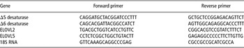
Total RNA was isolated from the hippocampus and liver tissue using TRIzol reagent (Invitrogen, Bleiswijk, The Netherlands). Reverse transcription of 1 μg of total RNA was performed in a 20-μl reaction using Superscript III reverse transcriptase (Invitrogen), dNTPs (Roche) and random hexamer primers (Roche, Almere, The Netherlands) for 1 h at 50°C according to the manufacturer's protocol (Invitrogen). Real-time PCR was performed on a LightCycler 2.0 Real-Time PCR System by using FastStart DNA Master SYBR Green I reagents (Roche). The primers used are presented in Table 3. All primer pairs, except for 18S RNA, were designed in such a way that they span an intron of their corresponding genomic sequence or that its sense or reverse primer anneals on an exon-intron junction. Templates were amplified after a preincubation for 10 min at 95°C, followed by amplification for 40 cycles (10 s at 95°C, 5 s at 60°C, 5 s at 72°C). PCR efficiencies for the genes were established to be between 97% and 100%. Expression levels of FADS1, FADS2, ELOVL2 and ELOVL5 were normalized using the corresponding values of 18S RNA. All reactions revealed a single product as determined using melting curve analysis and specificity of the primer sets were verified by sequencing of the generated amplicons.
Quantitative PCR primers

Statistical analysis
The trial was conducted as a 2 × 2 factorial arrangement of treatments with eight replicates per treatment. The individually housed pig served as the experimental unit. The results were analyzed by ANOVA using the software program SAS version 9.1 (Statistical Analysis Systems Institute Inc., Cary, NC, USA). The fatty acid concentrations in all tissues and enzyme RNA concentrations were tested using the following model:

where LA = daily LA intake i (i = low or high); ALA = daily ALA intake j (j = low or high); litter k (k = 1,…, 8).
For fatty acid concentrations in blood plasma, the initial concentrations, measured at the start of the experiment were included as a covariate.
Homogeneity of variance was tested using the Shapiro–Wilk test. When model residuals were not distributed normally (P < 0.05), the statistical analysis was performed on transformed data. The level of statistical difference was preset at P < 0.05.
Results
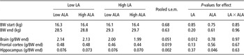
All pigs remained healthy throughout the study and rates of BW gain were within the normal range. The effects of LA and ALA intakes on BW and brain weight are presented in Table 4. The average BW at slaughter was 29 kg and did not significantly differ among treatments. Fresh brain weights (g/kg BW) were significantly (P = 0.012) lower for the pigs fed the high LA diet. The hippocampus weight was lower (P = 0.046) for the high ALA groups.
BW at the start and at the end of the trial and the effect of LA and ALA intakes on brain weight in growing pigs

BW = body weight; LA = linoleic acid; ALA = α-linolenic acid.
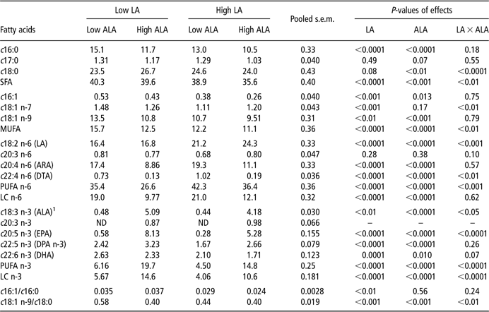
The effects of LA and ALA intakes on the content of liver fatty acids are presented in Table 5. The results of the dietary treatments on the composition of plasma fatty acids largely resembled the effects on liver fatty acid composition (data not shown). Nearly all fatty acid contents were affected by LA and ALA intakes. High LA pigs had a significant (P < 0.0001) higher content of LA and ARA in liver. The content of n-3 fatty acids such as ALA, docosapentaenoic acid (DPA; c22:5 n-3) and DHA were decreased (P < 0.01) in the high LA pigs. A high ALA intake resulted in an increased (P < 0.0001) ALA and EPA contents in liver. The proportion of DHA, however, was decreased (P = 0.01) by high intake of ALA. High ALA pigs had increased content of LA in the liver but ARA and docosatetraenic acid (DTA; c22:4 n-6) contents decreased (P < 0.0001) by approximately 50%.
Effect of LA and ALA intakes on the fatty acid profile (% of total fatty acids) of the liver in growing pigs

LA = linoleic acid; ALA = α-linolenic acid; SFA = saturated fatty acids; MUFA = monounsaturated fatty acids; ARA = arachidonic acid; DTA = docosatetraenic acid; PUFA = polyunsaturated fatty acids; LC = long chain (fatty acids with 20 or 22 C-atoms); EPA = eicosapentaenoic acid; DPA = docosapentaenoic acid; DHA = docosahexaenoic acid.
1P-value and s.e.m. are obtained from Y = log(1 + x) function.
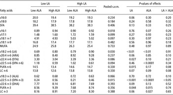
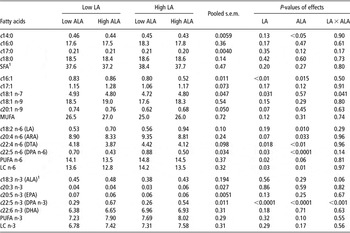
The fatty acid profiles of the frontal cortex and the hippocampus are presented in Tables 6 and 7, respectively. LA and ALA were hardly present in both brain tissues. ARA (9%), c22:4 n-6 (3% to 4%) and DHA (7% to 8%) were the main PUFA in the two brain tissues. High LA intake tended (P < 0.1) to increase the ARA content and significantly (P < 0.05) increased the proportion of c22:4 n-6 in both the hippocampus and frontal cortex. High LA intake resulted in a significant (P < 0.001) decrease of DPA and a tendency (P = 0.09) of a decreased DHA content in the frontal cortex. Increased ALA intake did not affect the ARA and c22:4 n-6 contents but lowered c22:5 n-6 in the frontal cortex. High ALA increased DPA, but did not affect DHA contents in the frontal cortex significantly. High LA intake resulted in a decreased content of DPA but did not affect that of DHA in the hippocampus. A high ALA intake significantly decreased all n-6 LC-PUFA, but increased the LA content in the hippocampus. High ALA intake increased the DPA content, but did not affect other n-3 fatty acids.
Effect of LA and ALA intakes on the fatty acid profile (% of total fatty acids) of the frontal cortex in growing pigs

LA = linoleic acid; ALA = α-linolenic acid; SFA = saturated fatty acids; MUFA = monounsaturated fatty acids; ARA = arachidonic acid; DTA = docosatetraenic acid; DPA = docosapentaenoic acid; PUFA = polyunsaturated fatty acids; LC = long chain (fatty acids with 20 or 22 C-atoms); DHA = docosahexaenoic acid.
Effect of LA and ALA intakes on the fatty acid profile (% of total fatty acids) of the hippocampus in growing pigs

LA = linoleic acid; ALA = α-linolenic acid; MUFA = monounsaturated fatty acids; ARA = arachidonic acid; DTA = docosatetraenic acid; DPA = docosapentaenoic acid; PUFA = polyunsaturated fatty acids; LC = long chain (fatty acids with 20 or 22 C-atoms); EPA = eicosapentaenoic acid; DHA = docosahexaenoic acid.
1P-value and s.e.m. are obtained from an inverse function.
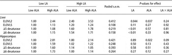
The change in mRNA expression levels of desaturase and elongase enzymes were measured in liver and hippocampus (Table 8). In liver, both LA and ALA intakes significantly (P < 0.05) increased the transcript level of ELOVL2. On the contrary, ELOVL5 mRNA was not significantly increased. In addition, both hepatic transcript levels of Δ5- and Δ6 desaturase were increased following high LA intake (P < 0.01). In hippocampus, high ALA intake significantly (P = 0.022) increased the mRNA expression of ELOVL2, but not of ELOVL5, Δ6- and Δ5 desaturase. In contrast, variation in LA intake had no effect on the mRNA expression of these four genes.
Effects of LA and ALA intakes on the mRNAs of D5- and D6 desaturases and elongase 2 and -5 in liver and hippocampus of growing pigs

LA = linoleic acid; ALA = α-linolenic acid.
mRNA levels were quantified by quantitative PCR and normalized to 18S ribosomal RNA. They are expressed relative to the Low LA/Low ALA diet group (1.00).
Discussion
The formulated diets contained LA and ALA and were free of other n-3 or n-6 LC-PUFA. Once consumed, LA and ALA can be converted to other LC-PUFA by desaturation and elongation. The route and assumed enzymes involved in the biosynthesis of LC-PUFA are given in Figure 1.
In this study, we supplied LA or ALA maintaining identical intakes of other nutrients, an approach that allows independent evaluation of the effects of LA and ALA in liver and brain. Importantly, we did not observe a difference in BW of the gilts among the experimental groups.
Liver
A high LA intake increased the content of LA and n-6 LC-PUFA such as ARA in liver lipid. This agrees well with other studies in pigs (Romans et al., Reference Romans, Wulf, Johnson, Libal and Costello1995; Schellingerhout, Reference Schellingerhout2002). Increasing the ALA intake by 1.3 g/kg BW0.75 per day increased the EPA content in liver lipid from less than 1% to 8%. In contrast to other n-3 LC-PUFA, the DHA content was decreased by high ALA intake. Interestingly, a similar effect was seen on the content of ALA, EPA and DHA in plasma phospholipids in humans (Goyens et al., Reference Goyens, Spilker, Zock, Katan and Mensink2006), indicating that the composition of these plasma lipids is a reflection of the lipid metabolism of the liver. Other earlier studies showed that plasma DHA level was not affected or marginally increased in humans by dietary ALA (Blank et al., Reference Blank, Neumann, Makrides and Gibson2002; Burdge and Calder, Reference Burdge and Calder2005). In contrast to our study, these authors compensated the higher intake of ALA with a decrease in the intake of other fatty acids. We believe that caution should be taken in choosing this exchange approach, as there is evidence that other lipids such as LA, oleic acid and myristic acid (c14:0) can alter hepatic desaturase activity as well (Bézard et al., Reference Bézard, Blond, Bernard and Clouet1994; Jan et al., Reference Jan, Guillou, D'Andrea, Daval, Bouriel, Rioux and Legrand2004; Portolesi et al., Reference Portolesi, Powell and Gibson2008; Rioux et al., Reference Rioux, Cathelina, Beauchamp, Le Bloc'h, Pedrono and Legrand2008). These fatty acids may contribute to the observed opposite effects on plasma DHA in their studies.
This study shows that high LA intake increased levels of both Δ5- and Δ6-desaturase mRNA in liver. Similar effects have been found in rats (Bézard et al., Reference Bézard, Blond, Bernard and Clouet1994) and piglets (Theil and Lauridsen, Reference Theil and Lauridsen2007). In contrast, in a human hepatoma cell line a suppression of Δ6-desaturase mRNA content was seen after the addition of LA or ALA (Portolesi et al., Reference Portolesi, Powell and Gibson2008). Increased ALA intake had no effect on Δ5- and Δ6 desaturase. The rate-limiting enzyme Δ6 desaturase has been reported to have a higher affinity for ALA than for LA (Rodriguez et al., Reference Rodriguez, Sarda, Nessmann, Boulot, Leger and Descomps1998; Sprecher, Reference Sprecher2002). Although not measured in our model, it is tempting to speculate that the surfeit of ALA in liver is preferentially converted above LA down the cascade (Figure 1) or alternatively catabolized (via β-oxidation) and that the LA-elevated expression of Δ6 (and Δ5)-desaturase may be necessary to metabolize the surplus of LA provided by the diet. Indeed, we found a strong increase in the EPA content in liver lipid in response to ALA intake, suggesting that the enzymatic activity of both desaturases and ELOVL5, at least for ALA, were not rate limiting.
In our study, high LA and high ALA intakes increased the transcript of ELOVL2 but not of ELOVL5 in the liver of growing piglets. These findings indicate that the conversion of ARA and EPA to longer elongation products became more efficient (see Figure 1). Indeed, we found an increased proportion of DPA and DTA in liver lipid in response to ALA and LA intakes, respectively. Unfortunately, we did not have the standards in order to establish whether this was paralleled by a positive effect on the content of c24:5 n-3 and c24:4 n-6 as well. Fatty acid-dependent changes in hepatic ELOVL2 expression have also been reported in rat and salmon (Igarashi et al., Reference Igarashi, Ma, Chang, Bell and Rapoport2007a; Morais et al., Reference Morais, Monroig, Zheng, Leaver and Tocher2009).
Interestingly, DHA levels were significantly reduced, whereas ELOVL2 mRNA expression was increased in response to ALA intake. This finding suggests a rate-limiting enzyme downstream of ELOVL2. As Δ6 desaturase has a higher affinity for ALA than for c24:5 n-3 (D'Andrea et al., Reference D'Andrea, Guillou, Jan, Catheline, Thibault, Bouriel, Rioux and Legrand2002; Portolesi et al., Reference Portolesi, Powell and Gibson2007), it is conceivable that Δ6 desaturation of c24:5 n-3 is inhibited when ALA is abundant, preventing DHA synthesis. We would suggest that in the liver of piglets, Δ6 desaturase rather than ELOVL2 is the rate-limiting enzyme downstream of EPA in the biosynthesis of DHA. Whether ALA has any inhibitory effect on the peroxisomal β-oxidation of c24:6 n-3 remains to be elucidated.
Increased intake of LA decreased the proportion of total n-3 LC-PUFA and a high intake of ALA decreased the total n-6 LC-PUFA in liver lipid. This confirms the general concept that competition between ALA and LA occurs because of sharing of the same desaturation and elongation enzymes. In addition, this study illustrates that for EPA and DHA, the effects of LA intake are stronger at high ALA intake and the effects of ALA intake are stronger at low LA intake (interaction LA × ALA for EPA, DHA, P < 0.001 and P = 0.07, respectively). The first concern is the effect of high LA as the Western diet is generally rich in LA and poor in n-3 fatty acids. High LA intake in our study decreased the proportion of EPA and DHA in liver fat by >35% and >20%, respectively. A similar effect was also observed in blood plasma fat. On the other hand, high ALA intake drastically decreased the proportion of ARA by approximately 50%. This inhibitory effect provides a strong indication that ALA and LA competes for the same desaturases and elongases.
Taken together, our experimental design allows a direct comparison of the effects of identical increments in daily intakes of LA and ALA (both 1.3 g/kg BW0.75), which is rarely found in literature. The effect of LA both as a substrate of the n-6 chain and as an inhibitor of the n-3 chain was much lower in comparison with that of ALA. Generally, the effect of ALA on LC PUFA appeared to be four times stronger than that of LA. Our findings in growing piglets are in agreement with human intervention studies showing a strong positive correlation of ALA intake and blood EPA, but weaker between LA intake and blood ARA (Mantzioris et al., Reference Mantzioris, James, Gibson and Cleland1995).
Brain
The timing of the pig brain growth spurt is similar to that in humans (Dobbing and Sands, Reference Dobbing and Sands1979; Pond et al., Reference Pond, Boleman, Fiorotto, Ho, Knabe, Merssmann, Savell and Su2000). Several trials with young piglets were conducted to study the effect of dietary fat on brain fatty acid composition (Arbuckle et al., Reference Arbuckle, Rioux, Mackinnon, Hrboticky and Innis1991; De la Presa-Owens et al., Reference De la Presa-Owens, Innis and Rioux1998; Arbuckle and Innis, Reference Arbuckle and Innis2003). These nutritional intervention studies were mainly focused on whole brain. In this study, we selected the frontal cortex, representing a rather large region of the brain, with primary functions (Ng and Innis, Reference Ng and Innis2003), and in addition, the hippocampus, which is involved in the memory storage and retrieval. Although it has been reported that deprivation of essential fatty acids causes a decrease in brain weight (Odutuga, Reference Odutuga1981), we found to our surprise a decreased brain weight in response to increased LA intake, irrespective of ALA intake. This is in contrast with the study by Hrboticky et al. (Reference Hrboticky, MacKinnon and Innis1990), showing that higher LA at the expense of oleic acid did not affect brain weight in young piglets. In our study, an increased LA intake tended (P < 0.1) to increase ARA and significantly increased c22:4 n-6 in fat extracts of both hippocampus and frontal cortex. Moreover, high ALA intake increased DPA, but not DHA concentrations in the fat of those brain tissues. Earlier studies in piglets demonstrated an increase in brain DHA by dietary ALA (Arbuckle et al, Reference Arbuckle, Rioux, Mackinnon, Hrboticky and Innis1991). The difference in the effect on DHA between the latter and our study may be due to their use of younger piglets, knowing that younger piglets respond stronger to dietary PUFA (Cheon et al., Reference Cheon, Huh, Lee, Park, Sohn and Chung2000). However, the lack of the effect on brain DHA was also found in adult rats in which ALA intake exceeded the low ALA groups in our study (Bourre et al., Reference Bourre, Dumont, Pascal and Durand1993). The elongation of n-3 fatty acids in brain astrocytes did not extend beyond DPA (Innis and Dyer, Reference Innis and Dyer2002). Although high ALA intake drastically increased the ALA content in plasma lipids (an increase of the proportion from ± 1% to 10%), which is a reflection of the increase of ALA content in liver (Table 4), the ALA and DHA contents in frontal cortex and hippocampus did not increase (Tables 5 and 6). Other studies in rat brain showed that ALA after uptake from the circulation across the blood-brain barrier will be mainly oxidized and only a small fraction is converted to DHA (DeMar et al., Reference DeMar, Ma, Chang, Bell and Rapoport2005; Igarashi et al., Reference Igarashi, DeMar, Ma, Chang, Bell and Rapoport2007b). The activity of desaturation and elongation are higher in liver in comparison with brain (Igarashi et al., Reference Igarashi, Ma, Chang, Bell and Rapoport2007a). This suggests that most DHA in the brain is derived from another source, such as liver, intestine (diet) and/or adipose tissue. Surprisingly, the DHA concentration in the pig liver lipid decreased with increasing ALA intake. High ALA intake resulted in a significant increase in ELOVL2 mRNA expression, which may relate to the strong increase in the content of DPA, a product of ELOVL2, in both frontal cortex and hippocampus. On the other hand, we did not observe an effect on mRNA expression of Δ6 and Δ5 desaturase and ELOVL5 in the hippocampus that agrees with studies in rat brain (Igarashi et al., Reference Igarashi, Ma, Chang, Bell and Rapoport2007a).
Similarly to liver, high dietary ALA intake increased the concentration of LA in fat extracts of both frontal cortex and hippocampus. The proportion of ALA, however, was not increased. The latter may be due to a high rate of oxidation in brain (DeMar et al., Reference DeMar, Ma, Chang, Bell and Rapoport2005), or elongation and desaturation, resulting in increased concentrations of n-3 LC PUFA in the frontal cortex. The higher LA concentration in high ALA diets might be due to competition for enzymes and sparing of LA oxidation in the presence of ALA. The rate of oxidation of ALA is higher than that of LA (DeLany et al., Reference DeLany, Windhauser, Champagne and Bray2000).
In conclusion, the effect of LA and ALA are important both as precursor and inhibitor for the synthesis of LC-PUFA. LA as a substrate is increasing mRNA expression of Δ6- and Δ5 desaturase and all n-6 PUFA in liver. High ALA intake did increase EPA in liver lipid but decreased the proportion of DHA. This study supports the idea that high dietary LA intake inhibits the conversion of ALA into n-3 LC-PUFA and that ALA inhibits the conversion of LA into n-6 LC-PUFA. The magnitude of the effect of ALA in liver is higher than that of LA. This suggests that manipulation of ARA and EPA availability by dietary interventions should be optimized varying both dietary LA and ALA. DHA in brain tissue is hardly affected by both dietary LA and ALA. mRNA expression of ELOVL2 was upregulated by dietary treatment and was, apart from Δ6 desaturase, identified as a potentially rate-limiting step.
Acknowledgements
This study was funded by the Dutch Ministry of Agriculture, Nature and Food quality. We thank Ronald P. Mensink for critical comments and Tamme Zandstra, Wouter Rijm, Robert Hovenier, Adriana Silveira de Souza, Marleen Scheer, Ilse van Kerkhof and people of ‘The Haar’ for their technical assistance.