

Introduction
Rigid oesophagoscopy was first reported in 1868 by Kussmaul, who used a rigid urethroscope to examine the oesophagus.Reference Kussmaul1 However, the first purpose-built, end-illuminated, rigid oesophagoscope was designed by Chevalier Jackson, whose design has been little changed in a century.Reference Jackson2
Teaching rigid oesophagoscopy has always been a challenging task for trainers, as the endoluminal view is only adequate for those performing the procedure. Trainees tend to learn the technique by observing the steps and then swapping places with the surgeon in order to get a ‘snapshot’ view through the oesophagoscope. Lack of a constant endoluminal view for the observer has previously made this procedure difficult for trainers to teach and for trainees to learn.
This report presents a simple method of achieving an endoscopic, endoluminal view of the rigid oesophagoscopy procedure, to aid teaching, video-recording and observation of trainees' performance. The accompanying, online video clip represents a valuable teaching resource.
Materials and Methods
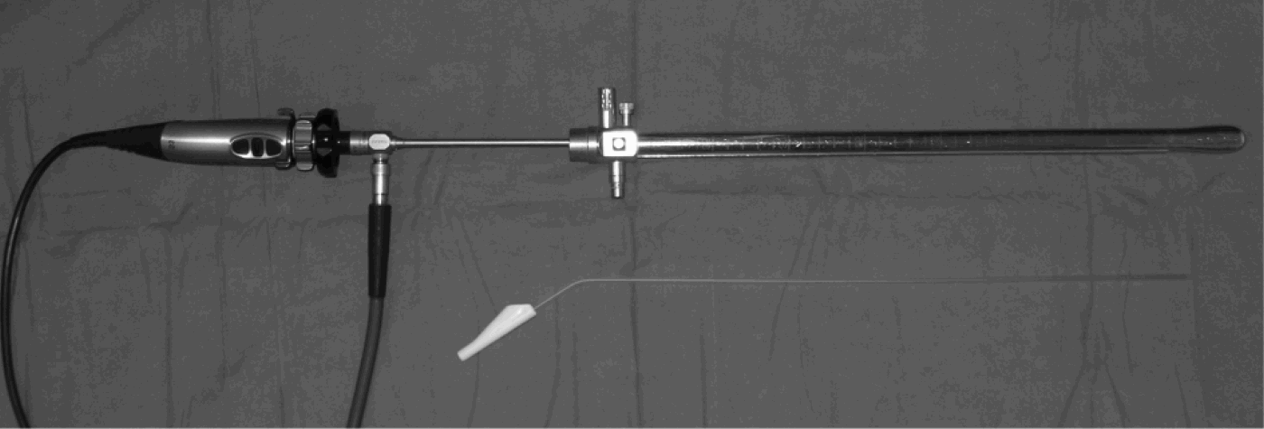
The following equipment is required (Figure 1): (1) a rigid oesophagoscope; (2) a 0° Hopkins rod lens endoscope™ (Karl Storz, Tuttlingen, Germany) (the endoscope should be longer than the length of the oesophagoscope); (3) a video camera capable of being mounted on the endoscope; (4) a flexible oesophageal suction catheter; and (5) Steri-StripTM adhesive strips (3M, St. Paul, USA).
Equipment required for endoscopic rigid oesophagoscopy.

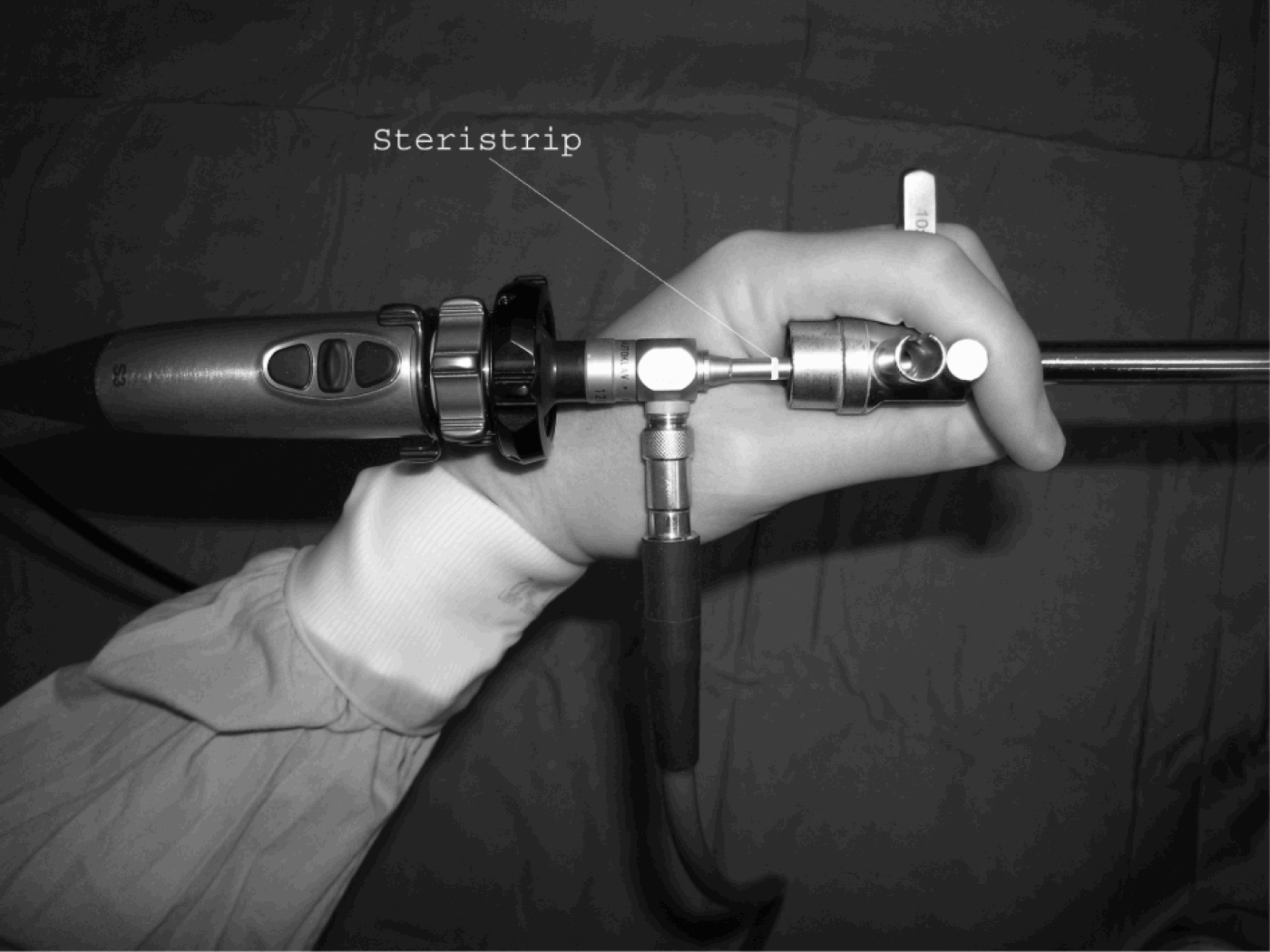
The camera is attached to the end of the Hopkins endoscope, and the endoscope is inserted through the rigid oesophagoscope so that the tip of the Hopkins endoscope does not protrude from the end of the oesophagoscope. The length of Hopkins endoscope required to fit inside the oesophagoscope can be marked by a piece of Steri-Strip (Figure 2).
The grip: the surgeon rests the Hopkins endoscope on their thumb web and secures it in place by adducting their thumb. In this view, the surgeon's thumb has moved from the grip position in order to demonstrate the Steri-Strip marker.

The surgeon holds the Hopkins endoscope so that it rests on their thumb web, and secures it in place by adducting their thumb. Rigid oesophagoscopy can now be performed under endoscopic vision. During the procedure, the assistant surgeon or nurse can introduce the flexible suction catheter when required.
The author performs rigid oesophagoscopy without using the handle of the rigid oesophagoscope. Use of the handle would make this method difficult, as the grip would be altered.
The accompanying online video clip (Appendix 1) demonstrates an endoscopic view of a rigid oesophagoscopy, and this can be used for teaching purposes.
Conclusion
This report presents a method for performing endoscopic rigid oesophagoscopy which can be used for teaching and assessment purposes.
The author also calls on manufacturers to design rigid oesophagoscopes that can incorporate a Hopkins rod-lens endoscope or similar rigid endoscope, to facilitate the above procedure.
Appendix 1. Supplementary video material
A video clip demonstrating an endoluminal view of a rigid oesophagoscopy procedure is available online on The Journal of Laryngology & Otology website, at www.jl.co.uk.