Introduction
Chronic kidney disease (CKD) includes a series of pathologies in which a progressive and irreversible alteration of the kidney structure and function is produced leading to end-stage renal disease (ESRD), condition that makes necessary dialysis or kidney transplantation (Ref. Reference Remuzzi1). CKD is a worldwide public health problem. The relevance of this disease is due to its high prevalence, given that 10–20% of the population suffer from any type of CKD, originating an elevated cost to the Health Care systems. Moreover, more than one million patients die per year due to CKD (Ref. Reference Boor and Floege2). In developing countries, the final stage of CKD in most cases equates to death, due to the limited access to renal replacement therapies (RRT), as it is initiated in less than 25% of patients with ESRD (Ref. Reference Anand, Bitton and Gaziano3). ESRD is defined as a glomerular filtration rate (GFR) persistently below 15 ml/min per 1·73 m2 (Ref. 4). Among those individuals without ESRD at age 40 years, the lifetime risk of ESRD was 2.66% for men and 1.76% for women. The risk was higher in persons with reduced kidney function: for GFR = 44–59 ml/min per 1.73 m2, the lifetime risk of ESRD was 7.51% for men and 3.21% for women, whereas men and women with relatively preserved kidney function (GFR = 60–89 ml/min per 1.73 m2) had lifetime risks of ESRD of 1.01 and 0.63%, respectively (Ref. Reference Turin5). In the USA, the prevalence of ESRD is increasing. The number of patients enrolled in the ESRD Medicare-funded programme has increased from approximately 10 000 beneficiaries in 1973, to more than 600 000 at the end of 2011 (United States Renal Data System – USRDS 2013 Annual Data Report: Atlas of Chronic Kidney Disease and End-Stage Renal Disease in the USA; National Institutes of Health; National Institute of Diabetes and Digestive and Kidney Diseases, Bethesda, MD, 2013). The growth of the number of patients included in ESRD programme has been partially explained by changes in the demographics of the population, differences in disease burden among racial groups and underrecognition of earlier stages of CKD and of risk factors for CKD (Ref. Reference Obrador, Pereira and Kausz6). Patients with ESRD consume a disproportionate share of healthcare resources. The total cost of the ESRD programme in the USA was approximately $49.3 billion in 2011. Medicare costs per person per year were more than $75 000 overall, ranging from $32 922 for transplant patients to $87 945 for those receiving haemodialysis therapy (United States Renal Data System. USRDS 2013 Annual Data Report: Atlas of Chronic Kidney Disease and End-Stage Renal Disease in the United States; National Institutes of Health; National Institute of Diabetes and Digestive and Kidney Diseases, Bethesda, MD, 2013). However, despite the magnitude of the resources committed to the treatment of ESRD and the substantial improvements in the quality of dialysis therapy, these patients continue to experience significant mortality and morbidity and a reduced quality of life. Each year about 440 000 patients worldwide start RRT, but 3 200 000 have no access to RRT and die prematurely (Ref. 4). Detailed mortality data are available only from registries in developed countries. Therefore, these data might not be representative of worldwide reality. Furthermore, information about patients with chronic kidney failure who do not receive RRT is scarce. The Global Burden of Disease 2010 study identified chronic kidney failure as one of the three causes of death with the greatest increase from 1990 to 2010. In some developing regions, such as Central America (Colombia, Costa Rica, El Salvador, Guatemala, Honduras, Mexico, Nicaragua, Panama and Venezuela) and Andean South American region (Bolivia, Ecuador and Peru), chronic kidney failure is the fifth most common cause of death (Institute for Health Metrics and Evaluation – GBD arrow diagram; available at http://www.healthmetricsandevaluation.org/gbd/visualizations/gbd-arrow-diagram; accessed June 8, 2014).
Irrespective of the initial causes of CKD, in final stages an increase of extracellular matrix (ECM) takes place in either the glomerulus (glomerulosclerosis), tubular interstitium (tubulointerstitial fibrosis) or both compartments (Ref. Reference Hewitson7). Interstitial inflammatory cell infiltration and tubular atrophy, also characterises this disease (Ref. Reference Lopez-Novoa8). Several conditions, especially diabetes and hypertension may favour the development of CKD. Besides, atherosclerosis, renal hypoxia, infections, autoimmune diseases and obstructive nephropathy may play an important role as well. Knowledge of molecular mechanisms leading to CKD allows us to find more specific treatments of this disease.
Given that renal parenchyma is composed mostly by tubules (90% approximately), tubulointerstitial fibrosis is better correlated with the outcome of renal damage than glomerular fibrosis (Ref. Reference Grande and Lopez-Novoa9). Numerous cells are involved in renal fibrosis, including inflammatory cells such as macrophages and leucocytes, endothelial cells, myofibroblasts, mesangial cells, tubular epithelial cells, etc. The main sources of ECM protein expression are mesangial cells in the glomerulus and myofibroblasts in the tubular interstitium.
The evolution of renal injury shares many features with other forms of interstitial renal disease and is very similar in virtually all progressive renal disorders.
ECM is a complex and intricate network within which molecules are precisely organised. Most of ECM molecules, collagens, glycoproteins and proteoglycans share similar structural domains. Laminin, types IV and V collagens, entactin and fibronectin are localised in the normal kidney, with different distribution in the glomerular basement membranes, the mesangial matrix and tubular basement matrix (Ref. Reference Border and Noble10). Tissue fibrosis is a result of the imbalance between enhanced production and deposition and impaired degradation of ECM components leading to excessive accumulation and the subsequent functional impairment of renal function.
Transforming growth factor beta (TGF-β) and renal fibrosis
TGF-β overexpression is a key mediator of fibrosis, as it has been demonstrated since 1990, when the fibrogenic effects of TGF-β were firstly demonstrated. These effects include synthesis of extracellular matrix, inhibition of its degradation and facilitation of cell–matrix interactions. In kidney diseases, TGF-β overexpression has been related to loss of glomerular and peritubular microvasculature (Ref. Reference Wang, Koka and Lan11). TGF-β also participates in endothelial and epithelial to mesenchymal transition, transdifferentiation of endothelial and epithelial cells into myofibroblasts, a major source of ECM proteins (Ref. Reference Lopez-Hernandez and Lopez-Novoa12). Renal TGF-β1 in CKD comes from renal parenchymal cells, inflammatory infiltrated cells (Refs Reference Yamamoto13, Reference Yamamoto14) and also from circulating TGF-β1 and from platelets (Ref. Reference Meyer15).
The TGF-β superfamily includes several proteins involved in the development and vascular homeostasis in mammals, such as TGF-βs, bone morphogenetic proteins (BMPs) and activins. There are three different isoforms of TGF-β in mammals (TGF-β1, TGF-β2 and TGF-β3), being TGF-β1 the one which plays the most important role in fibrogenesis. TGF-β1 enhances ECM synthesis, inhibits proteases secretion and induces synthesis of protease inhibitors, thus impairing ECM degradation (Ref. Reference Border and Noble10).
TGF-β plays a central role in the development of renal fibrosis (Ref. Reference Border and Noble10). TGF-β levels has been reported to be increased in several models of renal fibrosis (Refs Reference Pimentel16, Reference Rodriguez-Pena17, Reference Peters, Border and Noble18, Reference Ding19), and in human renal fibrosis from several origins (Refs Reference Nogare20, Reference Lim21). In human and experimental diabetic nephropathy, the expression of TGF-β is also elevated (Ref. Reference Yamamoto22). In addition, an increase in the type I receptor has been shown in obstructive nephropathy (Ref. Reference Sutaria23). Moreover, an increase in both the type II TGFβ receptor (Ref. Reference Cohen24), and one of its TGFβ auxiliary receptors, endoglin (Ref. Reference Prieto25) has been shown in experimental models of CKD. Thus, the purpose of the present paper is to describe the available data on the role of endoglin in the origin and development of renal fibrosis. As there are many excellent recent reviews on the role of TGF-β1 in renal fibrogenesis (Refs Reference Lopez-Hernandez and Lopez-Novoa12, Reference Meng, Chung and Lan26, Reference Munoz-Felix, Gonzalez-Nunez and Lopez-Novoa27, Reference Loeffler and Wolf28, Reference Garcia-Sanchez, Lopez-Hernandez and Lopez-Novoa29, Reference Lan30), we will only describe briefly the most important points necessary to understand the relationships between endoglin and TGF-β function.
The effect of increased TGF-β levels in renal fibrosis has been studied in several animal models. First studies showed tubulointerstitial fibrosis and glomerulosclerosis in transgenic mice overexpressing TGF-β (Ref. Reference Kopp31). It has been also described that a single intramuscular injection of human active recombinant TGF-β1 in rats and rabbits originate lesions in several organs, including the kidney where both glomerular alteration and tubular vacuolisation were observed leading to glomerular fibrosis (Ref. Reference Terrell32). In contrast, mice treated with TGF-β2 showed tubular alterations without glomerular affectation (Ref. Reference Ledbetter33). Indeed, the kidneys of these animals exhibited high levels of angiotensin II (Ang II) and endothelin-1. Ang II stimulates TGF-β1 synthesis in endothelial cells (Ref. Reference Zoja34), which suggests that TGF-β2 may collaborate in the profibrotic effect of TGF-β1 through Ang II production. Confirming the role of TGF-β in kidney diseases, several studies have evidenced that blocking TGF-β with neutralising antibodies reduces the progression of renal fibrosis in different animal models. For example, in the db/db mice model of type II diabetes treatment with anti-TGF-β antibodies avoids renal hypertrophy and ECM deposition, alleviating the factors that trigger renal failure in these animals (Ref. Reference Ziyadeh35). Consistently, rats that developed glomerulonephritis after treatment with antithymocyte serum, showed an overwhelming decrease of glomerular ECM production after administration of TGF-β antibodies compared with animals treated with placebo (Ref. Reference Border and Noble10). Moreover, treatment with an anti-TGF-β antibody (1D11) decreased tubular apoptosis, increased tubular cell proliferation and increased NO synthase activity after the unilateral ureteral obstruction (UUO) experimental model of tubulointerstitial fibrosis, contributing to kidney preservation (Ref. Reference Miyajima36). In a rat nephrosis model induced by puromycin aminonucleoside injection, it was reported that administration of anti-TGF-β antibodies induced a marked improvement in both renal structure and function (Ref. Reference Ma37).
Summarising the above-described data, the relevance of TGF-β in the origin and maintenance of glomerulosclerosis and of tubulointerstitial fibrosis has been clearly demonstrated. These effects of TGF-β on renal fibrosis could be a consequence of the direct effects of TGF-β on renal cells or of the indirect effects on cell proliferation and renal ECM synthesis and degradation (Ref. Reference Lopez-Hernandez and Lopez-Novoa12).
TGF-β1 signalling pathway and renal fibrosis
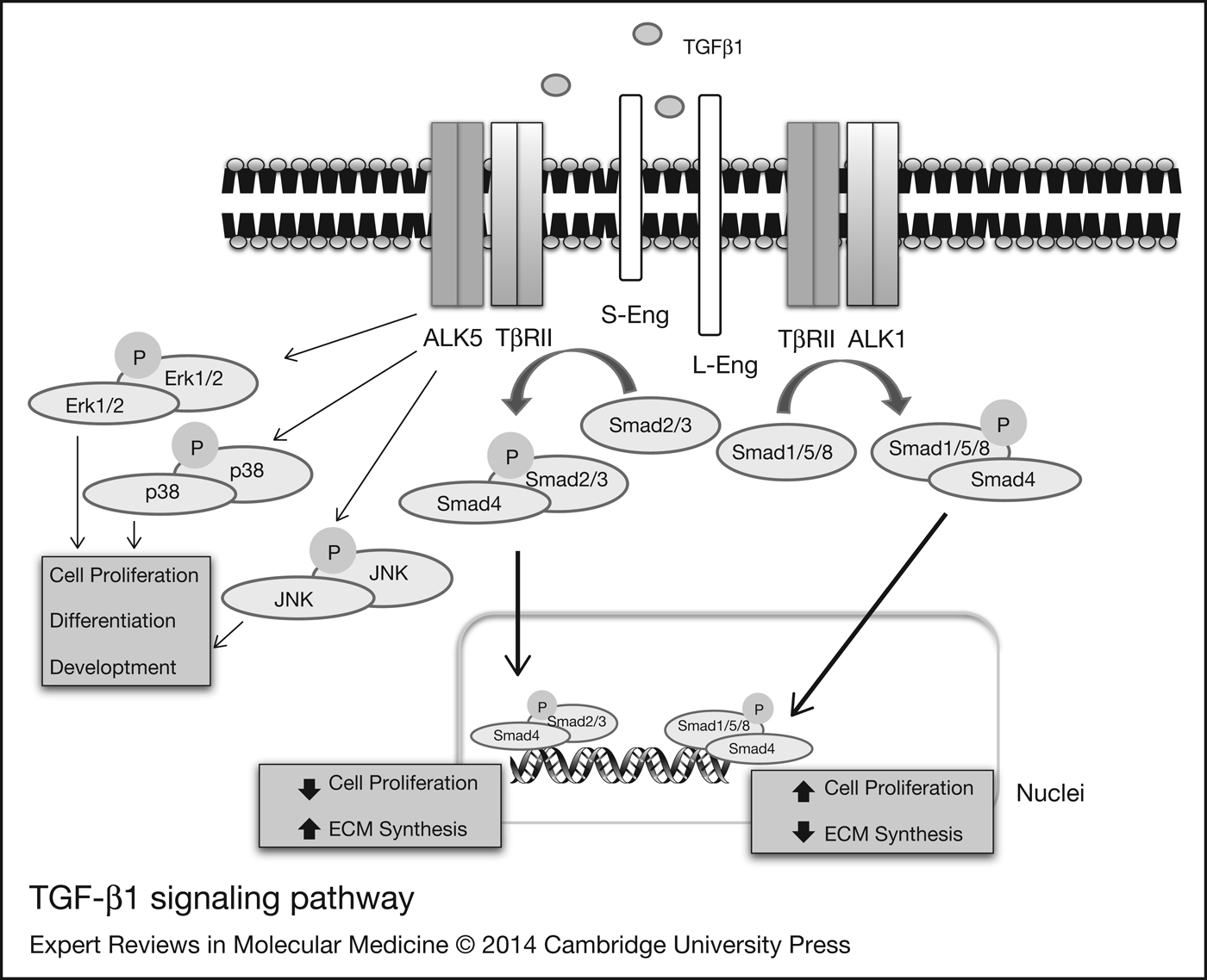
There are several recent reviews discussing extensively the TGF-β1-signalling pathway (Refs Reference Meulmeester and Ten Dijke38, Reference ten Dijke and Hill39, Reference Gonzalez-Nunez, Munoz-Felix and Lopez-Novoa40, Reference Sheen41). Thus, we will only review briefly the most relevant points in relationship with fibrosis and endoglin function. TGF-β1 signalling starts when this cytokine binds to the TGF-β type II receptor (TβRII) which in turn activates one of several TGF-β type I receptors (TβRI, hereafter named activin-like kinase or ALK). Both type I and type II receptors display serine/threonine kinase activity. The activated TGF-β1-TβRII–TβRI complex transduces the signal to the nucleus by recruitment and phosphorylation of receptor-activated Smad proteins (R-Smads). Depending on the TβRI recruited, different types of Smads become phosphorylated. Two different pathways have been described, the Smad 1/5/8 pathway, which is activated by ALK1, while the Smad 2/3 pathway is activated by ALK5 (Fig. 1). TGF-β has different affinities for the ALK receptors, being ALK5 the receptors with more TGF-β affinity (Ref. Reference Inman42). TGF-β signalling through ALK1 was described in vivo in endothelial cells (Ref. Reference Goumans43) but later bone morphogenetic protein 9 (BMP9) was identified as the most potent ligand for ALK1 (Ref. Reference David44). Moreover, in vivo studies relate that ALK1 pathway is likely activated by BMPs (Refs Reference Ricard45, Reference Levet46). Phosphorylated R-Smads dissociate from the receptor complex and bind common-mediated Smads (Co-Smads), mainly Smad4, and the heterodimer translocates into the nucleus where they interact with various transcriptional coactivators, corepressors and transcription factors to modulate target gene expression (Refs Reference ten Dijke and Arthur47, Reference Massague48). During the development of fibrotic processes TGF-β signalling has been reported to take place through the ALK5 receptor that activates Smad2/3 pathway, promoting the expression of genes involved in ECM synthesis (Ref. Reference Moon49). Furthermore, high levels of nuclear phosphorylated Smad2 (pSmad2) and Smad3 (pSmad3) have been reported in the obstructed kidney after UUO (Ref. Reference Fukasawa50), and in renal cortex of streptozotocin-induced diabetic mice (Ref. Reference Wang51), whereas down-regulation of the inhibitory Smad7 increased fibrosis in obstructed kidneys of the UUO model (Ref. Reference Fukasawa50). Targeted deletion of Smad3 markedly prevents from UUO-induced interstitial fibrosis (Refs Reference Flanders52, Reference Inazaki53), and glomerulosclerosis in the streptozotocin model of diabetes in mice (Ref. Reference Wang51) suggesting that most of the profibrotic activities of TGF-β are mediated by Smad3. Our research group have shown that heterozygous disruption of ALK1 leads to an increase in TGF-β/Smad3 signalling and its consequent tubulointerstitial fibrosis after UUO (Ref. Reference Munoz-Felix, Lopez-Novoa and Martinez-Salgado54). In addition, it has been demonstrated that the promoter areas of the genes COL1A2, COL3A1, COL5A1, COL6A1, COL6A3 and COL2A1 have Smad3-binding sequences and contribute to Smad3 mechanisms involved in ECM expression (Refs Reference Wang, Koka and Lan11, Reference Vindevoghel55). ALK1/ALK5 ratio has been shown by several authors that can play a role in ECM protein expression regulation, especially in in vitro models in human chondrocytes (Refs Reference Finnson56, Reference Finnson57) but also in in vivo studies (Ref. Reference Blaney Davidson58). ALK1 heterozygous mouse embryo fibroblasts (MEFs) express more ECM proteins such as collagen I, fibronectin and CTGF (Ref. Reference Munoz-Felix59). However, experiments performed by Abe et al. (Ref. Reference Abe60) reported the implication of Smad1 in collagen type IV synthesis, one of the main ECM components. Other study has demonstrated that ALK1 heterozygous mice develop less renal fibrosis after radiation, suggesting a possible role of ALK1 in renal fibrosis (Ref. Reference Scharpfenecker61). Moreover, Smad1 pathway seems to be implicated in systemic fibrosis (Ref. Reference Pannu62). Nevertheless, other authors have described the antifibrotic effect of BMP7 through the activation of the Smad1/5/8 pathway (Refs Reference Zeisberg and Kalluri63, Reference Manson64). These studies suggest that Smad1 play different roles that are dependent on the cell type and the ligand involved. Thus, Smad1/5 pathway may be antifibrotic as protects against EMT, whereas this pathway potentiates profibrotic response in scleroderma fibroblasts and in mesangial cells. The different roles of Smad1/5 signalling may be due to the different context where TGF-β acts (Ref. Reference Massague65). The possible involvement of ALK1/Smad1/5 pathway in fibrosis has been reviewed recently by our research group (Ref. Reference Munoz-Felix, Gonzalez-Nunez and Lopez-Novoa27). In addition of the Smad-dependent pathways TGF-β activates Smad-independent pathways, in which different proteins, including mitogen-activated protein kinases (MAPK), p38, Ras or integrin-linked kinases (ILK) are involved (Ref. Reference Martinez-Salgado, Rodriguez-Pena and Lopez-Novoa66). MAPK can in turn regulate the Smad activation (Ref. Reference Nakerakanti and Trojanowska67) (Fig. 1).
TGF-β1-signalling pathway. L-Eng and S-Eng modulate TGF-β1 cell response in a different way. L-Eng favours TGF-β/ALK1/Smad1/5/8 pathway promoting cell proliferation and inhibiting ECM synthesis. By contrast, S-Eng favours TGF-β/ALK5/Smad2/3 pathway, promoting ECM synthesis and hindering proliferation. These signalling pathways have been described mainly in endothelial cell.

Moreover, several studies have demonstrated that NADPH oxidase is necessary for TGF-β to induce myofibroblast maintenance of activated phenotype, ECM protein expression or apoptosis of epithelial cells, as it has been demonstrated in several in vivo studies in cardiac fibroblasts (Ref. Reference Cucoranu68). Several in vivo studies have shown the important involvement of NADPH oxidase in experimental fibrosis: inhibition of Nox4 – NADPH oxidase catalytic subunit – with siRNA lead to a decrease of bleomicyn-induced pulmonary fibrosis. Moreover, oral administration of GKT-136901 – selective inhibitor of Nox1 and Nox4 – lead to a reduction of liver fibrosis after bile duct ligation (Ref. Reference Jiang69). Furthermore, redox-dependent mechanisms may modulate TGF-β intracellular pathways (Ref. Reference Jiang70).
Endoglin
In addition to the TβRI y TβRII receptor types, TGF-β can also bind to co-receptors, also known as TβRIII, namely endoglin and betaglycan (Ref. Reference Goumans, Liu and ten Dijke71). Endoglin has been found to be up-regulated in biopsies from patients with glomerulonephritis (Ref. Reference Roy-Chaudhury, Simpson and Power72) and in several experimental models of renal fibrosis such as renal mass reduction (RMR) and UUO (Refs Reference Rodriguez-Pena17, Reference Rodriguez-Pena73). Moreover, endoglin is expressed in ECM-producing cells: mesangial cells and fibroblasts (Ref. Reference Rodriguez-Barbero74). The temporal changes of endoglin expression may be related to an increase of the abundance of ECM-producing cells. We propose that combined consideration of the temporal expression of endoglin in cells types involved in renal fibrosis and its possible function will allow us to understand whether endoglin is playing any role in regulating renal fibrosis.
Given that endoglin is expressed in ECM-producing cells, we can argue that the questions we propose as starting points to define in this review are the following:
• Does renal disease stimulate the increase of endoglin expression on ECM producing cells?
• Does endoglin modulate ECM gene expression in response to TGF-β stimulation?
• Which pathway can be modulated by endoglin: ALK5/Smad2/3 or ALK1/Smad1/5/8?
In the following paragraphs, we will analyse the possible function of endoglin in renal fibrosis regulation on the basis of its expression and role in ECM-producing cells.
Endoglin: structure and function
Endoglin or CD105 is a 180 KDa disulphide-linked homodimeric transmembrane protein (Ref. Reference Lopez-Novoa and Bernabeu75). It is expressed in several cell types, being its maximal expression not only in proliferating endothelial cells (Ref. Reference Bourdeau76) but also in other cell types, including macrophages (Ref. Reference Lastres77), bone marrow stromal cells (Ref. Reference Robledo78), vascular smooth muscle cells (Ref. Reference Conley79) or placenta – particularly in syncytiotrophoblast (Refs Reference St-Jacques80, Reference Gu, Lewis and Wang81). Of fundamental interest for this review is that endoglin is also expressed in almost all profibrogenic cells, including renal mesangial cells (Refs Reference Rodriguez-Barbero74, Reference Diez-Marques82), cardiac fibroblasts (Ref. Reference Chen83), skin fibroblasts (Ref. Reference Leask84), renal fibroblasts (Ref. Reference Rodriguez-Pena17), hepatic stellate cells (Ref. Reference Meurer85) and cultured portal fibroblasts (Ref. Reference Meurer86).
Mutations in the endoglin gene, that leads to changes in endoglin expression in the vasculature, cause a disease known as Rendu–Osler–Weber syndrome or hereditary haemorrhagic telangiectasia type 1, which is characterised by the presence of epistaxis, telangiectasias and arteriovenous malformations in different organs (Refs Reference McAllister87, Reference Abdalla and Letarte88).
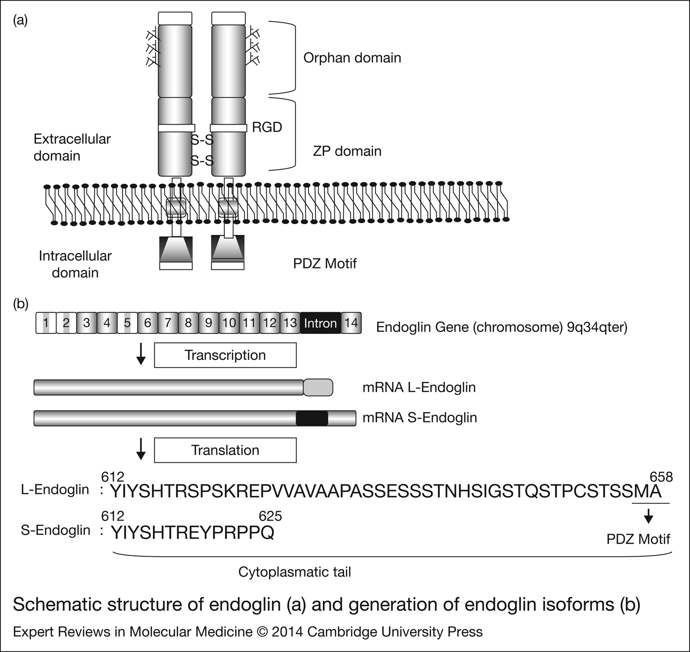
Endoglin structure has three domains (Fig. 2): (i) a large extracellular domain of 625 amino acids in humans that contains an Arg–Gly–Asp (RGD) sequence that may interact with integrins, an orphan domain and a zona pellucida (ZP). Besides, four potential N-linked glycosylation sites and a region of O-linked glycosylation rich in serine and threonine have been described in the extracellular domain (Ref. Reference Lopez-Novoa and Bernabeu75). (ii) A single transmembrane domain and (iii) a short cytoplasmic region without enzymatic activity but presenting several potential phosphorylation sites (Ref. Reference Lastres77). The analysis of the three-dimensional structure of the extra-cellular region of endoglin/CD105 by single-particle electron microscopy identified three well-defined domains for each monomer region, including the two ZP regions and one orphan domain (Ref. Reference Llorca89). ZP, divided in two subdomains (ZP-N and ZP-C, respectively), form about one-third of the extracellular region suggesting a crucial role in endoglin's function. Although the subdomain ZP-N is involved in the dimerisation of the protein, the subdomain ZP-C constitutes the region by which endoglin interact with TβRII and TβRI. This fact highlights the importance of ZP domain in endoglin–TGF-β signalling modulation. Even more, other proteins whose structure includes the ZP domain, that is, betaglycan, share with endoglin a number of characteristics, including the presence of a soluble form, high levels of glycosylation and the existence of a hydrophobic group at the C-terminal region (Ref. Reference Llorca89).
Schematic structure of endoglin (a) and generation of endoglin isoforms (b). Endoglin structure contains three domains: extracellular domain – that contains RGD sequence, single transmembrane domain and cytoplasmic region. S-Eng is generated by alternative splicing.
The cytoplasmic region differentiates two membrane endoglin isoforms, described in humans and mice but not in the rat. L-endoglin (L-Eng) is the longer and most abundant isoform and S-endoglin (S-Eng) the shorter isoform (Ref. Reference Lopez-Novoa and Bernabeu75). S-Eng is generated from the same gene that L-Eng, by intron retention, a type of alternative splicing in which the alternative splicing factor-2 (ASF/SF2) seems to be involved (Ref. Reference Blanco and Bernabeu90). During cell senescence ASF/SF2 is translocated to the cytoplasm where it binds to endoglin mRNA promoting the splicing alternative by the intron 14 retaining in the S-Eng mRNA. This intron introduces a premature codon stop and a shorter protein is generated. Thus, L-Eng and S-Eng have cytoplasmic tails of different lengths, being in human L-Eng 33 amino acids longer than S-Eng (Ref. Reference Lastres91). In mice, S-Eng is 12 amino acids shorter than L-Eng (Ref. Reference Perez-Gomez92). Both isoforms have identical extracellular domains and both are able to bind TGF-β with the same affinity but differ in how they modulate the TGF-β-dependent response. This difference may due to the lack of PDZ motive in S-Eng (Refs Reference Lastres77, Reference Bellon93). In the rat, there is a potential second isoform larger than the most abundant one, with a 100 aa cytoplasmic tail (49 aa inserted after aa 610) that diverges from L-Eng at the same aa than human S-Eng (GenBank Accession no. AA105861) which suggest that it is derived from the same intron retention.
The half-life of L-Eng has been estimated in 17 h (Ref. Reference Paquet94). The half-life of S-Eng is not known, but, taking into account its structure, it should be similar to that L-Eng.
In addition to these membrane isoforms, truncated form of Endoglin, known as soluble endoglin (sEng) has been described in plasma. Its plasma levels are increased in diseases such as preeclampsia (Ref. Reference Venkatesha95) and a wide variety of cancers and cardiovascular pathologies (Refs Reference Blazquez-Medela96, Reference Perez-Gomez97). sEng is the result of the cleavage of membrane isoforms (Ref. Reference Lopez-Novoa and Bernabeu75). Recently studies performed by Hawinkels et al., points at the membrane metalloprotease 14 or MMP14 (also named MT1–MMP) as being responsible for endoglin shedding at the G–L bond at 586 position (Ref. Reference Hawinkels98). Thus, MT1–MMP releases most of the extracellular region of endoglin to the plasma. In contrast to the membrane forms L-Eng and S-Eng, it has been proposed that sEng acts as a scavenger of TGF-β molecules reducing TGF-β-signalling pathway through its membrane receptors (Ref. Reference Lopez-Novoa and Bernabeu75) although other possible mechanisms such as forming complexes with TβRI and TβRI receptors cannot be ignored.
The expression of endoglin is up-regulated under conditions of cellular activation, for example, in endothelial cells during the process of angiogenesis, in which endoglin plays a major role (Refs Reference Lopez-Novoa and Bernabeu75, Reference Perez-Gomez97). This occurs both in physiological angiogenesis and in tumour angiogenesis (Ref. Reference Nassiri99). Endoglin up-regulation occurs also in the monocytes/macrophages transformation (Ref. Reference Lastres100), and under hypoxic conditions (Ref. Reference Sanchez-Elsner101). TGF-β1 and Ang II are also able to induce endoglin up-regulation in several cell types, including endothelial cells, mesangial cells and fibroblasts (Refs Reference Rodriguez-Barbero74, Reference Diez-Marques82, Reference Chen83).
Endoglin expression is mainly regulated by the transcription factor specificity protein 1 (SP1) (Ref. Reference Botella102), whereas a network of SP1, KLF-6, Smad3 and Hif-1α is involved under hypoxic or TGF-β1-stimulated conditions (Refs Reference Sanchez-Elsner101, Reference Botella103, Reference Botella104).
Endoglin and TGF-β1 signalling
The presence of endoglin modifies TGF-β-induced signalling, and this topic has been extensively discussed in several recent reviews (Refs Reference Lopez-Novoa and Bernabeu75, Reference ten Dijke, Goumans and Pardali105, Reference Weiskirchen and Meurer106, Reference Bernabeu, Conley and Vary107, Reference Bobik108, Reference Goumans and Mummery109). In brief, endoglin binds TGF-β1 and -β3 with high affinity in the presence of TβRII but does not bind TGF-β2 (Ref. Reference Bernabeu, Lopez-Novoa and Quintanilla110). Although endoglin is not a signalling receptor, it has been shown to associate with the TGF-β-signalling receptors (TβRII, ALK1 and ALK5) and to modulate their phosphorylation status as well as downstream Smad-dependent and Smad-independent signalling in endothelial cells (Ref. Reference ten Dijke, Goumans and Pardali105).
However, conflicting data on the role of endoglin in regulating TGF-β-mediated Smad1/5 and Smad2 signalling has been reported in several cell types. In endothelial cells, endoglin favours TGF-β1 signalling through the ALK1/Smad1/5/8 pathway and promotes proliferation and migration (Refs Reference Ray111, Reference Lebrin112). Conversely, using also endothelial cells Lee et al. (Ref. Reference Lee113) have reported that endoglin promotes TGF-β-mediated Smad1/5/8 signalling but inhibits endothelial cell migration. Endoglin overexpression was reported to increase phosphorylation (and total levels) of Smad2 (Ref. Reference Santibanez114), whereas another study reported that endoglin overexpression in myoblasts favours signalling through the TGF-β/ALK1/Smad1/5/8 pathway (Ref. Reference Velasco115). Another study using endoglin siRNA knockdown in human bone marrow stromal cells reported that endoglin acts as a positive regulator of both ALK1-induced Smad1 activation and ALK5-induced Smad2 activation (Ref. Reference O'Connor116). It has been reported that in human chondrocytes (Ref. Reference Parker, Goldring and Philip117) endoglin forms a heterodimeric complex with TβRII and TβRI (ALK1 and ALK5) (Ref. Reference Parker, Goldring and Philip117). Furthermore, endoglin enhances TGF-β1-induced Smad1/5 phosphorylation but inhibits TGF-β1-induced Smad2 phosphorylation, Smad3-driven transcriptional activity and ECM protein (type II collagen and PAI-1) expression (Ref. Reference Finnson57).
Taken together, the above-reported studies suggest that endoglin differentially regulates TGF-β signalling via ALK1/Smad1/5 and ALK5/Smad2 depending on cell-type and cellular context. In a recent review, Massagué concludes that TGF-β and BMP regulate several biological process being the signalling pathway activated (Smad1/5/8 or Smad2/3) the same but with different response, determined more by the context than the proteins of the signalling system which determine the response (Ref. Reference Massague65).
There are also some data suggesting that the two membrane endoglin play different roles regulating TGF-β signalling. Experiments performed by Velasco et al. (Ref. Reference Velasco115) in rat myoblasts overexpressing either L-Eng or S-Eng have shown that these isoforms differently modulate TGF-β signalling. Thereby, L-Eng favours signalling through the TGF-β/ALK1/Smad1/5/8 pathway, encouraging proliferation and endothelial migration, whereas S-Eng favours signalling by the TGF-β/ALK5/Smad2/3 pathway, promoting ECM synthesis and hindering proliferation and endothelial migration (Fig. 1). In contrast, sEng impairs TGF-β signalling by preventing its binding to the membrane receptors (Ref. Reference Venkatesha95).
In spite of the relevant role of endoglin in regulating TGF-β signalling, little is known about its role in the origin and development of renal fibrosis.
Increased levels of endoglin in fibrotic diseases
Endoglin plasma levels in patients with fibrotic diseases
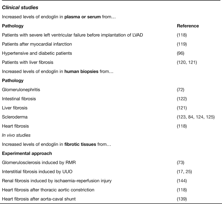
Plasma levels of endoglin, are significantly increased in individuals with severe left ventricular failure before implantation of the left ventricular assist device, whereas plasma levels on endoglin return to control levels in a separate cohort after left ventricular assist device placement (Ref. Reference Kapur118). Using human samples, these authors described that endoglin expression is elevated in human failing hearts and that the major source for plasma endoglin was the cardiac fibroblasts. We have previously reported that endoglin is an attractive biomarker of heart failure; thus, in a cohort of 183 patients with acute myocardial infarction, lower levels of plasma endoglin after acute myocardial infarction correlated with increased mortality (Ref. Reference Cruz-Gonzalez119). It should be noted that cardiac fibrosis is most commonly encountered accompanying acute myocardial infarction as the reparative processes promote collagen deposition and ECM remodelling, a fact that limits pump failure and myocardial dysfunction. Moreover in another study from our laboratory, it has been reported that plasma levels of endoglin are increased in patients with diabetes mellitus and in populations with hypertension as well (Ref. Reference Blazquez-Medela96). Plasma levels of endoglin were also found increased in patients with liver fibrosis (Ref. Reference Salem120) and in patients with hepatitis showing a correlation with hepatic fibrosis markers (Ref. Reference Clemente121).
Tissue or cells showing endoglin expression in organ fibrosis in patients
Roy-Chaudhury et al. have reported that endoglin was present within the glomerular mesangium and interstitium in normal kidneys. In diseased biopsies, there was a weak but significant correlation between staining for endoglin in the interstitium and the extent of chronic histological damage. This was supported by division of biopsies into those showing mild damage and those with moderate to severe damage, where the latter group had significantly increased interstitial staining for endoglin. However, there was no correlation between mesangial staining for endoglin and specific types of glomerular pathology, such as IgA nephropathy, suggesting that the interstitial expression of endoglin is associated with increased renal damage independent of the specific type of glomerular lesion which initiates the process (Ref. Reference Roy-Chaudhury, Simpson and Power72). Using human samples, Kapur et al. showed that the major source for cardiac endoglin in patients with left ventricular failure and some degree of fibrosis was not the cardiomyocytes but the cardiac fibroblasts. They also found a correlation between endoglin expression and disease outcome (Ref. Reference Kapur118).
Endoglin was found to be up-regulated in fibroblasts from patients suffering from intestinal fibrosis (Ref. Reference Burke122), in stellate cells from patients with hepatic fibrosis (Ref. Reference Clemente121), and in dermal blood vessels (Ref. Reference Dharmapatni123) and dermal fibroblasts of patients with scleroderma (Refs Reference Leask84, Reference Holmes124, Reference Morris125).
All the studies that show an increase in endoglin levels of plasma, serum or tissue are summarised in Table 1.
Summarising table with a complete list of clinical and experimental studies that shown increased levels of endoglin in serum, plasma or tissue in fibrotic-related pathologies
Endoglin and ECM regulation
Several studies have demonstrated that endoglin overexpression is associated with reduced expression of ECM proteins. Diez-Marques et al. (Ref. Reference Diez-Marques82) observed that human mesangial cells and fibroblasts overexpressing endoglin produced significantly lower amounts of collagen than control cells after TGF-β1 treatment. Moreover, studies performed in L6E9 rat myoblasts transfected with human endoglin showed that an increase in endoglin expression is associated with a reduced synthesis of ECM (Ref. Reference Obreo126). In the same experimental model, it has been observed that decreased collagen I expression mediated by endoglin overexpression is due to activation of the MAPK/Erk pathway (Ref. Reference Rodriguez-Barbero127). These data suggest that endoglin negatively regulates collagen production in vitro.
Finnson et al. (Ref. Reference Finnson56) have described the presence of both ALK1 and ALK5 in human chondrocytes, in which the balance between ALK1/ALK5 regulates the synthesis of ECM proteins, as it was described in endothelial cells. Further experiments performed in human chondrocytes overexpressing endoglin revealed that this protein favours the signalling through ALK1 thus inhibiting the Smad3-signalling pathway and, consequently, reducing ECM protein (type II collagen and PAI-1) expression. Moreover, endoglin knockdown in human chondrocytes was associated with increased Smad3 activation (Ref. Reference Finnson57).
Activated renal fibroblasts play a major role in renal tubulointerstitial fibrosis. Endoglin regulates the morphology, adhesion and migration of mouse fibroblasts. Thus, endoglin overexpression in a mouse fibroblast line leads to reduced migration and fibronectin synthesis (Ref. Reference Guerrero-Esteo128). Recently, Burke et al. (Ref. Reference Burke122) have studied the effect of either endoglin overexpression or endoglin suppression in intestinal fibroblasts. They observed that endoglin suppression increased the fibrogenic response to TGF-β1 in these fibroblasts. This response was associated with a higher Smad2/3 activation that promoted an increase in CTGF synthesis.
Pericacho et al. have demonstrated increased ECM protein expression (collagen I and fibronectin), proliferation and migration in Eng+/− mouse dermal fibroblasts, leading to an impaired wound healing characterised by an excessive accumulation of myofibroblasts and a higher phospho-Akt expression. Overexpression of human endoglin in NIH3T3 fibroblasts revert the phenotype observed in Eng+/− mouse dermal fibroblasts. Endoglin haploinsufficiency promotes fibroblast accumulation during wound healing through Akt activation (Ref. Reference Pericacho129). Other authors have demonstrated that this delayed wound healing observed in Eng+/− mice is due to a lower proliferation in Eng+/− keratinocytes and a lower NO bioavailability (Ref. Reference Perez-Gomez130). However, in scleroderma fibroblasts – which express highly endoglin in comparison with normal dermal fibroblasts – it has been described that knockdown of endoglin with siRNA decreased levels of collagen I and CTGF/CCN2, suggesting an important role of cytokine as a profibrotic molecule (Ref. Reference Morris125).
Studies in hepatic stellate cells (HSCs), the major profibrogenic cell type in the liver, reveals that TGF-β1 can activate both Smad2/3 and Smad1/5 pathways and that the presence of endoglin potentiates both pathways inducing ECM protein expression (Ref. Reference Meurer86). A recent study with immortalised mouse hepatic stellate cells and transfected with rat endoglin showed that overexpression of endoglin leads to a higher increase in phospho-Smad1/5/8 – but not phospho-Smad2/3 – and Erk1/2 leading to an increase of CTGF, α-SMA and vimentin (Ref. Reference Meurer131). In addition, expression, activation and transcriptional activity of Smad1 was increased during in vitro activation of HSC and in HSC isolated from carbon tetrachloride-intoxicated rats showing hepatic fibrosis (Refs Reference Shen132, Reference Shen133).
Wiercinska et al. (Ref. Reference Wiercinska134) demonstrated an increase in endoglin expression during HSCs differentiation into myofibroblasts. Moreover, an increase in endoglin levels has been observed in portal and septal myofibroblasts in biopsies from patients with advanced hepatic fibrosis, whereas in healthy patients endoglin expression was restricted to sinusoidal lining cells. These results suggest a correlation between Hepatitis C-virus-related chronic liver disease progression and endoglin levels (Ref. Reference Clemente121). All these data suggest an association between endoglin expression and liver fibrosis, but it is not clear whether endoglin overexpression causes fibrosis or if endoglin is overproduced as a mechanism of protection against fibrosis (Ref. Reference Meurer135).
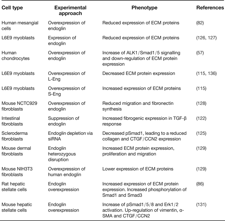
All these studies in different cell types in vitro (see tables 3 and 4), have concluded that endoglin modulate TGF-β responses in ECM-producing cells and cells involved in renal fibrosis. As most of these studies refer total endoglin and L-Eng is the largely expressed isoform, it is very likely that L-Eng is the main actor of these regulations of ECM in vitro. In L6E9 myoblasts, it has been described that overexpression of L-Eng leads to a decrease in ECM protein expression (Ref. Reference Scherner136), whereas overexpression of S-Eng leads to an increase in these proteins (Ref. Reference Velasco115).
Endoglin and renal fibrosis
The first study highlighting the possible involvement of endoglin in renal fibrosis described significant increases of endoglin in kidney biopsies from patients with chronic renal failure (Ref. Reference Roy-Chaudhury, Simpson and Power72). A few years later, Rodriguez-Pena et al. (Ref. Reference Rodriguez-Pena73) observed an increase in endoglin expression in rats after 5/6 RMR, an experimental model of glomerular fibrosis with the consequent renal function impairment. The increase in endoglin expression was suggested to be a protective mechanism against the fibrogenic effects of TGF-β. An increase in endoglin expression was also observed in a model of tubulointerstitial fibrosis induced by UUO (Ref. Reference Rodriguez-Pena17). This experimental model causes a marked renal tubulointerstitial fibrosis after 15 days of obstruction (Ref. Reference Bascands and Schanstra137).
The increase in endoglin in pathologies associated to renal fibrosis could be explained by three different mechanisms: (i) Increase of TGF-β levels. An increase in TGF-β levels has been reported in obstructed kidneys, (Ref. Reference Kaneto, Morrissey and Klahr138) and in rats with RMR (Ref. Reference Rodriguez-Pena73), and endoglin expression is induced by TGF-β, as previously described above. (ii) An increased renin–angiotensin–aldosterone system (RAAS) activity: Pimentel et al. (Ref. Reference Pimentel16) showed an overactivation of the RAAS activity after UUO that leads to TGF-β overexpression. This overexpression may contribute to endoglin expression. In fact, in vitro studies with cardiac fibroblasts have revealed that endoglin overexpression is mediated by increases in Ang II, which promotes an increased expression of profibrotic genes (Ref. Reference Chen83). (iii) TGF-β-independent mechanisms: They are some independent mechanisms such as microRNA-208a that increases endoglin expression leading to an increase in myocardial fibrosis (Ref. Reference Wang139). Furthermore, fibrotic processes take place with hypoxia as consequence of renal architecture loss (Ref. Reference Higgins140). This hypoxia acts along with TGF-β promoting endoglin expression as above explained. Sanchez-Elsner et al. (2002) have shown that HIF-1α, Smad3 and Sp1 form a multiprotein complex on the endoglin promoter, suggesting that hypoxia behaves as another TGF-β-independent endoglin up-regulation mechanism (Ref. Reference Sanchez-Elsner101).
To assess the contribution of endoglin in renal damage, Rodriguez-Pena et al. (Ref. Reference Rodriguez-Pena17) performed UUO in mice haploinsufficient for endoglin (Eng+/−). Kidneys were examined 15 days after the obstruction and no differences were found in interstitial fibrosis or in the expression of ECM proteins such as collagen or fibronectin between obstructed kidneys from Eng+/− and Eng+/+ mice. The relationship between RAAS and endoglin in chronic fibrosis has been studied by Prieto et al. (Ref. Reference Prieto141). These authors showed in rats that had undergone an RMR that after the treatment with the Ang II-converting enzyme (ACE) inhibitor trandolapril, kidneys showed a reduced fibrosis and ECM proteins expression compared with RMR rats without trandolapril treatment. However, there were no variations in endoglin levels, which suggest that the antifibrotic effect of this ACE inhibitor is independent of endoglin levels.
The role of endoglin in the inflammatory response-dependent renal fibrosis
Inflammation seems to play also a role in renal fibrosis. Most of the studies about the role of inflammation in renal fibrosis have been performed in the UUO model. UUO causes subacute renal injury characterised by tubular cell injury and cell death by apoptosis, interstitial inflammation and interstitial fibrosis. For this reason, UUO serves as a model both of irreversible acute kidney injury and of events taking place during human CKD progression. Experimental UUO has served to study the molecular mechanisms of apoptosis, inflammation and fibrosis, all three key processes in kidney injury of any cause (Ref. Reference Ucero142). Inflammation in renal fibrosis is promoted by several cytokines and growth factors, that are produced by tubular cells, infiltrated inflammatory cells – normally macrophages – and also interstitial myofibroblasts. Leucocyte accumulation in the renal interstitium is associated with renal fibrosis and injury progression. Proinflammatory cytokines such as TNF-α or IL-1β induce NF-κB activation, together with adhesion molecules, chemokines and others growth factors contribute to interstitial fibrosis (Ref. Reference Grande, Perez-Barriocanal and Lopez-Novoa143). Furthermore, some preliminary studies reveal that endoglin regulates macrophage infiltration in the kidney. Docherty et al. (Ref. Reference Docherty144) demonstrated that in an experimental model of renal ischaemia–reperfusion injury (I–R), kidneys from Eng+/− mice develop less renal injury and inflammation, less expression of adhesion markers such as ICAM-1 and VCAM-1, and less CD68 – a macrophage marker – following I–R than control mice (Ref. Reference Docherty144). Recently, Rossi et al. demonstrated that endothelial endoglin favours leucocyte adhesion and diapedesis into the tissue by interacting with the integrin α5β1 via its RGD motif (Ref. Reference Rossi145). These data suggest that endoglin is involved in the process of leucocyte adhesion and migration, and that this process is dependent on endoglin expression. It should be noted that endoglin is overexpressed in the endothelial cells of vessels in inflamed areas (Ref. Reference Lopez-Novoa and Bernabeu75). However, in an experimental model of renal injury-irradiation with 250 kV X-rays, Scharpfenecker et al. (Ref. Reference Scharpfenecker146) have evidenced that irradiated endoglin-heterozygous mice show a lover macrophage infiltration and interleukin 1b (Il1b) and 6 (Il6) expression than control mice. These authors have shown that bone marrow cells derived from endoglin heterozygous mice express less Il1b and Il6 than control mice (Ref. Reference Scharpfenecker146). At the present we cannot explain the reasons for this conceptual divergence, but it can be based in the differences between acute and chronic models in the mechanisms involved in the disease.
Role of endoglin in glomerulosclerosis
Glomeruloesclerosis is caused by an accumulation of ECM in the renal glomeruli, mainly in the glomerular mesangium resulting in the narrowing of glomerular capillaries, excessive protein ultrafiltration and a reduced GFR (Ref. Reference Lopez-Hernandez and Lopez-Novoa12). Mesangial cells are involved in glomerular capillary support and contribute to regulate glomerular capillary flow. The ability of mesangial cells to alter their morphology as well as other aspects of their biology as the synthesis of ECM components or the proliferation rate gives them a relevant role in this pathology (Ref. Reference Lopez-Novoa8). Thus, studies in mesangial cells have been very useful to analyse glomerulosclerosis and glomerular nephrotoxicity in vitro (Refs Reference Rodriguez-Barbero147, Reference Martinez-Salgado148). During glomerular injury, mesangial cells synthesise proteins such as collagens I, III, IV and osteopontin, who are not normally expressed in the mesangial matrix (Ref. Reference Abe60). One of the first evidences of the role of endoglin in glomerulosclerosis was revealed by Rodriguez-Barbero et al. (Ref. Reference Rodriguez-Barbero74), which showed that cultured human and rat mesangial cells express endoglin, and that treatment of these cells with TGF-β1 induced a significant increase in endoglin levels and collagen synthesis. Moreover, Diez-Marques et al. (Ref. Reference Diez-Marques82) observed that endoglin overexpression in human mesangial cells leads to a decrease in ECM proteins expression, both in basal conditions and after TGF-β1 treatment. These results seem to indicate that endoglin is involved in the negative regulation of TGF-β1-mediated ECM synthesis in the mesangial cells. The increase of endoglin in glomerulosclerosis should be considered as a protective mechanism against ECM protein expression, as endoglin regulates negatively this process.
With regard to the Smad-signalling pathway involved in glomerular sclerosis, kidneys from Smad3 knockout mice show less glomerulosclerosis, and less fibronectin and collagen IV expression than kidneys from wild-type mice after diabetic nephropathy induced by streptozotocin. This fact is transduced in a decrease in knockout mice and highlights a relevant role of Smad3 in glomerular pathology (Refs Reference Fujimoto149, Reference Wang150). However, other authors have described that an increase in Smad1 activation promotes collagen IV and alpha smooth muscle actin (α-SMA) synthesis by mesangial cells (Ref. Reference Abe151), as well as mesangial expansion (Ref. Reference Matsubara152).
Role of endoglin in tubulointerstitial fibrosis.
Tubulointerstitial fibrosis is characterised by the synthesis and accumulation of ECM in the peritubular, interstitial space, inflammatory cell infiltration, apoptosis of tubular cells, accumulation of fibroblasts/myofibroblasts and rarefaction of the microvasculature (Ref. Reference Zeisberg and Duffield153). Interstitial renal fibrosis is the final common pathological pathway of most progressive forms of renal disease regardless of the underlying disease or the originating compartment (Refs Reference Grande and Lopez-Novoa9, Reference Zeisberg154). Interstitial fibroblasts and myofibroblasts are considered the main cells involved in the synthesis and accumulation of ECM proteins in tubulointerstitial fibrosis (Refs Reference Grande and Lopez-Novoa9, Reference Strutz and Zeisberg155).
Tubulointerstitial fibrosis has been identified as one of the major secondary effects of radiation, in addition to vascular damage (Refs Reference Lara156, Reference Rodemann and Muller157). After radiation, Eng+/− mice showed less tubulointerstitial fibrosis than their respective controls. However, the explanation of this effect was not found in the ratio ALK1/ALK5, because a smaller activation of phospho-Smad1 was observed in these mice (Ref. Reference Scharpfenecker158). Recently it has been demonstrated that after radiation, Eng+/− mice show less deterioration of kidney function and ultrastructure than their controls littermates. Moreover, irradiated Eng+/− mice show a lower increase of PDGFR-β positive cells. These cells are pericytes that lose its contact with endothelial cells and undergo a transdifferentiation into myofibroblasts, the major ECM-producing cells. Thus, the reduced increase in tubulointerstitial fibrosis observed in Eng+/− mice may be due a lower formation of myofibroblasts (Ref. Reference Scharpfenecker159). However, the amount of ECM proteins in the obstructed kidneys of Eng+/− mice and in wild-type mice was similar after 15 days of UUO (Ref. Reference Rodriguez-Pena17).
In summary, during renal fibrosis, several TGF-β and ischaemia lead to an increase in endoglin expression in numerous ECM-producing cells, including mesangial cells, tubular cells and myofibroblasts. Endoglin in mesangial cells behaves as an antifibrotic protein, whereas in other cells such as myofibroblasts, endoglin may promote ECM protein expression. Moreover, endoglin acts as a chemoattractant molecule that favours inflammatory cells infiltration and adhesion.
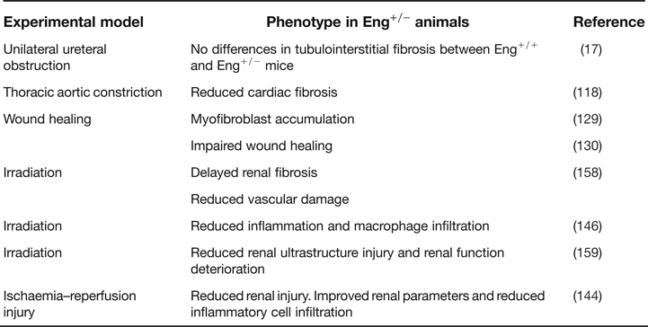
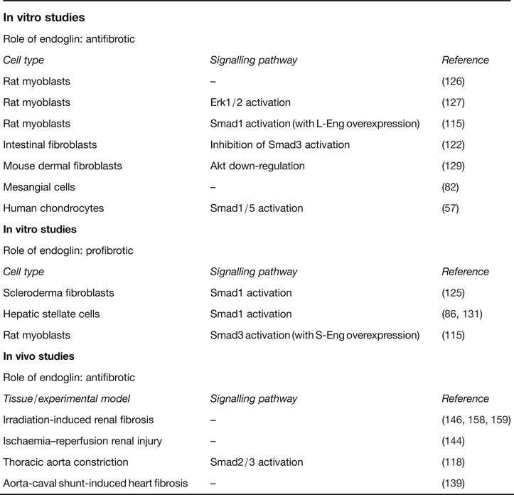
In order to clarify the profibrotic/antifibrotic role of endoglin we have summarised in Tables 2 and 3 all the studies in vivo and in vitro assessing the effect of endoglin in fibrosis regulation. Moreover, the numerous approaches to study the function of endoglin in different cells are summarised in Table 4.
Set of in vivo experimental studies with endoglin heterozygous mice (Eng+/−) and their respective controls (Eng+/+) to elucidate the possible role of endoglin in different models or contexts of fibrosis
Summarising table with a complete set of in vitro and in vivo studies to study endoglin role of fibrosis, its involvement (profibrotic or antifibrotic) and the signalling pathway involved in the phenotype
Complete list of in vitro studies made to understand the role of endoglin in ECM protein expression, with the experimental approach and the cell response or phenotype observed
Research in progress and outstanding research questions
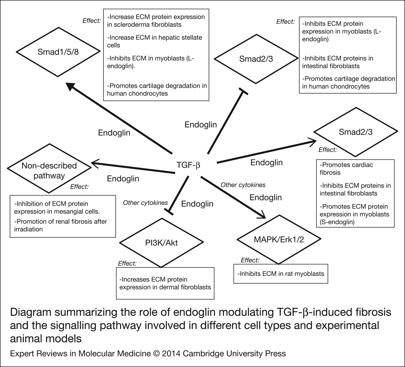
Although the available data are relatively few and fragmentary (Fig. 3), it can be suggested that endoglin plays an important role in several processes involved in renal fibrosis such as ECM protein synthesis in several cell types and inflammatory cell infiltration. However, further studies will be necessary in order to elucidate the precise role of endoglin in renal fibrosis and the specific pathways involved in this protection. The answer to the disparate role of endoglin in fibrosis will likely lie in various factors that influence the function of endoglin: (a) endoglin functions as co-receptor for different members of the TGF-β superfamily, of which individual members have been shown to have opposite functions in fibrosis (Refs Reference Hawinkels and Ten Dijke160, Reference Weiskirchen161); (b) different splice variants of endoglin and sEng can mediate different (even opposite) biological responses (Refs Reference Perez-Gomez92, Reference Velasco115, Reference Castonguay162), (c) the type of experimental model of fibrosis used (chronic or acute) and (d) the context in which endoglin operates in different cell types.
Diagram summarising the role of endoglin modulating TGF-β-induced fibrosis and the signalling pathway involved in different cell types and experimental animal models. Pointed arrows represent activator effect of endoglin in this signalling pathway, whereas flat-end arrows represent inhibitor effect of endoglin in this signalling pathway. In the boxes are described the effect of endoglin and the cell types or animal models in which these effects have been described.
More research tools such as conditional endoglin knock out, animal models using sEng and neutralising endoglin antibodies have recently become available (Refs Reference Castonguay162, Reference Mahmoud163, Reference Seon164) and they will give more light on the precise effects of endoglin on renal fibrosis an on the mechanisms involved in these effects.
The above-described role of endoglin as a major regulator of fibrosis and its relationship with TGF-β and BMPs signalling invites us to consider the possibility of a clinical use of endoglin as an important therapeutic target in renal fibrosis. In this respect, TRC105 (Tracon Pharmaceuticals) is a chimaeric human IgG1 antiendoglin antiboby, derived from a monoclonal antiendoglin antibody (Ref. Reference Uneda165). TRC105 has been shown to inhibit HUVEC cells proliferation and inhibits tumour metastasis and it is being studied against different tumour types (metastatic breast cancer, advanced solid tumour, renal cell cancer, glioblastoma multiforme, etc.) (Ref. Reference Bhatt and Atkins166). It is possible that the use of this antibody could give some light on the role of endoglin in fibrosis.
Other possible candidate for therapeutical target on endoglin-mediated fibrosis is sEng. Although the effects of sEng on TGF-β-activated signalling pathways are unclear, sEng seems to inhibit several cellular effects of TGF-β in the presence of full-length membrane endoglin: eNOS activation in endothelial cells (Ref. Reference Venkatesha95), angiogenesis (Ref. Reference Hawinkels and Ten Dijke160) and leucocyte adhesion (Ref. Reference Scherner136). Thus, performing studies in which sEng is administered in experimental models of kidney fibrosis or inducing kidney fibrosis in mice overexpressing sEng would be interesting to elucidate if sEng is able to counteract TGF-β-induced renal fibrosis and if sEng may be used as a therapeutic target. All these studies will ultimately lead to new therapies for fibrosis.
Acknowledgements
The authors thank to Dr Fernando Perez-Barriocanal and Dr Carlos Martinez-Salgado for critically reading the manuscript. Studies from the authors' laboratories have been supported by grants from Ministerio de Economía y Competitividad of Spain (SAF2007-61827, SAF2010-15881 and SAF2013-45784-R), Junta de Castilla y Leon (Grant GR100) and Red de Investigación Cooperativa en Enfermedades Renales (Red-inRen, RD06/0016/0013 and RD12/0021/0032 to JML-N). REDINREN is an initiative of the Instituto de Salud Carlos III (ISCIII) of Spain supported by FEDER funds. BO is supported by a fellowship from Ministry of Science and Innovation (BES-2008-005550) and JMMF is supported by a fellowship from Junta de Castilla y León and Fondo Social Europeo.