Case report
Percutaneous pulmonary valve implantation is widely used to treat native or post-surgical right ventricular outflow tract dysfunction. Reference Bonhoeffer, Boudjemline and Saliba1 Right ventricular outflow tract dysfunction leads to right ventricle dilation which in turn promotes dilation of the tricuspid annulus causing tricuspid regurgitation. If significant, tricuspid regurgitation can be surgically repaired with an annuloplasty ring. However, long-term results are not satisfactory, with more than 25% of patients developing moderate to severe regurgitation 5 years after surgical correction. For high-risk surgical patients, percutaneous implantation of a pulmonary valve in a tricuspid position, either valve-in-valve or valve-in-ring, is emerging as a lower risk solution. Reference Aboulhosn, Cabalka and Levi2 We report on a successful simultaneous percutaneous valve implantation at both locations. To the best of our knowledge, this is the first report describing the simultaneous of Melody® pulmonary valves during the same procedure in the pulmonary and tricuspid positions, the latter a valve-in-ring implantation.
Fifty-year-old female with congenital severe pulmonary valve stenosis and atrial septal defect, previously submitted to several surgical procedures, namely, surgical pulmonary valvotomy at 6 years of age, followed by, at the age of 21 years, right atrial reduction plasty, implantation of a 25 mm bioprosthetic pulmonary valve and a 33 mm Carpentier-Edwards incomplete ring annulloplasty. She also had type 2 diabetes mellitus, hypothyroidism, atrial fibrillation, and she was in New York Heart Association (NYHA) I–II functional class of heart failure when the intervention was planned. However, in the context of lower limb erysipelas, she developed a septic and cardiogenic shock, complicated with renal failure. At the time of the procedure, she had recovered from the infection, albeit, maintaining severely reduced functional capacity, as well as being dependent on daily haemodialysis.
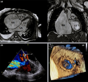
Multi-modality imaging examinations revealed mild right ventricular systolic dysfunction, severe right atrial dilation (300 ml/m2) (Fig 1a), right ventricular enlargement (end-diastolic volume of 155 mL/m2, end-systolic volume of 80 mL/m2), central torrential tricuspid regurgitation (Fig 1b), tissue dehiscence adjacent to the opening part of the ring (Fig 1c) and severe pulmonary valve stenosis (peak Doppler systolic gradient of 85 mmHg).
( a ) Cardiac magnetic resonance imaging, axial and coronal planes. Note the massive right atrium dilation. ( b ) 2D transesophageal echocardiography, four chambers view, colour Doppler mode. Carpentier ring in tricuspid position with massive tricuspid regurgitation through the ring. ( c ) 3D transesophageal echocardiography, mid-esophageal view. Carpentier ring seen from right atrium side. Note the tissue dehiscence adjacent to the opening part of the ring (arrow) (possibly overestimated due to very thin tissue drop-out artifact). Legend: Ao – Aorta; LA – left atrium; LV – left ventricle; PA – pulmonary artery; RA – right atrium; RV – right ventricle; TR – triscuspid regurgitation.

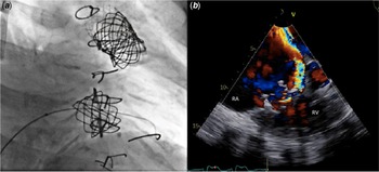
Left and right-heart catheterisation was carried out, and the right ventricle outflow tract angiogram confirmed severe bioprosthetic pulmonary valve stenosis with post-stenotic pulmonary artery dilation. The peak-to-peak pressure gradient was 50 mmHg, and the RV systolic pressure was greater than 75% of the left ventricle systolic pressure. We initially performed a percutaneous balloon pulmonary valvuloplasty with a 20/40 mm Atlas Gold balloon (BD®, United States of America). A 39 mm Bare CP Stent (NuMED®, United States) mounted on a 22/40 mm Atlas Gold balloon (BD®, United States) was then implanted. In order to further secure this stent and create a safe landing zone, an AndraStent XXL 48 mm (Andramed®, Germany) mounted on a 24/60 mm Andraballoon (BD®, United States of America) was also implanted. This was followed by successful implantation of a 22 mm Melody valve (Medtronic®, United States of America) in pulmonary position. Despite the immediate optimal result, with no pulmonary regurgitation, tricuspid regurgitation remained significant as confirmed by transesophageal echocardiography. As the mechanism of tricuspid regurgitation was considered to be due to a major coaptation gap with leaflet tethering mainly through the tricuspid ring, we then proceeded with a valve-in ring implantation with a 22 mm Melody valve (Medtronic®, United States of America) in the tricuspid position under 3D-echocardiography guidance and balloon sizing, with good overall result (Fig 2a). Nonetheless and as expected, a moderate paravalvular leak at triscuspid position was observed post-implantation (Fig 2b). The patient was discharged home 1 week later with progressive improvement of the heart failure functional class (NYHA I 3 months later).
( a ) Catheterisation fluoroscopy, anteroposterior view. Two 22 mm Melody® valves implanted in the pulmonary position (valve-in-valve, upper valve) and in tricuspid position (valve-in-ring, lower valve). ( b ) 2D transesophageal echocardiography colour doppler mode, four chambers view. Melody® valve implanted successfully inside carpentier ring with moderate paravalvular leakage. Legend: * – paravalvular leakage; RA – right atrium; RV – right ventricle.

Discussion
Congenital severe pulmonary valve stenosis leads to right ventricular outflow tract dysfunction with right ventricular dilation and systolic dysfunction. As the ventricle enlarges, the tricuspid annulus also dilates and tricuspid regurgitation occurs, which in turn further worsens the dilation of the right chambers. Surgical annuloplasty ring is a solution to reduce the annulus size but the long-term results are not satisfactory.
Our patient presented with both right ventricular outflow tract dysfunction and torrential tricuspid regurgitation, and was a high surgical risk candidate due to the other comorbidities. When we first implanted the Melody valve (Medtronic®, United States of America) in the pulmonary position, we expected that the reduction of the end-diastolic right ventricular pressure would also reduce the tricuspid regurgitation. However, little impact was observed, and thus we opted to proceed with the valve-in-ring implantation as described above. As reported, the worldwide experience of percutaneous tricuspid valve implantation is scant, Reference Webb, Wood and Ye3 and less so in valve-in-ring contexts. The valve-in-valve percutaneous tricuspid valve implantation in the setting of a dysfunctional bioprosthesis is more feasible because the bioprosthesis calcification creates a suitable and safer landing zone to the device implantation. However, an annuloplasty ring, specially an incomplete ring, as in our case, represents a greater challenge to device implantation, as it tends to dilate, creating a larger and irregular landing zone. Furthermore, the risk of paravalvular leak is more frequent with incomplete rings. In this case, the tricuspid anatomy, including ring size was carefully assessed using different imaging techniques (cardiac computed tomography scan, 3D-transesophageal echocardiography during procedure and balloon sizing) and the risk of paravalvular leak was taken into account. Based on the data and the patient’s clinical condition, we decided on valve-in-ring implantation. Despite the paravalvular leak, the patient improved significantly. Additional device closure of the paravalvular leak is being considered for a later stage.
The transcatheter implantation of both valves in one single procedure with a tricuspid valve-in-valve implantation has already been previously reported. Reference Jux, Akintuerk and Schranz4 As far as we know, this is the first reported case of a simultaneous two valves implantation in the setting of a valve-in-ring.
We highlight the multi-modality of imaging examinations prior and during the procedure and the multi-disciplinary team at the cath lab in order to achieve a good result. 2D and colour Doppler transthoracic echocardiography, cardiac magnetic resonance and cardiac computed tomography scan were important in the decision-making process prior to the intervention. 2D and 3D transesophageal echocardiography played an essential role during the procedure, as it highlighted morphological and functional issues which guided our management options.
Conclusion
Transcatheter valve-in-ring device implantation for tricuspid valve regurgitation is feasible using currently available valvular devices used in the context of pulmonary percutaneous valve implantation. The simultaneous percutaneous implantation of both valves is possible, potentially avoiding a second procedure.
Acknowledgements
The authors would like to thank all the other health staff involved in the case.
Financial support
This research received no specific grant from any funding agency, commercial or not-for-profit sectors.
Conflicts of interest
None.