
Introduction
In medical devices, biofouling is an undesirable process of bacterial colonization that occurs spontaneously, in temporary (e.g., contact lenses, endotracheal tubes as well as urinary and central venous catheters) and in permanently implanted devices (e.g., cardiac valves, vascular grafts and joint replacements), leading to biomaterial-associated infections.[Reference Costerton, Stewart and Greenberg1] Biofilms grown on biomaterials are responsible for 75% of human bacterial infections[Reference Miquel, Lagrafeuille, Souweine and Forestier2] and among these, urinary tract infections are very common, affecting around 150 million people each year worldwide.[Reference Stamm and Norrby3] Escherichia coli is the predominant uropathogen responsible for about 80% of all hospital-acquired infections and can adhere to biomedical surfaces and consequently, develop biofilms.[Reference Koseoglu, Aslan, Esen, Sen and Coban4–Reference Shunmugaperumal6] Delaying the biofilm onset can reduce the need for antimicrobial treatment and also the costs for replacing the infected materials.
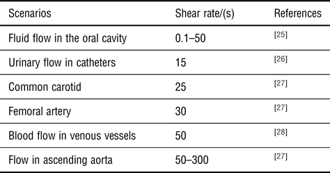
Antifouling surfaces have been extensively developed by altering the physical and/or chemical surface properties, mimicking natural antifouling surfaces, and enhancing the surface resistance to biologic contamination.[Reference Nir and Reches7] Great efforts in surface engineering have been made in order to prevent or mitigate bacterial adhesion and biofilm development using nanoparticles, polymer brushes, antifouling self-assembly monolayers, and hydrogels.[Reference Kirschner and Brennan8–Reference Azeredo, Azevedo, Briandet, Cerca, Coenye, Costa, Desvaux, Di Bonaventura, Hébraud and Jaglic15] Maity, Nir, Zada, and Reches[Reference Maity, Nir, Zada and Reches14] reported the self-assembly of a tripeptide into a functional coating that interferes with the first step of biofouling. The peptide design includes the amino acid, 3,4-dihydroxy-L-phenylalanine, a constituent of mussel adhesive proteins, which promotes adhesion to any substrate. Diphenylalanine is the element that directs the self-assembly and modification of the benzene rings of phenylalanine with fluorine atoms provides the antifouling function. This peptide configuration displayed a reduction of up to 90% of the attached bacteria (Pseudomonas aeruginosa and E. coli) under static conditions.[Reference Maity, Nir, Zada and Reches14] Nevertheless, previous reports demonstrated that bacterial adhesion may not only be affected by the surface properties, but also by the shear forces existing in the system.[Reference Moreira, Simões, Melo and Mergulhão12, Reference Absolom, Lamberti, Policova, Zingg, van Oss and Neumann16, Reference Liu and Zhao17] In the biomedical field, the fluid behavior is dependent on specific physiological conditions and can affect cell attachment, biofilm development, morphology, and antimicrobial susceptibility.[Reference Gomes, Silva, Simoes, Melo and Mergulhao18–Reference Baier, Meyer, Natiella, Natiella and Carter22] One important parameter that describes the shear effects is the shear rate which is the derivative of the velocity in the perpendicular direction from the surface and it is also an indicator of the frequency at which cells contact the surface. Table I lists several biomedical scenarios where the shear rate is within the range tested here (7–80/s). Testing was performed using a parallel plate flow chamber (PPFC), which is a widely used platform for studying biofilms under controlled hydrodynamic conditions.[Reference Azeredo, Azevedo, Briandet, Cerca, Coenye, Costa, Desvaux, Di Bonaventura, Hébraud and Jaglic15, Reference Moreira, Araújo, Miranda, Simões, Melo and Mergulhão23, Reference Busscher and van der Mei24]
Wall shear rates found in the human body and in medical devices.

The main goal of this study was to assess the antifouling performance of a peptide coating under a broad range of hydrodynamic conditions that can be found in many biomedical applications. The surface coating was characterized by various techniques and the effect of the surrounding medium composition was evaluated. This work follows a previous report in which this antifouling surface was tested under static conditions.[Reference Maity, Nir, Zada and Reches14]
Materials and methods
Preparation and characterization of surfaces
Sodium dodecyl sulfate (SDS) was obtained from Merck™ (Israel). Ethanol and methanol were obtained from Bio-lab (Israel). Glass microscope slides (W × D × H; 76 × 26 × 1 mm, VWR) were dipped in 2% SDS (w/w) for 30 min. They were then rinsed with triple-distilled water (TDW), transferred to methanol for an additional 30 min, dipped in ethanol for 30 min and dried under nitrogen. Clean slides were then placed in a ventilated hood and coated with peptide solution (0.5 mg/mL in ethanol) using a dual action spray airbrush (DuraPowers, CA, USA). Each slide was sprayed three times.
Attenuated total reflectance (ATR), Fourier transform infrared (FTIR) analysis
FTIR (Nicolet 6700 model, Thermo Scientific, MA, USA) with a Ge-ATR arrangement (VariGATR™, Harrick Scientific, NY, USA) recorded the ATR spectra. The spectra were collected with an applied force of 350 N at 4/cm resolution with 3000 scans averaged signal and an incident angle of 65°. OMNIC™ analysis program (Thermo Scientific, MA, USA) determined the absorbance peaks. Applying the second derivative on the spectrum enhanced the signal of the peaks.
Quartz crystal microbalance with dissipation monitoring (QCM-D) analysis
Peptide adhesion onto SiO2 surfaces was studied using QCM-D (Q-sense, Biolin Scientific, Sweden). Specifically, we used a flow module Explorer system and SiO2 sensors with a fundamental resonant frequency of 5 MHz. Prior to each experiment, SiO2 sensors were cleaned with Oxygen/Plasma (Atto, Diener Electronic, Germany), followed by washing with 2% SDS and TDW and finally dried under nitrogen. The studied solutions were injected into the sensor crystal chamber at 0.1 mL/min. The peptide was dissolved in ethanol to a concentration of 0.5 mg/mL. Data were analyzed using QTools software. The frequency was fitted to the Sauerbrey equation and the adsorbed mass and layer thickness were calculated according to the linear relation[Reference Marx29]:
 $$\Delta m = - \displaystyle{{C \cdot \Delta f} \over n},$$
where Δm is the adsorbed mass, Δf is the frequency change during peptide adsorption, C is the sensitivity constant characteristic of the quartz crystal (17.7 ng/cm2·Hz) and n is the overtone number. The reported values were averaged for overtones 5 and 7.
Ellipsometry
α-SE spectroscopic ellipsometer measured the thickness of the peptide-based coating (J.A. Woollam, NE, USA). Measurements were carried out at wavelengths ranging from 380 to 900 nm at a 70° angle of incidence. The optical properties of the substrate were fitted using SiO2. The thickness of the layers and refractive indices were fitted according to the Cauchy model[Reference Tompkins and McGahan30] and an angle offset was permitted. Then, the parameters were allowed to be fitted to determine values that are more accurate.
Atomic force microscopy (AFM) analysis
AFM analysis was carried out using the AC mode with a Si3N2 tip (spring constant of 3 N/m, NanoWizard 3, JPK Instruments, Germany). Data were processed using JPK data processing software (JPK Instruments, Germany). Arithmetic roughness (Ra) and peak-valley values were calculated from a cross-section.
Bacterial culture
E. coli JM109(DE3) supplied by Promega (Madison, WI, USA) was used in this work has it was shown to adhere to different surfaces in a diversity of platforms operated under different shear conditions.[Reference Gomes, Silva, Simoes, Melo and Mergulhao18, Reference Moreira, Ponmozhi, Campos, Miranda and Mergulhão31, Reference Teodósio, Simões, Melo and Mergulhão32] Its genotype is [endA1, recA1, gyrA96, thi, hsdR17 ( $r_k^ - $,
 $m_k^ + $), relA1, supE44, λ − Δ(lac-proAB), [F”, traD36, proAB, lacIq ZΔM15], λ(DE3)][Reference Teodósio, Simões and Mergulhão33] and this strain has shown a similar adhesion behavior when compared with a clinical isolate, E. coli CECT 434.[Reference Gomes, Moreira, Teodósio, Araújo, Miranda, Simões, Melo and Mergulhão19] Motility studies show that E. coli JM109(DE3) and CECT 434 have similar swarming and twitching motilities although the later has a higher swimming motility (not shown) indicating that flagella are active[Reference Neidhardt34] in both strains.
A cell suspension was prepared by inoculating a glycerol stock in 0.2 L of inoculation medium. This medium consisted of 5.5 g/L glucose (Chem-Lab nv, Belgium), 2.5 g/L peptone (Oxoid, UK), and 1.25 g/L yeast extract (Merck KGaA, Germany) in phosphate buffer (1.88 g/L KH2PO4 and 2.60 g/L Na2HPO4; Chem-Lab nv, Belgium), pH 7.0.[Reference Teodósio, Simões, Melo and Mergulhão32] The culture was incubated at 37 °C, overnight with agitation (160 rpm; IKA KS 130 basic, Staufen, Germany). Cells were harvested by centrifugation (for 10 min at 3200g), and washed twice either in 0.05 mol/L of citrate buffer (CB; Sharlau, Spain), pH 5.0[Reference Moreira, Ponmozhi, Campos, Miranda and Mergulhão31] or in enriched nutrient medium (EM), pH 7.0. EM is a 1:100 dilution of the inoculation medium where the phosphate buffer concentration is maintained.[Reference Moreira, Fulgêncio, Alves, Machado, Bialuch, Melo, Simões and Mergulhão35, Reference Moreira, Gomes, Simões, Melo and Mergulhão36] EM was chosen since it was reported that higher nutrient levels can favor cell adhesion[Reference Gomes, Moreira, Teodósio, Araújo, Miranda, Simões, Melo and Mergulhão19, Reference Frias, Ribas and Lucena37]; however, the inoculation medium was still diluted to decrease cell division and to enable us to work with cells that would be in a more uniform physiological state. The pellet was resuspended in the corresponding medium and the cellular suspensions were adjusted to a final concentration of 7.6 × 10⁷ cells/mL, assessed by optical density at 610 nm (OD = 0.1).
Surface hydrophobicity and free energy of adhesion
The hydrophobicity of the bare glass and peptide-coated glass was evaluated using the Lifshitz-van der Waals acid-base approach.[Reference Van Oss38, Reference Van Oss, Good and Chaudhury39] Contact angles were measured (OCA 15 Plus; Dataphysics, Filderstadt, Germany) using three reference liquids: water and formamide (polar solvents) and α-bromonaphthalene (nonpolar solvent) (Sigma-Aldrich Co., Portugal).[Reference Janczuk, Chibowski, Bruque, Kerkeb and Caballero40] Surface tension components were taken from the literature.[Reference Janczuk, Chibowski, Bruque, Kerkeb and Caballero40] Measurements of at least 10 drops on each surface with each liquid were performed at room temperature. The surface energy components of a solid surface (s) were determined by measuring the contact angles (θ) with three reference liquids (l) with known surface tension components, followed by solving three equations of the type[Reference Van Oss38]:
 $$(1 + {\cos}\theta)\gamma_l^{\rm Tot} = 2\left({\sqrt {\gamma_s^{LW} \times \gamma_l^{LW}} + \sqrt {\gamma_s^{+} \times \gamma_l^{-}}+ \sqrt {\gamma_s^{-} \times \gamma_l^{+}}}\right).$$
From the model proposed by van Oss,[Reference Van Oss41] the total surface energy  $\lpar {\gamma_l^{{\rm Tot}}} \rpar $ of a pure substance can be expressed as:
 $$\gamma _l^{{\rm Tot}} = \gamma ^{LW} + \gamma ^{AB},$$
where γ LW and γ AB are the Lifshitz-van der Waals components of the surface free energy and Lewis acid-base components, respectively.
The polar AB component comprises the electron acceptor (γ +) and electron donor (γ −) parameters, given as:
 $$\gamma ^{AB} = 2\sqrt {\gamma ^ + \times \gamma ^ -}. $$
The hydrophobicity of a surface is expressed as the free energy of interaction ΔG TOT between two entities of the surface immersed in a polar liquid like water (w). In this approach, the ΔG value can be determined using the surface tension components using the following equation:
 $$\eqalign{& \Delta G^{{\rm TOT}} = - 2\left( {\sqrt {\gamma_s^{LW}} - \sqrt {\gamma_w^{LW}}} \right)^2 \cr & \quad + 4\left( {\sqrt {\gamma_s^ + \times \gamma_w^ -} + \sqrt {\gamma_s^ - \times \gamma_w^ +} - \sqrt {\gamma_s^ + \times \gamma_s^ -} - \sqrt {\gamma_w^ + \times \gamma_w^ -}} \right).} $$
If ΔG < 0 mJ/m2, the material is considered hydrophobic because the interaction between the two entities is stronger than the interaction of each entity with water. Conversely, if ΔG > 0 mJ/m2, the material is hydrophilic.
The free energy of adhesion between a surface (s) and bacteria (b) immersed in water, is expressed as the total interaction energy, (ΔG Adh), and can be calculated by:
 $$\eqalign{& \Delta G^{{\rm Adh}} = \gamma _{sb}^{LW} - \gamma _{sw}^{LW} - \gamma _{bw}^{LW} \cr & \quad + 2\left[ {\sqrt {\gamma _w^ + } \left( {\sqrt {\gamma _s^ - } + \sqrt {\gamma _b^ - } - \sqrt {\gamma _w^ - } } \right) + \sqrt {\gamma _w^ - } \left( {\sqrt {\gamma _s^ + } } \right.} \right. \cr & \quad + \sqrt {\gamma _b^ + } - \sqrt {\gamma _w^ + } ) - \sqrt {\gamma _s^ + \times \gamma _b^ - } - \sqrt {\gamma _s^ - \times \gamma _b^ + } ].} $$
Thermodynamically, if ΔG Adh < 0 mJ/m2, adhesion is favored, whereas adhesion is not expected to occur if ΔG Adh > 0 mJ/m2. The contact angle measurements and the subsequent hydrophobicity and thermodynamic analysis was performed on the pristine surfaces (Table II) and also after 30 min incubation of the surfaces in CB or EM (Table S1, Supporting Information).
Contact angles with water (θ w), formamide (θ F), and α-bromonaphthalene (θ B); surface tension parameters (γ LW,γ +, and γ −), hydrophobicity (ΔG) and free energy of adhesion (ΔG Adh) between E. coli and each surface.

Values are means ± SDs of three independent experiments.
Parallel plate flow chamber assays
A PPFC was used to follow E. coli adhesion to both bare glass and peptide-coated glass, as previously described.[Reference Moreira, Araújo, Miranda, Simões, Melo and Mergulhão23] Briefly, the PPFC was coupled to a jacketed tank connected to centrifugal pumps. This system was sterilized by recirculating a sodium hypochlorite solution (3% v/v; 3 cycles of 15 min each). Subsequently, it was washed with sterile water and placed inside a laminar flow chamber for UV sterilization (30 min). The PPFC contained an opening area for introducing the test surfaces as previously described.[Reference Moreira, Araújo, Miranda, Simões, Melo and Mergulhão23] All the surfaces were sterilized by spraying with absolute ethanol for 5 min. The surfaces were then placed in the PPFC and the system was properly mounted in a microscope stage to monitor cell attachment. The bacterial suspension was recirculated through the PPFC system at different flow rates (from 1 to 8 mL/s) that correspond to shear rate values between 7 and 80/s.[Reference Moreira, Araújo, Miranda, Simões, Melo and Mergulhão23] Cell attachment was monitored in real-time for 30 min. At 60 s intervals, a camera (Nikon DS-Ri 1, Japan) connected to a microscope (Nikon Eclipse LV100, Japan) was used for image acquisition. A recirculating water bath was connected to the tank jacket to enable temperature control at 37 °C to mimic human body conditions. Imaging conditions (e.g., exposure time, light intensity, magnification, condenser height, and approximate positioning of the slide) were maintained between experiments. Three independent experiments were performed for each surface and condition tested.
The microscopic images were analyzed by ImageJ (version 1.38e) software to determine the number of adherent cells per square centimeter at each time point for each shear rate tested. Initial adhesion rates (IAR) were obtained from linear regression of the data presented in Fig. 2.
Statistical analysis
The data presented in Figs. 2 and 3 represent an average of three independent results for each tested condition. To verify the statistical significance, the data were analyzed using GraphPad Prism® 6 (La Jolla, CA, USA). Paired t-test analysis was also performed for confidence levels of 95% (differences are reported as significant for P values < 0.05 and are denoted by  ) and 90% (differences are reported as significant for P values <0.1 and are denoted by
 ).
Results and discussion
Characterization of the peptide-based coating
The glass was coated since it is a flat transparent inexpensive surface that has been previously used with this flow system.[Reference Moreira, Simões, Melo and Mergulhão12, Reference Moreira, Araújo, Miranda, Simões, Melo and Mergulhão23]
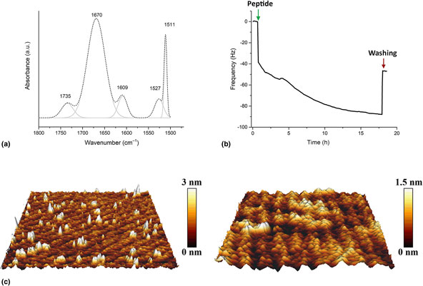
The formulation evaluated here consists of a low-molecular-weight peptide that interferes with the first step of biofouling.[Reference Maity, Nir, Zada and Reches14] Unlike the previous work where the peptide self-assembled on the surface by dip coating,[Reference Maity, Nir, Zada and Reches14] here the peptide-coated the surface by spraying and rapid evaporation of the solvent. ATR-FTIR results [Fig. 1(a)] showed that despite the different coating process, the peptide preserved its surface orientation, mostly consisting of anti-parallel beta sheets with peaks at 1609 and 1670 cm−1, as previously demonstrated.[Reference Maity, Nir, Zada and Reches14]
Characterization of the peptide-coated surface. (a) ATR-FTIR spectrum of the amide region: original measurement (the dashed line) and the second derivative fit (the grey line). (b) Real-time QCM-D measurement. A fifth overtone is presented. (c) The surface topography of bare glass (on the left) and peptide-coated glass (on the right) (2 µm × 2 µm scan).

QCM-D measurement followed the peptide adsorption onto a SiO2 sensor, which is used as the model system for glass.[Reference Ojovan42] Fig. 1(b) depicts the QCM-D real-time monitoring of the process. Upon adding the peptide, the frequency decreased instantly to about −40 Hz. Then, it decreased at a more moderate rate, changing again after 5 h. At the end of the run, the sensor was washed with clean ethanol, the non-adsorbed peptide was removed, and the overall frequency change was ~45 Hz. From Fig. 1(b), it can be concluded that the adsorption process can be divided into three steps, where the peptide bonding to SiO2 decreases in each step. This may be related to the formation of the layers in which the first peptide layer formed immediately, resulting in a high-frequency change [Fig. S1(phase 1), Supporting Information]. Then, owing to the change in the sensor–solvent interface, the adsorption process slowed down and the amount of peptide adsorbed decreased [Fig. S1(phase 2), Supporting Information]. Eventually, all binding sites were saturated and the peptide was adsorbed by a different mechanism, probably via non-specific interactions [Fig. S1(phase 3), Supporting Information]. However, these interactions are weaker and the peptide was washed away at the end of the run. By fitting the frequency change to the Sauerbrey equation [eq. (1)], it was possible to calculate the adsorbed mass. The loaded mass per surface area was found to be 901 ± 166 ng/cm2 and the layer thickness was 9 ± 1 nm [Fig. S2, Supporting Information]. This thickness value is similar to the one determined by ellipsometry (10.7 ± 0.5 nm).
AFM was used to characterize the nano- and micro-scale topographical structures of the bare glass and peptide-coated glass [Fig. 1(c)]. The images revealed that after deposition of the peptide onto the glass slide, the surface seems smoother and more homogeneous. Additionally, the average roughness (Ra) decreased from 490 ± 40 pm for the bare glass to 232 ± 1 pm for the peptide-coated glass.
A thermodynamic analysis of the pristine surfaces and E. coli cells was performed, based on contact angle measurements (Table II). It was observed that the bare glass and E. coli cells are hydrophilic (ΔG > 0 mJ/m2), whereas the peptide coating is hydrophobic (ΔG < 0 mJ/m2). Regarding the γ + and γ − components, the bare and coated glass are both polar surfaces, being mostly electron donors with a small electron acceptor component. From a thermodynamic point of view, E. coli adhesion to both surfaces was not expected to occur (ΔG Adh > 0 mJ/m2). Nevertheless, just considering the analysis of the pristine surfaces, E. coli adhesion to glass is predicted to be thermodynamically less favorable than to peptide because ΔG Adh glass >ΔG Adh peptide.
Bacterial adhesion assays
The performance of an antifouling peptide-coated glass was evaluated under different hydrodynamic conditions (shear rates between 7 and 80/s) using two different media and E. coli as a model organism. The peptide formulation used here was the best configuration for biofilm reduction, as demonstrated in a previous study.[Reference Maity, Nir, Zada and Reches14] In that work, biofilms of P. aeruginosa and E. coli were formed under static conditions on bare titanium surfaces and titanium surfaces coated with different formulations of peptides. The peptide containing fluorinated phenylalanine enabled a 93 and 74% reduction in the biofilm biomass of P. aeruginosa and E. coli, respectively, as assayed by crystal violet staining. Hydrodynamics are known to affect biofilm formation on medical devices as a result of the fluid flow, which can modulate cell adhesion to the surface.[Reference Moreira, Araújo, Miranda, Simões, Melo and Mergulhão23, Reference Busscher and van der Mei24, Reference Teodósio, Silva, Moreira, Simões, Melo, Alves and Mergulhão43] To evaluate the performance of the same peptide under different hydrodynamic conditions, a PPFC was used.
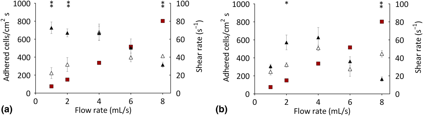
Figure 2 presents the initial adhesion results obtained for each surface (glass and peptide coating) for 30 min on the PPFC, from which the IARs were calculated (Fig. 3). The number of adherent bacteria increased with time for both surfaces and culture media (Fig. 2). In general, higher bacterial adhesion was obtained for glass (particularly in CB), which is the most hydrophilic surface material (Table II). Previous reports have shown that E. coli adhesion is higher on hydrophobic surfaces than on hydrophilic surfaces.[Reference Fletcher and Loeb44, Reference Cerca, Pier, Vilanova, Oliveira and Azeredo45] However, a correlation between bacterial adhesion and surface hydrophobicity was not found here probably due to other effects (biologic and physical), which may be comparatively more important.[Reference Moreira, Simões, Melo and Mergulhão12, Reference Oliveira, Oliveira, Teixeira, Azeredo, Henriques and Oliveira46] In order to assess the effect of the assay media on surface hydrophobicity, the contact angles were measured and the subsequent thermodynamic analysis was also performed after 30 min incubation of the surfaces in CB or EM without the presence of bacterial cells (Table S1, Supporting Information). We found that the hydrophobicity of glass increased after incubation in CB and the predicted energy of adhesion decreased. A similar trend was observed for the peptide coating after it was immersed in EM; however, no significant changes were detected in these two parameters when glass was immersed on EM or the peptide was incubated with CB. Thus, it seems that the model can predict a higher adhesion on glass after immersion in CB (a higher hydrophobicity and a lower energy of interaction) when compared with EM, but it fails to predict the observed behavior in the peptide coating. Apparently, the thermodynamic model is thus better suited to handle simple surfaces like glass[Reference Moreira, Simões, Melo and Mergulhão12] and fails to predict the results on more complex surfaces, such as the peptide coating where other interactions can be significant.[Reference Bos, Van der Mei and Busscher47]
Adhesion of Escherichia coli on bare glass (●) and peptide-coated glass (○) for 30 min under different shear rates: (a) 7/s, (b) 15/s, (c) 30/s, (d) 50 and (e) 80/s. Two different culture media were used: citrate buffer (CB) and enriched medium (EM). The means ± SDs for three independent experiments are illustrated. Statistically significant differences obtained between the surfaces are indicated by for P < 0.05 or for P < 0.1 at the corresponding shear rate values.

Initial adhesion rates for bare glass (▲) and peptide-coated glass (Δ) at each shear rate ( )/(s). (a) Citrate buffer (CB) and (b) enriched medium (EM). Statistically significant differences obtained between the surfaces are indicated by for P < 0.05 or for P < 0.1 at the corresponding shear rate values.

The IARs were higher on glass using CB (particularly at lower shear rates), exhibiting a decreasing trend as the shear rate increased [Fig. 3(a)]. With peptide-coated glass, the IARs increased until a shear rate of 30/s was reached and then decreased at higher shear rates [Fig. 3(a)]. Regarding the EM [Fig. 3(b)], similar results were observed between the glass and the peptide coating and the IAR evolution with increasing shear rates; this was similar to what was obtained for the coated peptide in CB. The best results were attained for the lower shear rates (7.5 and 15/s) using CB (about 70 and 50% of adhesion reduction, compared with bare glass, P < 0.05), showing that the antifouling action of the peptide is enhanced when compared with glass under low nutrient conditions and lower shear rate values. Using EM, a maximum reduction of 40% was attained at a shear rate of 15/s on the peptide-coated surfaces, compared with bare glass [Fig. 3(b)]. The number of adherent cells represents a balance between the cells that arrive at the surface and can initiate a reversible adhesion process, and those that are detached from the surface due to shear forces.[Reference Moreira, Araújo, Miranda, Simões, Melo and Mergulhão23] A reduction in cell adhesion was only obtained for the glass surface in CB at the highest shear rates tested [Fig. 3(a)]. This was probably because the higher shear forces contribute to higher cell detachment (or hamper the process of initial reversible adhesion).[Reference Moreira, Araújo, Miranda, Simões, Melo and Mergulhão23, Reference Bakker, Van der Plaats, Verkerke, Busscher and Van der Mei25] The performance of the peptide on CB was the opposite of what was determined for glass at low shear rates (7–30/s). It is likely that the detrimental effects on adhesion caused by the increased shear forces may be counterbalanced by an increased number of contacts with the surface per unit time. The relative importance of these two factors seems to be different for both surfaces under low nutrient conditions. Interestingly, when using EM [Fig. 3(b)] both surfaces display the same behavior, with the beneficial effects of higher contact frequency being observed from 7.5 to 30/s and the deleterious effects of the increased shear forces becoming dominant at higher shear rates. The similar behavior of the two surfaces may be related to the fact that very few materials can effectively resist non-specific protein adsorption particularly from complex media and meet the challenges of medical implants.[Reference Kirschner and Brennan8, Reference Graham, Mosier, Kiehl, Kaloyeros and Cady48]
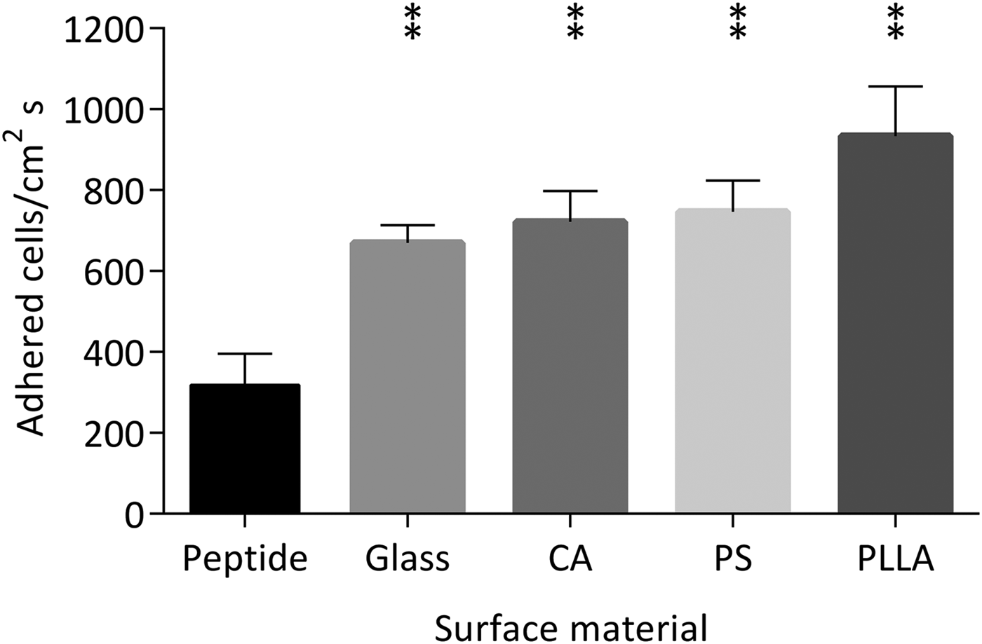
In order to assess the performance of the peptide-based antifouling coating, we plotted the IAR obtained in this study with those obtained from a previous study.[Reference Moreira, Simões, Melo and Mergulhão12] That study was performed in the same PPFC and with the same strain and surrounding fluid (CB) at a shear rate of 15/s (which can be found in urinary catheters). Importantly, the comparison showed that the peptide coating is superior to other polymers such as polystyrene, poly-L-lactic acid and cellulose acetate (Fig. 4), which are commonly used to manufacture different biomedical devices.
Bacterial adhesion rates on peptide, bare glass and on three polymeric surfaces: cellulose acetate (CA), polystyrene (PS) and poly-L-lactic acid (PLLA) obtained in the PPFC for 30 min at a shear rate of 15/s using CB as the surrounding fluid. Results for polymeric surfaces were extracted from.[Reference Moreira, Simões, Melo and Mergulhão12] Statistically, significant differences obtained between each surface and the peptide are indicated by for P < 0.05.

Concluding remarks
This study shows the importance of evaluating the antifouling properties of new surfaces under hydrodynamic conditions that are relevant for a particular application. In addition, the importance of the chemical composition of the surrounding fluid was highlighted because it can affect the performance of the surface. This is particularly important for biomedical applications where fluid compositions and flow dynamics can vary significantly. Although the results indicate considerable adhesion reduction under the conditions tested, longer-term studies under controlled hydrodynamic conditions are needed in order to evaluate the effects of surface chemistry and topography on bacterial adhesion and biofilm formation, using a surrounding fluid that is relevant to a particular biomedical application. The tests performed so far support the beneficial properties of the peptide coating, and this may be considered as an initial screening step in evaluating in vitro surface performance for a broad range of biomedical applications.
Supplementary material
The supplementary material for this article can be found at https://doi.org/10.1557/mrc.2018.160.
Acknowledgments
This work was supported by project NORTE-01-0145-FEDER-000005 – LEPABE-2-ECO-INNOVATION from NORTE 2020, under the Portugal 2020 Partnership Agreement, through the European Regional Development Fund (ERDF) and by the Rosetrees Trust. Patrícia Alves acknowledges the receipt of a Ph.D. grant from the Portuguese Foundation from Science and Technology (FCT) (PD/BD/114317/2016). Sivan Nir acknowledges the support of the Israeli Water Authority. The authors also acknowledge support from the EU COST Action iPROMEDAI TD1305.
Authors’ contributions
Reches M and Mergulhão F conceptualized and designed the study. Alves P performed all the adhesion assays and the surface hydrophobicity analysis. Nir S characterized the peptide coating and surface. All authors contributed to the data interpretation. All the authors reviewed the manuscript and approved the final manuscript.