Introduction
There is a large amount of published data concerning myringoplasty success rates.Reference Glasscock1–Reference Kotecha, Fowler and Topham7
The current study investigated a consecutive series of patients undergoing myringoplasty using the underlay technique and utilising temporalis fascia. The study aimed to determine whether the cause of tympanic membrane perforation influenced the myringoplasty graft ‘take’ rate.
This question has not been well addressed by earlier studies, with the exception of those investigating welding injuries to the tympanic membrane, which are known to heal poorly.Reference Vartiainen and Nuutinen8 In 2008, Sckolnick stated, ‘To date no large single institution study has been conducted to look at comparative success rates for multiple etiologies of perforation and multiple techniques of closure’.Reference Sckolnick, Mantle, Li and Chi9
The current study also investigated the effect of the following factors, previously identified as important for myringoplasty graft healing: surgical method, surgeon experience, and the perforation size and site.Reference Glasscock1, Reference Aggarwal, Saeed and Green3, Reference Albera, Ferrero, Lacilla and Canale5, Reference Bhat and De6
Materials and methods
We collected data on primary myringoplasties conducted in the Swedish cities of Linköping and Norrköping. All surgical ear procedures performed in Linköping from 1994 to 2004 were recorded on the Medlog® database; 230 myringoplasties were recorded. In Norrköping, data were documented on the Medlog database from 2002 to 2004; 26 myringoplasties were recorded. The current series thus included a total of 256 myringoplasty procedures.
Myringoplasty is frequently included as a minor component of various ear surgical procedures. However, the current series addressed operations in which only myringoplasty was performed. Cases requiring ossiculoplasty for a disrupted ossicular chain were excluded. Fat graft myringoplasty was not performed.
An underlay technique with dry temporalis fascia was used consistently; this is a reliable and well reported surgical technique.Reference Glasscock1, Reference Packer, Mackendrick and Solar10 Our myringoplasty technique and underlying philosophy are described below. In most cases (n = 217), a retroauricular approach was used, although some cases were operated upon endaurally or via the external ear canal through an ear speculum (n = 26). The latter technique provides a poorer view of the operative field, and we termed this type of surgery ‘mini-myringoplasty’. In most mini-myringoplasties, a tympanomeatal flap was raised to enable the best possible view of the surgical field. A simplified solution was chosen in two cases: after cleaning the perforation edges, the fascia was placed without lifting the fibrous anulus. In three cases, no post-operative hearing gain was expected, since the operation was done to facilitate the use of a hearing aid.
Our series comprised a total of 256 ears undergoing myringoplasty, representing 245 patients. Thirteen patients did not attend their scheduled post-operative appointment. Bilateral surgery was performed in 11 patients. Thus, we were able to collect data for 243 ears (136 right and 107 left) in 232 patients (106 females and 126 males). The mean patient age was 29 years (median 21 years). One hundred and thirteen operations were performed in children (i.e. aged less than 18 years of age) and 130 operations in adults. The youngest patient was aged three years and the oldest 82 years. Of the seven participating surgeons, three were senior and four junior.
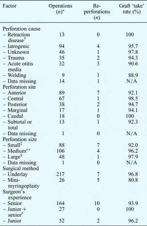
Tympanic membrane perforations were sized as follows: small = <25 per cent of the total tympanic membrane area; medium = 25–50 per cent; and large = >50 per cent. Using this sizing system, 88 (36 per cent) tympanic membrane perforations were small, 106 (44 per cent) medium and 48 (20 per cent) large; data were missing for one patient. Six categories of perforation site were used: anterior, posterior, caudal, central, marginal, and subtotal or total (Table I).
Myringoplasty patients' perforation cause, site and size, and surgical method and surgeon's experience

*Total n = 243. †Primary retraction with secondary perforation. ‡<25%; **25–50%; §>50%. #Senior surgeon present in supervisory capacity. N/A = not applicable.
Patient selection
Patients selected for myringoplasty needed to be able to cooperate with post-operative care. Children were considered for myringoplasty when they had passed the age of greatest risk of otitis media and secretory otitis (i.e. pre-school age), and when they could perform a Valsalva manoeuvre of their own. All selected patients had good eustachian tube function, indicated by normal status of the contralateral ear. In cases of bilateral perforation, the patient was required to be able to perform a Valsalva manoeuvre, before being offered surgery. We imposed no upper age limit for myringoplasty.
Pre-operative preparation and management
The ENT surgeons at both hospitals agreed that myringoplasty was preferably performed in a dry ear. In cases with infection, surgery was postponed and the patient given medical treatment, with the aim of achieving a two- to four-week infection-free interval prior to surgery.
Pre- and post-operative audiometry
Pre- and post-operative air and bone conduction audiometry was conducted, together with pre-operative speech discrimination scoring. In 52 ears, only air conduction hearing was assessed post-operatively. The pure tone average (PTA) for 500, 1000, 2000 and 4000 Hz was used for both pre- and post-operative assessment. Comparison of the pre- and post-operative air–bone gap (ABG) (measured at the above four frequencies) was possible in 175 ears.
Prophylactic antibiotics
In most cases, patients were given prophylactic antibiotics. In the absence of bacterial culture results, this comprised cefuroxim 1.5 g given as three doses at 8 hourly intervals.
Surgical technique
Two important general principles of surgery are (1) achieving a clear view of the surgical field and (2) maintaining good haemostasis. In our series, most myringoplasties were performed via a retroauricular incision. A meatoplasty was performed when needed to prevent post-operative stenosis.
In order to secure healing, it is important to clean the perforation edges and to thoroughly remove keratinising epithelium on the inside of the tympanic membrane as well as mucosal epithelium on the outside. This procedure was facilitated by excising the edge of the perforation with scissors, before raising the fibrous anulus and the tympanic membrane remnant. Myringosclerosis was removed without excessive sacrifice of the tympanic membrane remnant.
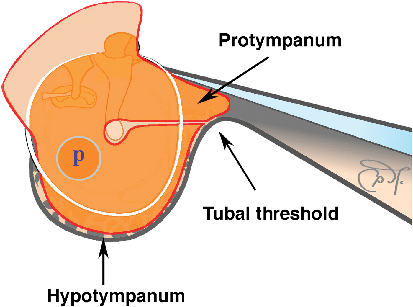
To secure healing of the fascial transplant, the mucosa was thoroughly cleaned from the lateral wall of the protympanum and hypotympanum. The bony septae in the hypotympanum and at the ‘tubal threshold’ (i.e. the threshold of cellular bone located between the hypo- and protympanum) were removed to make way for the fascia, and to ensure good overlap (see Figure 1).
Fascia profile used for a small perforation (p) that does not reach the malleus. The tympanic membrane is dissected from the malleus shaft. A short horizontal slit is made in the fascia and the malleus tip is passed through. The inferior half of the manubrium will thus be located on the lateral surface of the fascial transplant. Bony septa are removed at the tubal threshold as well as the hypotympanum, to achieve a good overlap.
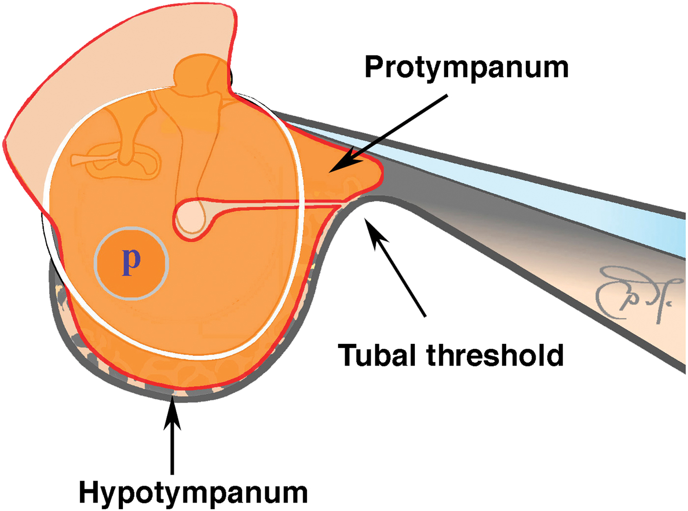
It is important that the fascial transplant is large enough to cover the whole of the tympanic membrane remnant, with good margins. The fascia should reach the bottom of the hypotympanum and also reach into the protympanum. The transplant graft was fashioned with scissors from dried fascia which was slightly re-hydrated before placement. Two different outlines of the fascial transplant were used, both equipped with an upper extension which would cover part of the external ear canal.
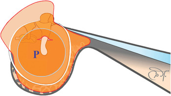
In the majority of larger perforations, the eardrum was released along the entire length of the malleus handle. To avoid lateralisation of the eardrum, a small, central slit was made in the fascia and the distal end of the malleus handle was passed through this slit. This meant that the distal half of the malleus handle came to rest on the lateral side of the fascial transplant (Figure 2).
Fascia profile used for a large perforation (P). The tympanic membrane is dissected from all of the malleus shaft except for a small area at the tip. A horizontal slit is made in the fascia from the anterior edge to the centre. The fascia is placed with one ‘tail’ above and the other below the umbo. The preserved attachment at the umbo prevents lateralisation of the transplant.

However, in smaller perforations that were not situated close to the umbo, we kept the eardrum fixed to the tip of the malleus and made an anterior slit in the fascia. The two fascial ‘tails’ were then passed on either side, one above and one below the umbo (Figure 1). This provided a good attachment to the malleus, thus avoiding lateralisation.
In cases in which much of the middle-ear mucosa required removal because of abundant adhesions due to chronic inflammation, we use a silicone sheet to cover denuded areas of the promontory, in order to avoid the formation of post-operative adhesions.
In patients with a history of long-lasting infection, some surgeons also drilled a small opening through the mastoid cell system to enable flushing with saline.
Follow up
Dressings were removed one week post-operatively. Patients then used eardrops containing hydrocortisone, oxytetracycline and polymyxin B, until the third post-operative week.
A second check was performed after three weeks. At this stage, the ear had usually healed, with near-complete epithelialisation of the transplant, and the patient was encouraged to perform Valsalva manoeuvres to facilitate aeration.
Air conduction hearing levels were tested three to four weeks post-operatively. If hearing was satisfactory at that time, and if the middle ear was aerated, a final follow-up appointment was scheduled one year post-operatively.
Data analysis
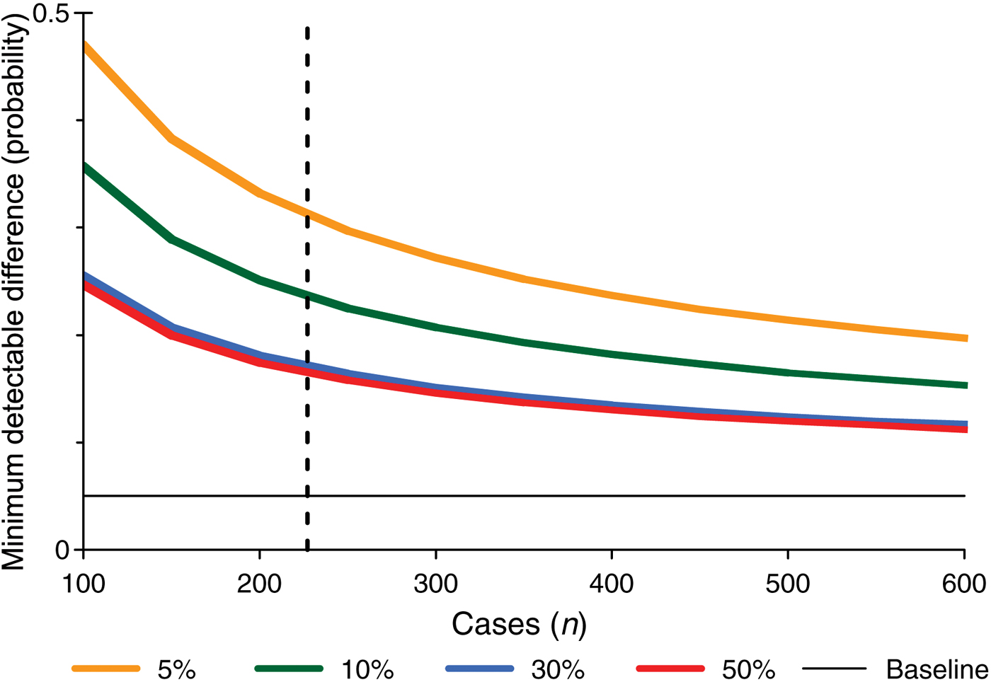
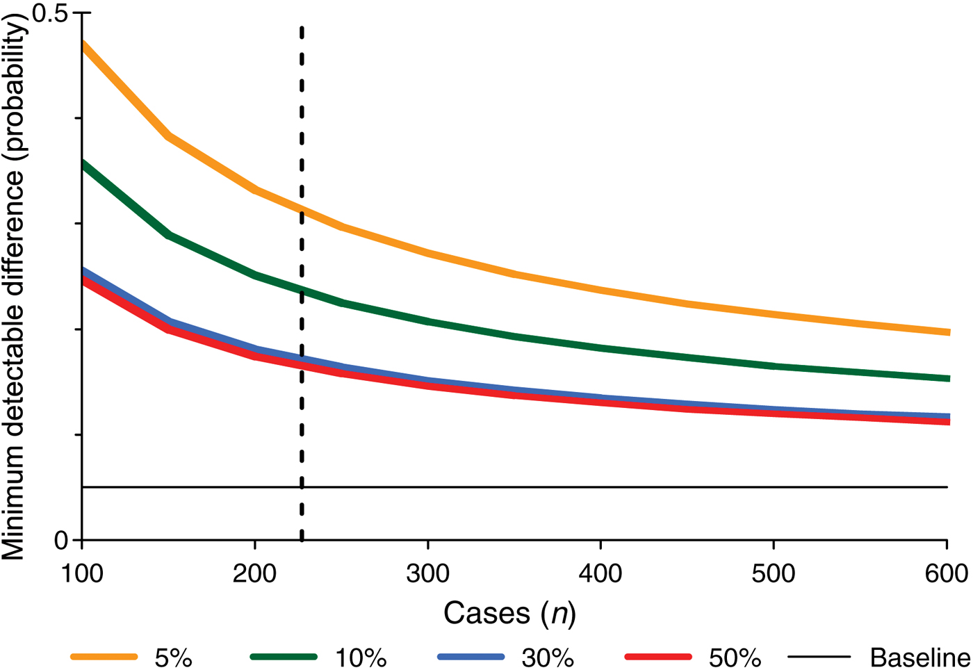
We used logistic regression analysis to assess the relationship between tympanic membrane healing and perforation cause, perforation size and site, surgical method, and surgeon's experience. After excluding cases with missing information, 227 cases were available. All factors entered the model as dummy variables. In addition, we performed power calculations to assess the minimum detectable difference for different factors; this indicated which factors had enough observations to detect a moderate effect (Figure 3). We calculated the minimum detectable difference for a logistic regression,11 assuming a statistical power of 80 per cent and a 5 per cent significance level, and using different levels of frequency for the occurrence of a binary independent factor (i.e. 5, 10, 30 and 50 per cent of n).
Minimum detectable difference (MDD) in probability of re-perforation, calculated using logistic regression analysis. The MDD for an independent (binary) factor is related to how often it occurs; thus, graph plot lines represent the MDDs for different percentage occurrences in the population. The MDDs are related to a hypothetical expected re-perforation occurrence of 5 per cent (baseline). All calculations assume an 80 per cent power level and 5 per cent significance level (two-tailed). The dashed line exemplifies n = 227 cases, as in the current study. For example, 11 per cent of patients (approximating to the green line) underwent mini-myringoplasty. A true probability of re-perforation after mini-myringoplasty of 0.236 or more would have an 80 per cent chance of being detected at a 5 per cent significance level. The observed probability was 0.208 (i.e. five of 24 ears undergoing mini-myringoplasty had re-perforation).
Results
Frequency of follow up
The total series comprised 256 operated ears. Ideally, and in agreement with the inclusion criteria, the status of the tympanic membrane would have been assessed one year post-operatively. However, in reality some patients were examined later and some earlier. Thus, we report results for ears in which the status of the tympanic membrane was checked one to five years post-operatively. We included as a separate category ears with documented status from six months post-operatively.
Two hundred and twenty-one ears underwent a documented post-operative follow-up check between one and five years post-operatively, giving a follow-up frequency of 86 per cent. When we added those ears with a documented post-operative follow-up check at six months (giving a total of 243 ears), the follow-up frequency was 95 per cent. A more detailed description of the latter group follows.
Thirteen of the operated ears had no follow-up information from the specified post-operative period. Of these 13 ears, five underwent follow up which was too early, and five underwent follow up which was too late. In 10 of these 13 ears, a healed tympanic membrane was observed. One of the 13 patients showed a minimal perforation three months post-operatively. For a further two of these 13 ears, no follow-up information was available.
Cause of perforation
The most common cause of tympanic membrane perforation was previous treatment with transmyringeal ventilation tubes (i.e. iatrogenic); this applied to 94 ears (39 per cent of all cases) (see Table I). Most such cases involved the use of straight 7 mm Teflon tubes (n = 63 ears; 66 per cent), although 18 ears (19 per cent) had been treated with T-tubes specifically designed for long term treatment. Armstrong, Parapella and metal tubes had also occasionally been used.
Of those ears with an unknown cause of perforation, the majority were classified as chronic otitis. Hence, the case history was unclear, and the time and origin of perforation unknown.
Surgeon's experience and operative time
Senior surgeons performed 164 procedures, while 27 were performed by a junior surgeon in a training capacity together with a senior surgeon. The remaining 52 procedures were performed independently by a junior surgeon.
The mean operative time for the whole series was 146 minutes. The mean operative time for senior surgeons was 128 minutes, that for junior surgeons in training positions (working together with a senior surgeon) was 166 minutes, and that for junior surgeons working independently was 205 minutes.
Graft take rate and post-operative re-perforation
Following the 243 operations, there were 12 cases of re-perforation (5 per cent), equating to a 95 per cent overall graft take rate (Table I). For ears having a follow-up check one to five years post-operatively, the graft take rate was also 95 per cent. For ears operated upon via a retroauricular approach, the graft take rate was 97 per cent.
Four cases of late perforation were detected. In these cases, a healed tympanic membrane was noted at the scheduled post-operative follow-up appointment but re-perforation was subsequently observed, at 2.5 years post-operatively in one ear, at three years in two ears, at four years in one ear and at 4.5 years in one ear.
None of the ears with re-perforation had had acute infection at the time of surgery. In four of these cases, no prophylactic antibiotic had been given. In one case, no documentation of antibiotic prophylaxis (or lack thereof) was available.
In seven of the 12 cases with re-perforation, we found no particular circumstances that might explain the outcome. In the remaining five cases, records indicated: difficulty in reaching the anterior angle because of exostoses; early post-operative infection (in two cases; in one, surgery had been performed without antibiotics); early swimming; or playing on a waterslide.
Statistical analysis
Logistic regression analysis was conducted to test for an association between the graft take rate and the initial perforation cause, the surgical method, the surgeon's experience, and the perforation size and site. The only factor associated with a significantly poorer outcome was performance of a mini-myringoplasty, compared with an ordinary underlay technique (226 degrees of freedom, null deviation = 88.0; mini myringoplasty: estimate = 2.08, z value = 2.63, p = 0.008).
Power calculations showed that moderate to large increases in probability were required to detect the cause of re-perforation in a logistic regression (between 0.16 and 0.31 at n = 227). Thus, no factor other than performance of a mini-myringoplasty was likely to have had any significant effect on tympanic membrane healing. The effect of other factors would probably have required a much larger sample size in order to be detectable (Figure 3).
Hearing results
Data on post-operative PTA were available for 227 ears, and data on post-operative ABG for 175. An air conduction hearing improvement of 8.4 dB PTA (standard deviation (SD) 10.7) was seen in 79 per cent of the operated ears, while improvements in bone conduction hearing and ABG were seen in 43 and 85 per cent, respectively. The post-operative ABG closure was 8.6 dB (SD 10.2). In 84 per cent of ears, the post-operative air conduction, measured as PTA, was ≥30 dB, a level known to be adequate for daily life. Post-operative results for air conduction, bone conduction and ABG were unchanged in 16, 43 and 11 per cent of ears and worsened in 4.8, 13 and 4.6 per cent, respectively. Post-operative deafness was not seen in any of the cases reviewed.
We did not expect to see a hearing gain in all patients. In three patients, the indication for surgery was to obtain a healed tympanic membrane, in order to facilitate the use of hearing aids to compensate for significant sensorineural hearing loss. We found one case of stapes fixation caused by otosclerosis; this was discovered at the time of surgery and was later corrected by stapediotomy. Four patients had no documented hearing results as the indication for surgery was reduction in the risk of infection. One patient had Down's syndrome, which made bone conduction hearing assessment difficult. One patient was congenitally deaf. Two patients were autistic, making it impossible to perform hearing tests.
Discussion
Over a 10-year period from 1994 to 2004, we collected data for primary myringoplasties performed by both experienced and inexperienced surgeons.
The initial cause of the perforation had no significant effect on the outcome of surgery. The expected poorer surgical outcome for perforations caused by welding beadsReference Vartiainen and Nuutinen8 was not seen in our study; however, the statistical power was low due to our small number of such cases.
In summary, our post-operative results were good, with a 95 per cent overall graft take rate – 97 per cent for ears operated upon via a retroauricular approach. Earlier publications have reported healing rates of between 65.9 and 96 per cent.Reference Glasscock1–Reference Kotecha, Fowler and Topham7 A graft take rate of 56 per cent has previously been reported for selected cases with large perforations.Reference Lee, Kelly and Mills12 A 2006 review reported graft take rates of 35–94 per cent for children and 66–96 per cent for adults.Reference Aggarwal, Saeed and Green3 Various data have been published comparing the success rates of children and adults. Some authors have reported poorer results in children compared with adults, apparently reflecting widespread conventional belief.Reference Vartiainen and Nuutinen8, Reference Gersdorff, Garin, Decat and Juantegui13 Other authors have reported equally good healing rates in children and adults,Reference Albera, Ferrero, Lacilla and Canale5, Reference Bhat and De6, Reference Singh, Sidhu, Sharma and Singh14 in agreement with our own results.
We wish to emphasise that our series contained all myringoplasty procedures conducted within a 10-year period in the Swedish cities of Linköping and Norrköping. These procedures ranged from easy to difficult cases, which could be expected to have a negative effect on the success rate. The patients' subjective experience was not part of our analysis, nor did we consider post-operative complications (e.g. possible lateralisation of the eardrum and myringitis). Even though post-operative retractions were observed in 17 of the operated ears, no cases of post-operative cholesteatoma or atelectasis were observed.
Perforation causes and patient age
We observed a multiplicity of causes for tympanic membrane perforation, rather than a single pathogenesis for one well delineated disease. Little has previously been published concerning the causes of tympanic membrane perforation and their possible effect on myringoplasty success rates. One study reported poor healing in patients with chronic perforations and perforations of unknown or traumatic aetiology, compared with perforations due to active tube extrusion with immediate placement of Gelfoam®, a paper patch or a fat graft.Reference Sckolnick, Mantle, Li and Chi9 These results are difficult to compare with our study findings, since the selection of cases and the surgical techniques were very different.
In our series, the dominant cause for tympanic membrane perforations was iatrogenic, following treatment with transmyringeal ventilation tubes (Table I). In most such cases, straight, 7 mm Teflon tubes had been used. A small proportion of patients had been treated with ventilation tubes designed for long-term use (e.g. T-tubes); perforations after insertion of these tubes were not followed by a significantly greater number of re-perforations. Therefore, we propose that there is no need to restrict ventilation tube insertion in children for fear that any subsequently required myringoplasty will be unsuccessful. This proposal is in agreement with an earlier publication by our last author.Reference Hydén15
When using ventilation tubes, the surgeon should be aware of the risk of permanent perforation of the tympanic membrane. Since children are prone to develop secretory otitis media, it is important to instruct such patients in how to inflate their middle ears, by nose-blowing, performing the Valsalva manoeuvre or blowing up an Otovent® balloon. Such practices, performed regularly during ‘watchful waiting’ periods, may clear the problem, and should be undertaken before the decision is made to insert ventilation tubes.
In one case of re-perforation, the initial perforation had been caused by a welding bead. This kind of perforation is known to have a poor surgical outcome.Reference Vartiainen and Nuutinen8 Our power analysis indicated that the number of cases of welding bead perforation was too small to be interpreted as significant.
Surgeon's experience and operative time
We did not observe poorer healing rates for myringoplasty operations performed by junior trainee surgeons. Other studies have reported both conflictingReference Palva and Virtanen2, Reference Vartiainen and Nuutinen8 and supportiveReference Bhat and De6, Reference Blanshard, Robson, Smith and Maw16 findings. Our results indicate that surgeons at all levels of experience can have high myringoplasty success rates, as regards both tympanic membrane healing and hearing improvement. Training and support by an experienced colleague is of great importance. We believe that our high success rates were due to a reliable, standardised surgical process suitable for both experienced and trainee surgeons. The time of disposal for surgery is likely to be contributing to a good result. No restrictions were placed on the operative time; the surgeon thus had adequate time to complete each individual procedure.
Perforation size and site
The size of perforations did not influence the myringoplasty healing rate. This is in agreement with some earlier studiesReference Karela, Sandeep, Watkins and Phillipps4, Reference Vartiainen and Nuutinen8, Reference Blanshard, Robson, Smith and Maw16 and in disagreement with others.Reference Lee, Kelly and Mills12 In a review of articles on this subject, higher success rates were found for smaller perforations,Reference Aggarwal, Saeed and Green3 in contrast to our results.
There was no statistically significant relationship between perforation site and myringoplasty healing rate. Ambiguous results have been published earlier on this matter.Reference Aggarwal, Saeed and Green3 Some authors have not found any correlation between perforation site and healing rate,Reference Karela, Sandeep, Watkins and Phillipps4, Reference Sckolnick, Mantle, Li and Chi9 whereas others have.Reference Albera, Ferrero, Lacilla and Canale5, Reference Bhat and De6
In our study, the majority of perforations were situated anteriorly; this may be explained by the predominantly anterior placement of ventilation tubes.
Hearing results
Like other authors, we found that hearing improvement could usually be expected after myringoplasty.Reference Karela, Sandeep, Watkins and Phillipps4, Reference Bhat and De6 Our finding that 84 per cent of operated ears had a post-operative air conduction level of 30 dB or better is comparable to hearing results reported by other authors (i.e. 66–88 per cent).Reference Blanshard, Robson, Smith and Maw16
• Myringoplasty is a reliable surgical procedure
• By standardising the surgical technique, the same good results may be achieved by junior as well as senior surgeons
• In this series, a retroauricular approach gave better results than an endaural approach
• The cause of perforation did not influence the myringoplasty success rate
Mini-myringoplasty
In our series, re-perforations were seen most frequently in the small number of patients undergoing mini-myringoplasty – i.e. surgery performed via an endaural approach or via an ear speculum. Among the 26 mini-myringoplasty operations (11 per cent), there were five re-perforations (19 per cent). This procedure is evidently an inferior method. A retroauricular approach is thus the myringoplasty method of choice to ensure better healing and improved post-operative hearing, irrespective of the primary cause of the perforation.
Conclusion
We present a 10-year series of patients undergoing primary myringoplasties conducted between 1994 and 2004, who were operated upon by experienced and trainee ENT surgeons. Our overall success rate (i.e. graft take rate) was 95 per cent. A retroauricular approach was used in 97 per cent of ears, and was found to be superior to simpler surgical methods. We conclude that our favourable results were due to the employment (in most cases) of this reliable, standardised surgical method, which is suitable for surgical trainees as well as experienced surgeons. Post-operative tympanic membrane healing rates were unaffected by the primary cause of perforation. Thus, regardless of the cause of perforation, high rates of post-myringoplasty healing and hearing improvement can be expected.