Introduction
In vitro maturation (IVM) of oocytes is a key step in the in vitro production of embryos (Paramio and Izquierdo, Reference Paramio and Izquierdo2016; Sasaki et al., Reference Sasaki, Hamatani, Kamijo, Iwai, Kobanawa, Ogawa, Miyado and Tanaka2019). However, during IVM there is an increase in the production of reactive oxygen species (ROS), which are natural byproducts of cellular metabolism. The excessive production of these species can be triggered by factors such as atmospheric conditions, temperature and the composition of the culture medium, promoting oxidative stress and, consequently, damage to both mitochondrial and nuclear DNA in oocytes (Li and Zhao, Reference Li and Zhao2019). In this context, studies have shown that supplementation of IVM media with antioxidants can exert a protective effect against ROS-induced damage, contributing to the maintenance of oocyte quality and embryonic development (Tripathi et al., Reference Tripathi, Nandi, Gupta and Mondal2023).
Among the antioxidants used, N-acetylcysteine (NAC) stands out as a precursor of cysteine that reduces oxidative stress by restoring intracellular levels of reduced glutathione (GSH) (Barrozo et al., Reference Barrozo, Silva, Paulino, Barbalho, Nascimento, Costa, Batista, Lopes, Rodrigues and Silva2022; Silva and Silva, Reference Silva and Silva2023). NAC also exerts a direct antioxidant effect by interacting with free radicals through its free thiol group (Pei et al., Reference Pei, Liu, Yang, Yang, Jiao, Tay and Chen2018), in addition to supplying cysteine, an essential amino acid for GSH synthesis (Lim et al., Reference Lim, Ali, Liao, Nguyen, Ortiz, Reshel and Luderer2020). Furthermore, this compound influences several cellular signalling pathways, including those involved in apoptosis, angiogenesis, cell proliferation and cell cycle arrest, as well as the regulation of gene expression mediated by the redox state in response to oxidative stress (Andrade et al., Reference Andrade, Moura, Dos Santos, De Araujo, De Farias Santos and Goulart2015; Moura et al., Reference Moura, Andrade, Araújo, Nunes-Souza, Santos, Rabelo and Goulart2016).
In cattle, oocytes matured in the presence of 1 mM NAC or 0.6 mM L-cysteine showed increased intracellular levels of reduced GSH and decreased levels of ROS (Sun et al., Reference Sun, Jang, Park, Oh, Lee, Hwang, Xu, Hwang and Lee2021). Moreover, NAC has been applied in different stages of reproductive biotechnology, including in vitro follicular growth, IVM and oocyte cryopreservation, contributing to reduced ROS levels and improved resumption of meiosis (Barrozo et al., Reference Barrozo, Silva, Paulino, Barbalho, Nascimento, Costa, Batista, Lopes, Rodrigues and Silva2022). In studies involving mouse oocytes exposed to bisphenol A, supplementation with 100 μM NAC during IVM resulted in higher nuclear maturation and fertilization rates, as well as improved blastocyst formation and reduced ROS levels (Li et al., Reference Li and Zhao2019).
Despite the benefits described above, studies evaluating the use of NAC in the IVM of sheep oocytes remain limited. Therefore, the present study aimed to evaluate the effect of NAC supplementation on oocyte IVM, as well as on the subsequent in vitro production of sheep embryos.
Materials and methods
Media, reagents and study location
The media and reagents used for the maturation and in vitro fertilization of oocytes, as well as for the in vitro culture (IVC)) of embryos, were acquired from Sigma Aldrich Brazil®. The experiment was conducted at the Laboratory of Physiology and Biotechnology of Animal Reproduction (LAFIBRA) and at the Laboratory of Cell Biology, Cytology and Histology, both located at the Agricultural Sciences Campus of the Federal University of Vale do São Francisco (UNIVASF) in Petrolina, Pernambuco, Brazil.
Obtaining ovaries and harvesting and selecting oocytes
The sheep ovaries were obtained from females of reproductive age, between 1 and 5 years old, at the Geomarco Coelho de Souza Municipal Public Slaughterhouse in Petrolina/PE, Brazil. Following the slaughter of ewes without a defined breed standard, the ovaries were collected and transported to the laboratory within two hours in a 0.9% NaCl solution containing 0.05 g of pentabiotic/L (composed of basic streptomycin, basic dihydrostreptomycin, benzathine benzylpenicillin, procaine benzylpenicillin and potassium benzylpenicillin) at a temperature of 33°C to 34°C in a thermos. In the laboratory, the ovaries were washed three times in prewarmed transport solution (33°C to 34°C) and then kept in a water bath, immersed in 300 mL of the transport solution at 34°C. Next, cumulus–oocyte complexes (COCs) derived from antral follicles measuring 3.0 to 6.0 mm in diameter were collected by follicular aspiration using a vacuum pump set at 20 mmHg (or a flow rate of 5 mL/min) connected to an 18 G Jelco catheter. The recovery medium consisted of TCM-199 supplemented with 25 mM HEPES, 500 IU penicillin, 0.5 mg streptomycin, 1.25 μg amphotericin B, 1% (v/v) fetal bovine serum (FBS) and 5 IU/mL heparin sodium. Soon after collection, the COCs recovered from the follicular fluid and aspiration medium were transferred to 60 mm Petri dishes, evaluated and classified under a stereomicroscope. COCs presenting a compact and multilayered cumulus with finely granulated oocyte cytoplasm (Grade I), or those with one to three layers of cumulus cells and finely granulated cytoplasm (Grade II), were selected for IVM (Freitas and Melo, Reference Freitas and Melo2010).
In vitro maturation
After selection, the Grade I and II, an average of 692 oocytes were randomly divided into four treatment groups: the control group (CRT), in which the maturation medium was composed of TCM-199 supplemented with 500 IU of penicillin, 0.5 mg of streptomycin, 1.25 μg of amphotericin B, 0.2 mM of sodium pyruvate, 10% (v/v) FBS and 10 IU/mL of eCG (equine chorionic gonadotropin); and the experimental groups NAC1, NAC1.5 and NAC2.0, in which the maturation medium was the same as that of the CON group, supplemented with 1, 1.5 and 2.0 mM NAC, respectively. An average of 10 to 15 COCs were placed in 75 μL drops of IVM medium for each treatment in 60 mm Petri dishes under mineral oil and then incubated at a constant temperature of 38.5°C in a humidified atmosphere with 5% CO2 for 24 h. The NAC concentrations used were based on previous studies, which showed that NAC improves the maturation rate and reduces ROS levels in the IVM of vitrified porcine oocytes (Whitaker and Knight, Reference Whitaker and Knight2010) and sheep oocytes (Ahmadi et al., Reference Ahmadi, Shirazi, Shams‐Esfandabadi and Nazari2019).
Evaluation of the degree of expansion of cumulus cells
Soon after the maturation (692 oocytes) process, the COCs were assessed for cumulus cell expansion. Additionally, the degree of expansion was estimated, classifying them as unexpanded, partially expanded or fully expanded (Marei et al., Reference Marei, Wathes and Fouladi-Nashta2010).
Chromatin configuration assessment
All oocytes subjected to IVM were assessed, and only those exhibiting cumulus cell expansion (73 oocytes for group, n = 292 oocytes) were selected for further analysis. Oocytes from the four treatment groups were denuded by pipetting, then washed three times in 50 μL drops of phosphate-buffered saline (PBS) and incubated in 50 μL drops containing 5 μL/mL of Hoechst 33342 in PBS for 15 min at room temperature and in the absence of light. Slides (Coleman®, Roster, Brazil) were prepared for further analysis under an epifluorescence microscope (Nikon E200®, Nikon, Japan) at a magnification of 400x. Following the methodology of Rajabi-Toustani et al. (Reference Rajabi-Toustani, Motamedi-Mo, Mohamma, Roostaei-Ali and Reihaneh2013), the oocytes subsequently underwent chromatin evaluation to verify the different meiotic stages of each cell structure: germinal vesicle (GV), germinal vesicle breakdown (GVBD), metaphase I (MI) and metaphase II (MII).
Assessment of intracellular levels of mitochondrial activity, ROS and GSH
After IVM, mature oocytes from the control, 1 mM and 1.5 mM NAC groups (n = 70 per group; total = 210 oocytes) with higher MII rates were pipetted and incubated in the dark for 30 min at 37°C in PBS (PBS Buffer®, Bio-Rad Laboratories Ltda., Portugal) supplemented with 10 μM of 4-chloromethyl-6,8-difluoro-7-hydroxycoumarin (CellTracker Blue®; CMF2HC;® Invitrogen Corporation, Carlsbad, USA), 10 μM 2’,7’-dichlorodihydrofluorescein diacetate (H2DCFDA®, Invitrogen Corporation) and 100 nM MitoTracker Red (MitoTracker Red®, CMXRos, Molecular Probes, Melbourne, Victoria, Australia) to detect intracellular levels of GSH, ROS and mitochondrial activity through blue, green and red fluorescence, respectively (Lins et al., Reference Lins, Cavalcante, Santos, Menezes, Barros, Barberino, Bezerra, Macedo and Matos2016). Next, the oocytes were washed with PBS (PBS Buffer®, Bio-Rad Laboratories Ltda., Portugal) and fluorescence was observed under an epifluorescence microscope (E200®; Nikon, Tokyo, Japan), with 370, 460 and 579–599 nm UV filters for GSH, ROS and mitochondrial activity, respectively. The oocyte fluorescence intensities were analysed using Image J (version 1.47®; National Institutes of Health®, Bethesda, MD, USA).
DNA fragmentation assessment
Following the methodology of Macedo et al. (Reference Macedo, Barros, Monte, Gouveia, Bezerra, Cavalcante, Barberino, Menezes and Matos2017), subsets of mature oocytes from the control and 1 mM NAC groups (n = 70 oocytes), which showed the best results in previous chromatin configuration and ROS analyses, were evaluated for DNA fragmentation using the TUNEL assay, in which the enzyme terminal deoxynucleotidyl transferase catalyses the incorporation of dUTP nucleotides at the free ends of fragmented DNA. Briefly, oocytes were fixed in 4% paraformaldehyde for 1 h at room temperature. Subsequently, they were permeabilized in 0.1% (v/v) Triton X-100 in 10 mM PBS for 3 h at room temperature. The treatment groups were divided into positive and negative controls: the positive control (10 oocytes) was incubated with RNase-free DNase at 37°C for 1 h and then washed three times in PBS supplemented with polyvinylpyrrolidone (PVP), while the negative control (10 oocytes) was processed similarly but without DNase. Both control and treatment groups were incubated with the TUNEL reaction mixture (In Situ Cell Death Detection Kit, Fluorescein; Boehringer Mannheim/Roche Diagnostics) for 1 h at 37°C. After incubation, oocytes were washed three times in PBS-PVP and stained with Hoechst 33342 for 15 min at room temperature in the dark. Approximately 50 oocytes per treatment were analysed using an epifluorescence microscope. Oocytes exhibiting fragmented chromatin showed green fluorescence.
In vitro fertilization
A total of 400 oocytes that reached maturity after IVM were subjected to IVF, with 10 to 15 COCs allocated per treatment group. Semen was obtained from a ram with confirmed fertility using the artificial vagina technique, as described by Gonzales et al. (Reference Gonzales, Soares, Gomes and Sousa2002). After collection, 1 mL of semen was layered onto a 45%/90% Percoll discontinuous gradient and centrifuged at 700 g for 15 min to separate motile spermatozoa. The precipitate was resuspended in 2 mL of IVF medium (SOF®, supplemented with 500 IU/mL penicillin, 0.5 mg/mL streptomycin, 1.25 μg/mL amphotericin B and 10 μg/mL hypotaurine) and washed again by centrifugation (100g; 5 min). The final precipitate was resuspended in 2 mL of fertilization medium, and the sperm concentration was adjusted to 1 × 10⁶ spermatozoa/mL. The sperm were then incubated with the COCs in 50 μL drops of IVF medium under mineral oil in a 60 mm Petri dish for 20 h in a culture incubator at 38.5°C in a humidified atmosphere with 5% CO2.
In vitro culture
The potential zygotes were denuded by pipetting and subsequently cultured in vitro for 48 h in an IVC medium composed of SOF supplemented with 3 mg/mL of bovine serum albumin (BSA), 500 IU of penicillin, 0.5 mg of streptomycin and 1.25 μg of amphotericin B. The zygotes were arranged in Petri dishes under mineral oil, with 10 to 15 structures per 50 μL drop of SOF medium. The dishes were incubated at 38.5°C with 5% CO2, and the number of embryos was recorded at the end of the 2-day (D2) cultivation period. Embryo quality, colour, morphology, number of blastomeres and characteristics of the zona pellucida were used as evaluation criteria.
Statistical analysis
The degrees of expansion of cumulus cells, chromatin configuration, DNA fragmentation and formation of presumed zygotes were expressed as percentages and compared using the chi-square test in Epi Info (Epi Info 7.2.5, Atlanta, GA, USA, 2021). Data for GSH levels, mitochondrial activity and ROS underwent the D’Agostino–Pearson normality test. Subsequent analyses, including the Kruskal–Wallis and Student–Newman–Keuls tests, were performed using SAS OnDemand for Academics 9.04.01 online statistical program (SAS® 3.8 Enterprise Edition, USA, 2020). Four independent replicates were conducted for each analysis. Differences were considered statistically significant when P < 0.05.
Results
Expansion of cumulus cells
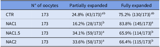
Initially, all groups exhibited some degree of cumulus cell expansion (Figure 1). In the analysis of partial expansion, the 1 mM of NAC showed a lower expansion rate compared to the 1.5 mM and 2 mM of NAC (P < 0.05), although it did not differ significantly from the control (Table 1). Regarding complete expansion, the 1 mM NAC group presented a higher rate compared to the 2 mM of NAC (P < 0.05), with no significant difference observed in relation to the control group (Table 1).
Expansion of cumulus cells after in vitro maturation without the addition of antioxidant (CTR) and with the addition of 1 mM (NAC1), 1.5 mM (NAC1.5) and 2.0 mM (NAC2) of NAC. The fully expanded cumulus cells is indicated by an arrow (↑).

Evaluation of cumulus cell expansion in ovine oocytes matured in vitro in the absence (CTR) or presence of N-acetylcysteine at different concentrations (1.0, 1.5 and 2.0 mM; NAC1, NAC1.5 and NAC2, respectively)

Lowercase letters indicate differences between lines (P < 0.05).
Chromatin configuration
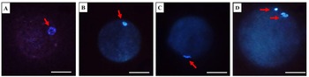
After evaluating the chromatin configuration of the matured oocytes between the different treatments (n = 73 oocytes per group; Figure 2), it was observed that the concentration of 1 mM NAC resulted in significantly fewer oocytes in the GV phase compared to the control and 2 mM of NAC (P < 0.05), with no difference in relation to the NAC of 1.5 mM (P > 0.05) (Table 2). Relative to the GVBD stage, 1 mM NAC had a lower proportion of oocytes compared to 1.5 mM and 2 mM NAC (P < 0.05) but had a similar percentage to that observed in the control. In the metaphase I (MI) stage, the 1 mM concentration of NAC showed a lower rate compared to the other treatments (P < 0.05). When analysing metaphase II (MII) stage, NAC concentrations of 1 mM and 1.5 mM showed higher rates of oocytes reaching MII (P < 0.05) compared to control and 2 mM of NAC (Table 2).
Chromatin configuration by Hoechst 33342 of oocytes matured in the absence of antioxidant (CTR) and in the groups plus NAC. (A) Germinal vesicle of the CTR group. (B) Germinal vesicle breakage in the NAC1,5 group. (C) Metaphase I in the NAC1,5 group. (D) Metaphase II in the NAC1 group. Scale bar 50 μm.

Chromatin configuration after in vitro maturation of sheep oocytes matured in the absence (CTR) or presence of N-acetylcysteine at different concentrations (1.0, 1.5 and 2.0 mM; NAC1, NAC1.5 and NAC2, respectively)

Lowercase letters indicate differences between lines (P < 0.05). GV: Germinal Vesicles; GVBD: Germinal Vesicular Breakage; MI: Metaphase I; Metaphase II.
Mitochondrial activity, GSH and ROS
Fluorescence analyses were quantified based on the pixel intensity of mature oocytes (n = 70 per group) in the CTR, NAC1 and NAC1.5, which showed higher MII rates compared to the other treatments. Analysis of mitochondrial activity revealed no significant difference between the control and 1 mM NAC groups (P > 0.05); however, both exhibited higher activity compared to the 1.5 mM NAC (Figure 3A,B). Additionally, the 1 mM NAC group showed significantly lower ROS levels compared to the other groups (P < 0.05). In contrast, the 1.5 mM NAC exhibited significantly higher ROS levels and lower GSH levels compared to the other treatments. No significant difference was observed between the control and 1 mM NAC (P > 0.05) regarding GSH levels.
Intracellular levels of mitochondrial activity, ROS and GSH (pixel/oocyte) of oocytes matured in the absence of antioxidant (CTR) and the plus groups of 1 mM (NAC1) and 1.5 mM (NAC1.5). (A). Pixel intensity percentage per oocyte. (B). Representative fluorescence images of oocytes. a,b,c, different letters indicate a significant difference between treatments (P < 0.05). Scale bar 50 μm.

DNA fragmentation
Following TUNEL analysis (Figure 4), the 1 mM NAC group showed a lower percentage of DNA fragmentation than the control group (P < 0.05).
DNA fragmentation analysis, by TUNEL, of matured sheep oocytes in the absence of antioxidant (CTR) and in the presence of 1 mM NAC (NAC1). (A) DNA fragmentation percentage. (B) Representative fluorescence images of oocytes. CTR+: positive control group, which is performed with the aid of DNase to identify whether there was TUNEL marking and CTR-: control negative, which does not receive DNase. a,b different letters indicate a significant difference between treatments (P < 0.05). Scale bar 50 μm.

Embryonic development
The percentage of cleaved embryo following the IVF of mature oocytes treated with NAC is shown in Table 3 and Figure 5. The NAC1 group exhibited a significantly higher cleavage percentage (P < 0.05) than the other treatment groups. Additionally, the NAC1 group produced a greater number of embryos with more than 8 cells after 2 days of culture, compared to the other NAC concentrations (P < 0.05). It is worth noting that a 7-day culture period, which would be necessary to evaluate later stages of embryonic development, was not performed.
Embryo after in vitro fertilization of matured oocytes in the group without the use of antioxidants (CTR) and the groups plus 1 mM (NAC1), 1.5 mM (NAC1.5) and 2.0 mM (NAC2) of NAC. The embryo is indicated by an arrow (↑). Scale bar 50 μm.

Effect of N-acetylcysteine supplementation during in vitro maturation on cleavage rate following in vitro fertilization of ovine oocytes. Antioxidant free (CTR) or presence of N-acetylcysteine at different concentrations (1.0, 1.5 and 2.0 mM; NAC1, NAC1.5 and NAC2, respectively)

Lowercase letters indicate differences between lines (P < 0.05). PZ: potential zygotes.
Discussion
This study demonstrated the effect of adding different concentrations of NAC to the IVM medium of sheep oocytes and on in vitro embryo production. The inclusion of 1 mM NAC in the IVM medium increased cumulus cell expansion and the metaphase II (MII) rate. In our investigation, a 100% cumulus cell (CC) expansion rate was observed in all evaluated groups. When assessing the degree of expansion among NAC-supplemented groups, the 1 mM NAC concentration showed a higher percentage of total expansion compared to the 1.5 mM and 2 mM NAC groups. Increased antioxidant levels may reduce CC expansion, possibly due to a pro-oxidant effect (Yusuflu and Ülküm, Reference Yusuflu and Ülküm2022).
When evaluating chromatin configuration, oocytes matured with 1 and 1.5 mM NAC showed a higher percentage of oocytes at the MII stage (54.7% in both) compared to the control and the 2 mM NAC group. Previous studies demonstrated that adding 1.5 mM NAC to the IVM medium improved the maturation of porcine oocytes subjected to oxidative stress for 8 h (Hu et al., Reference Hu, Cheng, Wang, Luo, Zhao, Tian and An2020). Similarly, in mice, 1.5 mM NAC enhanced in vitro embryo production from vitrified/warmed oocytes subjected to IVM and IVF (Yue et al., Reference Yue, Zhang, Wang, Sun, Xing, Wen and Zhou2016). Studies by Wang et al. (Reference Wang, Wang, Ji, An, Wang, Ju, Li, Ruan, Zhao, Cao and Chen2025) showed that 1.5 mM NAC supplementation in goat oocyte IVM improved MII rates and embryo production. However, in our study, increasing NAC concentration to 2 mM significantly reduced the percentage of oocytes reaching MII (26%), with no significant difference compared to the control. Likewise, 1 mM NAC increased maturation rates in bovine oocytes during an 8-hour IVM period, whereas higher concentrations reduced MII rates. This effect may be associated with inhibition of the nuclear factor kappa B (NF-κB) pathway during oocyte maturation (Sun et al., Reference Sun, Jang, Park, Oh, Lee, Hwang, Xu, Hwang and Lee2021).
When evaluating mitochondrial activity and GSH levels, 1 mM NAC resulted in higher levels compared to 1.5 mM NAC and also reduced ROS levels. In contrast, 1.5 mM NAC showed higher ROS levels compared to other treatments. Increased ROS production can damage mitochondrial activity and reduce GSH levels, potentially leading to meiotic arrest or apoptosis. NAC acts as a precursor of GSH, which explains the intracellular antioxidant balance observed with 1 mM NAC (Pei et al., Reference Pei, Liu, Yang, Yang, Jiao, Tay and Chen2018). Intracellularly, NAC is deacetylated to form L-cysteine, which combines with other amino acids to enhance GSH synthesis (Sun et al., Reference Sun, Jang, Park, Oh, Lee, Hwang, Xu, Hwang and Lee2021).
Studies with bovine oocytes matured in vitro with 1 mM NAC demonstrated increased intracellular GSH levels and reduced ROS levels (Sun et al., Reference Sun, Jang, Park, Oh, Lee, Hwang, Xu, Hwang and Lee2021). Other studies using 0.5 mM NAC in in vitro follicle culture and oocyte maturation reported reduced ROS levels, transient changes in mRNA and cyclin B expression, and increased meiotic resumption (Barrozo et al. Reference Barrozo, Silva, Paulino, Barbalho, Nascimento, Costa, Batista, Lopes, Rodrigues and Silva2022).
Additionally, 100 μM NAC mitigated oxidative stress caused by mycotoxins such as zearalenone in porcine oocytes by regulating ROS production and increasing GSH levels (Lai et al., Reference Lai, Ma, Liu, Wang, Cheng, Sun, Li, Li, Nyachoti and Shen2015). Similarly, 100 μM NAC reduced damage in mouse oocytes exposed to bisphenol A by lowering ROS levels, increasing intracellular GSH and promoting meiotic resumption (Li and Zhao, Reference Li and Zhao2019). In this context, our study demonstrated that 1 mM NAC reduces ROS production while maintaining intracellular GSH levels and mitochondrial activity in sheep oocytes, contributing to improved oocyte quality and maturation. It is important to note that the optimal NAC concentration may vary depending on species, cell type and specific IVM or IVC conditions.
We also demonstrated that 1 mM NAC prevents oxidative stress and reduces DNA fragmentation. Previous studies showed that 3.5 mM NAC reverses spindle disorganization in porcine oocytes matured in vitro (Hu et al., Reference Hu, Cheng, Wang, Luo, Zhao, Tian and An2020) and inhibits extracellular signal-regulated kinase (ERK) signalling (Barrozo et al., Reference Barrozo, Silva, Paulino, Barbalho, Nascimento, Costa, Batista, Lopes, Rodrigues and Silva2022). Wang et al. (Reference Wang, Wang, Ji, An, Wang, Ju, Li, Ruan, Zhao, Cao and Chen2025) reported that NAC reduces cellular oxidative stress by upregulating antioxidant genes (SIRT1, MITF and GGCT) and energy-related genes, while downregulating apoptotic genes (CASP8 and FOS), thereby improving oocyte quality. In porcine oocytes, NAC has also been shown to repair epigenetic alterations, such as histone methylation errors, by inhibiting the expression of genes like KDM6A, correcting the H3K27me3 epigenetic mark and reducing DNA fragmentation and apoptosis (Hu et al., Reference Hu, Cheng, Wang, Luo, Zhao, Tian and An2020). Consistent with the literature, our findings indicate that 1 mM NAC promotes higher nuclear maturation rates, reduces oxidative stress and protects DNA integrity.
In this study, oocytes matured with 1 mM NAC showed higher cleavage rates and a greater proportion of embryos with more than 8 cells compared to other NAC-supplemented groups, indicating improved embryonic development. Similarly, vitrified/warmed murine oocytes subjected to IVF with 0.25 and 0.50 mM NAC yielded 2-cell embryo rates of 80.5% and 85.9%, respectively (Mukunoki et al., Reference Mukunoki, Takeo and Nakagata2019). Additionally, 1 mM NAC improved blastocyst production rates in murine oocytes (Matilla et al., Reference Matilla, Martín-Cano, González-Fernández, Sánchez Margallo, Álvarez and Macías-García2019).
In conclusion, supplementation of the IVM medium with 1 mM NAC improves MII rates, reduces ROS levels, maintains GSH levels and preserves mitochondrial activity. Furthermore, it decreases DNA fragmentation, contributing to higher cleavage rates and improved embryonic quality.
Acknowledgements
This research has not received any specific grants from public, commercial or non-profit funding agencies.
Funding
The authors would like to thank the Foundation for the Support of Science and Technology of Pernambuco (FACEPE) for granting a scholarship to Sueli de Oliveira Lima. M.H.T. Matos is supported by a grant from the National Council for Scientific and Technological Development (CNPq, Brazil; n° 317582/2021-6).
Competing interests
The authors declare no conflicts of interest.
Ethical standards
The present study was submitted to and approved by the Ethics Committee on the Use of Animals (CEUA) at the Universidade Federal do Vale do São Francisco (UNIVASF), under protocol number 0003/310122.