Introduction
Congenital hearing impairment affects approximately 3 in every 1000 live births and is the most frequent birth defect in developed countries.Reference White1 Children born with hearing impairment encounter challenges in speech development, education and language acquisition. This results in decreased opportunities for the individual and a potential burden on society. It has been estimated that the lifetime social costs of untreated hearing loss can reach up to US $1.1 million per individual. Treatment and early intervention could decrease these costs by 75 per cent.Reference Keren, Helfand, Homer, McPhillips and Lieu2
More than 50 per cent of congenital sensorineural hearing impairment is hereditary and caused by genetic mutations.Reference Smith, Bale and White3 Hearing loss can either be syndromic, as part of multiple anomalies throughout the body, or non-syndromic, being restricted to the inner ear. Non-syndromic hearing loss can be further categorised by mode of inheritance. In the largest fraction of cases (80 per cent), inheritance is autosomal recessive (often labelled ‘DFNB’). This type of hearing loss is generally congenital, although some forms may present later in life. In the other 20 per cent of cases, inheritance of hearing loss is autosomal dominant (‘DFNA’). In this type there is usually a delayed onset of hearing loss. The rest of non-syndromic hearing loss is either X-linked or mitochondrial (less than 1 per cent).Reference Morton4
Insight into the genes involved in congenital hearing loss and the underlying mechanisms may enable targeted genetic counselling and treatment. Screens for specific genetic mutations can be performed based on clinical features such as mode of inheritance, morphological appearance, and onset and progression of hearing loss. Genes that are a common cause of hearing loss, such as GJB2, SLC26A4 and OTOF, are frequently included in diagnostic tests.Reference Hilgert, Smith and Van Camp5 The results can be used to counsel parents about the prognosis of hearing loss in their child, the chance of recurrence in future offspring and the predicted outcomes of therapeutic options like cochlear implantation. For example, the outcome of cochlear implantation in syndromic hearing loss is less predictable than in non-syndromic hearing loss. Patients with isolated mutations in the gene GJB2 have speech outcomes that are better than average, whereas patients with cochleovestibular dysplasia fare worse than average.Reference Pakdaman, Herrmann, Curtin, Van Beek-King and Lee6, Reference Black, Hickson, Black and Perry7 The presence of auditory nerve malformation or neuropathy also predicts inferior results.Reference Rance and Barker8, Reference Breneman, Gifford and Dejong9 A better understanding of underlying pathophysiology will enable us to better assess cochlear implant candidacy.
More importantly, there is the potential to exploit knowledge of underlying genetic mutations to prevent or ameliorate hereditary hearing loss. Gene therapy utilises viral vectors delivered to the inner ear to replace the defective gene with a normal copy of the gene. There are promising early results from animal studies: adenovirus-delivered SLC17A8 (VGLUT3, vesicular glutamate transporter 3) restores hearing in mice lacking this gene.Reference Akil, Seal, Burke, Wang, Alemi and During10 It may also be possible to use gene therapy to recover hair cell degeneration: delivery of the ATOH1 gene product has been found to induce hair cell development and regeneration.Reference Lustig and Akil11
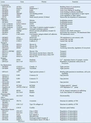
This review provides an overview of the currently known genes involved in non-syndromic hereditary hearing loss (Table I).
Genes associated with non-syndromic hearing loss

DFNA = non-syndromic deafness, autosomal dominant; DFNB = non-syndromic deafness, autosomal recessive; TM = tectorial membrane; Ca2+ calcium ion; K+ = potassium ion
Hearing mechanism
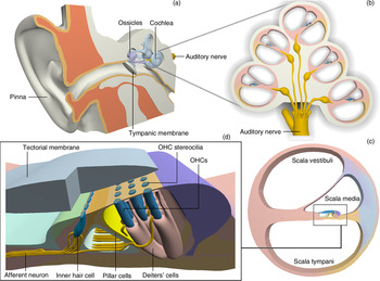
We begin with a brief review of the mechanism of human hearing. The cochlea is the auditory portion of the inner ear and is a spiral structure containing three compartments: the scala vestibuli and scala tympani, which are filled with perilymph, and the scala media which is filled with endolymph (Figure 1c). The organ of Corti is located within the middle compartment, the scala media, and includes the inner hair cells, which detect sound (Figure 1d). Airborne sound waves are transmitted through the external and middle ear to the oval window of the cochlea (Figure 1a). Displacement of the oval window causes a wave in the fluids of the cochlea (Figure 1b), leading to displacement of the basilar membrane. Inner hair cells attached to the basilar membrane carry on their apical surface numerous finger-like projections called stereocilia, which will deflect upon physical vibration and move relative to one another. This movement is thought to cause the opening of specific ion channels, a mechanism called mechanotransduction.Reference Dror and Avraham12 Opening of these channels initiates an influx of potassium and calcium ions which depolarises the hair cell. This leads to calcium-dependent exocytosis of neurotransmitter vesicles at the cell's basolateral surface in an area called the synaptic ribbon.Reference Hudspeth13 The release of neurotransmitters excites adjacent auditory neurons which signal to auditory centres in the brain, contributing to the perception of sound.
Anatomical diagrams of the human ear, showing: (a) the outer ear, middle ear and inner ear; (b) a cross section of the cochlea; (c) the three compartments of the cochlea; and (d) the organ of Corti. OHC = outer hair cell

The organ of Corti also contains outer hair cells; the stereocilia of these hair cells are connected to the overlying tectorial membrane.Reference Legan, Lukashkina, Goodyear, Lukashkin, Verhoeven and Van Camp14 Whereas the inner hair cells function as sensory players, capturing information about the frequency, intensity and timing of sound, the outer hair cells function as cochlear amplifiers, changing the sensitivity and selectivity to sound.Reference Brownell, Bader, Bertrand and de Ribaupierre15
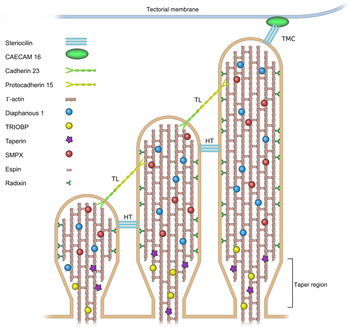
The structure and physiology of the inner ear is in many ways unique and unmatched at other anatomical locations. This explains why so many genes are thought to be involved in inner-ear function and why the ear is so sensitive to mutation at these loci. Mutations in genes that control the cytoskeleton of hair cells, the adhesion of hair cells, intracellular transport, neurotransmitter release or ionic homeostasis can all lead to malfunction of the cochlea (Figure 2).
Diagrammatic representation of important structural proteins in mature hair cell stereocilia. CEACAM16 = carcinogenic antigen-related cell adhesion molecule 16; TRIOBP = trio-binding protein; SMPX = small muscle protein, X-linked; TMC = tectorial membrane attachment crown; TL = tip link; HT = horizontal top connector

Cytoskeleton
As described in the introduction, hair cells have a characteristic shape, with linear, microvilli-like projections called stereocilia arising from their apical surface. The bundles are aligned in a ‘V’ shape and ranked in increasing height. A number of the genes involved in the organisation of the cytoskeleton can cause non-syndromic hearing loss. These include ACTG1 (γ-actin), DIAPH1 (diaphanous 1), TRIOBP (trio-binding protein), TPRN (taperin), SMPX (small muscle protein, X-linked), ESPN (espin) and RDX (radixin).
It is known that γ-actin functions as the building block of hair cell stereocilia. These stereocilia are constantly undergoing actin polymerisation at the tip and depolymerisation at the base.Reference Rzadzinska, Schneider, Davies, Riordan and Kachar16 Mutations in ACTG1 can interfere with this process and cause autosomal dominant hearing loss, DFNA20/26.Reference van Wijk, Krieger, Kemperman, De Leenheer, Huygen and Cremers17, Reference Zhu, Yang, Wei, DeWan, Morell and Elfenbein18 Other proteins are important in this constant remodelling process. For instance, diaphanous 1 regulates the polymerisation and reorganisation of actin monomers into polymers, and has been associated with autosomal dominant hearing loss (DFNA1).Reference Lynch, Lee, Morrow, Welcsh, Leon and King19 The organisation and binding of γ-actin at the base (the so-called ‘taper region’) of hair cell stereocilia is regulated by two alternative splice isoforms of the TRIOBP gene.Reference Kitajiri, Sakamoto, Belyantseva, Goodyear, Stepanyan and Fujiwara20 Mutations in these isoforms, TRIOBP4 and TRIOBP5, form the origin of DFNB28.Reference Riazuddin, Khan, Ahmed, Ghosh, Caution and Nazli21, Reference Shahin, Walsh, Sobe, Abu Sa'ed, Abu Rayan and Lynch22 Another protein localised at this taper region is the regulating protein taperin, which is associated with DFNB79.Reference Rehman, Morell, Belyantseva, Khan, Boger and Shahzad23 The X-linked gene SMPX (DFN4) encodes a protein suggested to have a function in stereocilial development and maintenance in response to the repetitive mechanical stress that these stereocilia are subjected to.Reference Huebner, Gandia, Frommolt, Maak, Wicklein and Thiele24, Reference Schraders, Haas, Weegerink, Oostrik, Hu and Hoefsloot25
The protein espin acts as a bundling protein, providing stability to the stereocilial cytoskeleton.Reference Bartles, Wierda and Zheng26 In ‘jerker’ mice that lack espin, stereocilia shorten and merge from postnatal day 11, which is simultaneous with the onset of hearing. The hair cells further degenerate over time, and after three months the whole organ of Corti is degraded, indicating the importance of a well-organised cytoskeleton.Reference Zheng, Sekerkova, Vranich, Tilney, Mugnaini and Bartles27 In humans, mutations in ESPN cause DFNB36Reference Boulouiz, Li, Soualhine, Abidi, Chafik and Nurnberg28, Reference Naz, Griffith, Riazuddin, Hampton, Battey and Khan29 and autosomal dominant hearing loss.Reference Donaudy, Zheng, Ficarella, Ballana, Carella and Melchionda30 More stability is provided by radixin, which is present along the length of the stereocilia and links actin filaments to the plasma membrane.Reference Pataky, Pironkova and Hudspeth31 Mutations in RDX cause recessive deafness, DFNB24.Reference Khan, Ahmed, Shabbir, Kitajiri, Kalsoom and Tasneem32
Adhesion proteins
Throughout their existence, stereocilia are interconnected and linked to the tectorial membrane by a set of different adhesion proteins. During maturation of the hair bundle in the mouse embryo, a set of temporary links maintain stability. These include transient lateral links (or shaft connectors) and ankle links. These links probably function to provide hair bundle integrity during maturation and/or induce signalling complexes needed for growth regulation and arrangement.Reference Goodyear, Marcotti, Kros and Richardson33 In the mature hair bundle, stereocilia are connected by tip links, horizontal top connectors and tectorial membrane attachment crowns (Figure 2). To date, several genes crucial for the linking apparatus have been identified. These include: DFNB31 (WHRN (whirlin)),Reference Mburu, Mustapha, Varela, Weil, El-Amraoui and Holme34 DFNB18 (USH1C (harmonin)),Reference Ouyang, Xia, Verpy, Du, Pandya and Petit35, Reference Ahmed, Smith, Riazuddin, Makishima, Ghosh and Bokhari36 DFNB66/67 (TMHS (tetraspan membrane protein)),Reference Shabbir, Ahmed, Khan, Riazuddin, Waryah and Khan37 DFNB84 (PTPRQ (tyrosine phosphate receptor Q)),Reference Schraders, Oostrik, Huygen, Strom, van Wijk and Kunst38 DFNB16 (STRC (stereocilin)),Reference Verpy, Masmoudi, Zwaenepoel, Leibovici, Hutchin and Del Castillo39 DFNA4 (CEACAM16 (carcinogenic antigen-related cell adhesion molecule 16)),Reference Zheng, Miller, Yang, Hildebrand, Shearer and DeLuca40 DFNB22 (OTOA (otoancorin)),Reference Zwaenepoel, Mustapha, Leibovici, Verpy, Goodyear and Liu41 DFNB23 (PCDH15 (protocadherin 15))Reference Ahmed, Smith, Riazuddin, Makishima, Ghosh and Bokhari36 and DFNB12 (CDH23 (cadherin 23)).Reference Bork, Peters, Riazuddin, Bernstein, Ahmed and Ness42
Whirlin and harmonin are scaffolding proteins that regulate the formation of the link complexes. Through their PDZ domain binding sites (i.e. binding sites for other proteins), scaffolding proteins fulfil their role in organising multi-protein aggregates and assembling signalling complexes.Reference Michalski, Michel, Caberlotto, Lefevre, van Aken and Tinevez43 Mutations in whirlin and harmonin cause autosomal recessive hearing loss. A third scaffolding protein is Sans, which is associated with the complex syndromic hearing loss of Usher syndrome. USH2a and VLGR1b, two other genes associated with Usher syndrome, are part of the stereocilial ankle link.Reference Michalski, Michel, Bahloul, Lefevre, Barral and Yagi44
Cadherin 23 and protocadherin 15, as well as PTPRQ and TMHS, are presumably part of the transient lateral link. They prevent fusion of stereocilia by keeping them at a fixed distance from each other during development.Reference Goodyear, Legan, Wright, Marcotti, Oganesian and Coats45–Reference Michel, Goodyear, Weil, Marcotti, Perfettini and Wolfrum47 In the mature hair cell, cadherin 23 and protocadherin 15 become the main components of the tip link. These provide stability and gate the mechanotransduction channel; these tip links provide stability and gate the mechanotransduction channel, which plays a central role in auditory function.Reference Kazmierczak, Sakaguchi, Tokita, Wilson-Kubalek, Milligan and Muller48 TMHS co-localises with protocadherin 15 and is a proposed subunit of the mechanotransduction channel.Reference Shabbir, Ahmed, Khan, Riazuddin, Waryah and Khan37, Reference Xiong, Grillet, Elledge, Wagner, Zhao and Johnson49
Stereocilin is an extracellular protein that is thought to make up both horizontal top connectors and tectorial membrane attachment links. The latter, combined with the so-called attachment crown, attach the tallest stereocilia of the outer hair cell stereocilia bundle to the tectorial membrane.Reference Verpy, Leibovici, Michalski, Goodyear, Houdon and Weil50 This tectorial membrane attachment crown is probably formed by CEACAM16. In a similar way, otoancorin is thought to attach non-sensory cells to the tectorial membrane.Reference Zwaenepoel, Mustapha, Leibovici, Verpy, Goodyear and Liu41
Transport proteins
Motor proteins can be used to transport different proteins to target sites in the cell. In the inner ear, the proteins used for transport are all part of the unconventional myosin family. These proteins can bind to the actin cytoskeleton and move forward along actin filaments by using energy derived from ATP. The carboxyl-terminal tails of the transport protein contain binding sites for the proteins they will carry.Reference Friedman, Dror and Avraham51 Seven unconventional myosins have been associated with hereditary hearing loss: myosin Ia (DNFA48),Reference Donaudy, Ferrara, Esposito, Hertzano, Ben-David and Bell52 myosin IIIa (DFNB30),Reference Walsh, Walsh, Vreugde, Hertzano, Shahin and Haika53 myosin VI (DFNA22/DFNB37),Reference Melchionda, Ahituv, Bisceglia, Sobe, Glaser and Rabionet54, Reference Ahmed, Morell, Riazuddin, Gropman, Shaukat and Ahmad55 myosin VIIa (DFNA11/DFNB2),Reference Liu, Walsh, Tamagawa, Kitamura, Nishizawa and Steel56, Reference Weil, Kussel, Blanchard, Levy, Levi-Acobas and Drira57 non-muscle myosin heavy chain IX (DFNA17)Reference Lalwani, Goldstein, Kelley, Luxford, Castelein and Mhatre58 and XIV (DFNA4),Reference Donaudy, Snoeckx, Pfister, Zenner, Blin and Di Stazio59 and myosin XVa (DFNB3).Reference Wang, Liang, Fridell, Probst, Wilcox and Touchman60 These proteins all have their own unique transport function in the inner-ear hair cells.
Synapse
Otoferlin (OTOF) acts with one of the myosins, myosin VI, at the synaptic cleft of the inner hair cell. The protein is thought to be involved in the (calcium-dependent) fusion of synaptic vesicles to the plasma membrane. As a result, the neurotransmitter glutamate is released into the synaptic cleft with subsequent excitation of the afferent neuron. In OTOF-mutant mice, a reduction in exocytosis is detected.Reference Heidrych, Zimmermann, Kuhn, Franz, Engel and Duncker61 A number of allelic variants of OTOF that cause DFNB9 in humans have been identified.Reference Yasunaga, Grati, Cohen-Salmon, El-Amraoui, Mustapha and Salem62
Another player at the inner hair cell synapse is VGLUT3, a member of the vesicular glutamate receptors. VGLUT3 is encoded by SLC17A8 and associated with autosomal recessive hearing loss DFNA25.Reference Ruel, Emery, Nouvian, Bersot, Amilhon and Van Rybroek63 The protein is thought to regulate the endocytosis and exocytosis of glutamate. Both OTOF and SLC17A8 knockout mice show a reduction in the number of postsynaptic ganglion cells, indicating that these proteins are crucial for the development and preservation of normal hearing.Reference Seal, Akil, Yi, Weber, Grant and Yoo64
Ion homeostasis
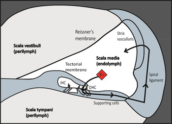
The cochlea contains two types of fluids, both different in ion composition. Perilymph is high in sodium and low in potassium, whereas endolymph is high in potassium and low in sodium. This contributes to a highly positive potential (+80 mV) called the endocochlear potential. A potassium influx from the endolymph into the hair cell causes depolarisation of the cell. Immediately after depolarisation, the hair cell repolarises, shifting cations via neighbouring structures back into the endolymph (Figure 3). This process of ion homeastasis involves claudin 14 (CLDN14), tricellulin (MARVELD2/TRIC), tight junction protein 2 (TJP2), a number of connexins (GJB's), KCNQ4 (KCNQ4), ATP2b2 (ATP2b2/PMCA2), Barttin (BSND) and pendrin (SLC26A4) all of which are related to hereditary hearing loss.
Diagrammatic representation of potassium circulation within the cochlea: the opening of ion channels in the hair cell apical membrane allows a potassium influx from the endolymph into the hair cell; potassium is then moved to supporting cells and pumped back into the endolymph via spiral ligament and stria vascularis gap junction networks. K+ = potassium ion; IHC = inner hair cells; OHC = outer hair cells

Tight junctions guard the border between endolymph and perilymph compartments. By generating a seal between two adjacent cells, a barrier is created that restricts the free diffusion of ions. In this way, the apical side of the outer hair cells and supporting cells are exposed to the endolymph, and the basolateral surface is bathed in cortilymph, a fluid which is similar to perilymph and fills up the so-called space of Nuel. This space of Nuel, which surrounds the basolateral surface of outer hair cells, might change in electric potential when the tight junction protein claudin 14 is absent or dysfunctional, as in DFNB29.Reference Ben-Yosef, Belyantseva, Saunders, Hughes, Kawamoto and Van Itallie65, Reference Wilcox, Burton, Naz, Riazuddin, Smith and Ploplis66 In a similar way, tricellulin, encoded by MARVELD2/TRIC, is presumed to function as tight junction that connects three cells together and causes DFNB49 when mutated.Reference Riazuddin, Ahmed, Fanning, Lagziel, Kitajiri and Ramzan67 TJP2 acts as a scaffolding protein, binding tight junctions to the actin cytoskeleton. Duplication with over-expression of the protein causes DFNA51.Reference Walsh, Pierce, Lenz, Brownstein, Dagan-Rosenfeld and Shahin68 This process occurs as a result of another role of the TJP2 protein, namely its involvement in the nucleus' signaling pathways that regulate the cell cycle. Over-expression of TJP2 disturbs the balance between pro-apoptotic and anti-apoptotic genes and will induce apoptosis.Reference Lenz and Avraham69
A network of gap junctions (channels that extend over two adjacent membranes) in the cochlea enables the exchange of various small molecules and ions. These gap junctions are made up of specialised proteins called connexins, which are expressed in the supporting cells of the organ of Corti and the connective tissue of the spiral ligament.Reference Hoang Dinh, Ahmad, Chang, Tang, Stong and Lin70 This gap junction network is associated with the recycling of potassium ions needed for normal hearing. The first identified gene and most common cause of non-syndromic hearing loss is GJB2, which encodes connexin 26 (DFNA3a/DFNB1a).Reference Kelsell, Dunlop, Stevens, Lench, Liang and Parry71 Mutations in the GJB2 gene account for 30–50 per cent of all cases of childhood deafness, and 1–4 per cent of the average human population are estimated to be carriers.Reference Hoang Dinh, Ahmad, Chang, Tang, Stong and Lin70 Other connexins associated with non-syndromic hearing loss are connexin 31 (GJB3, DFNA2b/DFNB91)Reference Xia, Liu, Tang, Pan, Huang and Dai72, Reference Liu, Xia, Xu, Pandya, Liang and Blanton73 and connexin 30 (GJB6, DFNA3b/DFNB1b).Reference Grifa, Wagner, D'Ambrosio, Melchionda, Bernardi and Lopez-Bigas74, Reference del Castillo, Villamar, Moreno-Pelayo, del Castillo, Alvarez and Telleria75
KCNQ4 encodes a protein that forms a voltage-gated potassium channel. This gene is expressed in the outer hair cells of the cochlea and is mutated in a dominant form of non-syndromic hearing loss, DFNA2a.Reference Kubisch, Schroeder, Friedrich, Lutjohann, El-Amraoui and Marlin76 KCNQ4 is thought to aid repolarisation of the outer hair cells. Furthermore, KCNQ4 is proposed to regulate sensitivity to sound by changing the resting membrane potential of the outer hair cells.Reference Heidenreich, Lechner, Vardanyan, Wetzel, Cremers and De Leenheer77
On their apical surface, in the stereocilial membrane, hair cells express ATP2b2/PMCA2, which is a modifier of DFNB12.Reference Schultz, Yang, Caride, Filoteo, Penheiter and Lagziel78 The protein product PMCA2 is a calcium pump that uses energy from ATP to function. Calcium, which is used (in addition to potassium) to excite the cell, is constantly pumped back into the endolymph by PMCA2, thereby ensuring a stable concentration of this ion.Reference Bortolozzi, Brini, Parkinson, Crispino, Scimemi and De Siati79 PMCA2 fulfils a similar function in the synaptic region on the basolateral surface of the hair cell.
BSND and SLC26A4, which encode barttin and pendrin respectively, are genes involved in both syndromic and non-syndromic hearing loss. Barttin is a chloride channel subunit. Most mutations in BSND cause Bartter syndrome, which comprises hearing loss and renal abnormalities. The molecular basis of DFNB73 has been attributed to a mutation in BSND, which causes non-syndromic deafness.Reference Riazuddin, Anwar, Fischer, Ahmed, Khan and Janssen80 The anion exchanger pendrin plays a major role in maintaining a constant acid–base balance. Both syndromic hearing loss (Pendred's syndrome, associated with goitre) and non-syndromic hearing loss (DFNB4) have been described in relation to this, the occurrence of which depends on the extent of the mutation in SLC16A4.Reference Li, Everett, Lalwani, Desmukh, Friedman and Green81, Reference Everett, Glaser, Beck, Idol, Buchs and Heyman82
Electromotility
The outer hair cells have a unique feature of altering sensitivity and selectivity to sound. The protein Prestin is thought to be responsible for this by introducing a process called electromotility. Prestin changes its configuration in reaction to changes in membrane potential, enabling the outer hair cell length to be altered. In this way, the cylindrical outer hair cell becomes shorter on depolarisation and longer on hyperpolarisation, thereby amplifying its sensitivity to sound.Reference Brownell, Bader, Bertrand and de Ribaupierre15 Prestin, encoded by SLC26A5, was first identified by Zheng et al. in 2000.Reference Zheng, Shen, He, Long, Madison and Dallos83 In homozygous Prestin null mice, cochlear thresholds were found to be 40–60 dB higher than in wild-type mice.Reference Liberman, Gao, He, Wu, Jia and Zuo84 In humans, mutations in SLC26A5 are the cause of DFNB61 hearing loss.Reference Liu, Ouyang, Xia, Zheng, Pandya and Li85
Others
Other important groups of genes involved in hereditary hearing loss that will not be discussed further are extracellular matrix proteins, including TECTA (α-tectorin), COL11A2 (type XI collagen α2) and COCH (cochlin), and a number of transcription factors, including POU4f3 (class 4 POU), POU3f4 (class 3 POU), EYA4 (eyes absent 4), MIR96 (microRNA96), ESRRB (oestrogen-related receptor β) and GRHL2 (grainyhead-like 2).
Conclusion
In the last two decades, our understanding of the basis of hereditary hearing loss has advanced significantly. This has been powered by major developments in human and mouse genetics. Improved genotyping and mapping has enabled the study of human families with multi-generational hereditary hearing loss. This has led to the identification of many genetic mutations that can cause syndromic or non-syndromic deafness. The mouse has proven to be an excellent genetic model for human hearing loss and deafness because of its structural, physiological and genetic similarities. Programmes for the systematic mutationReference Brown, Hardisty-Hughes and Mburu86 and phenotypingReference Hardisty-Hughes, Parker and Brown87 of mice have enabled the discovery of many new genes involved in cochlear embryology or function, and many genetic loci have subsequently been found to underlie human deafness.
In this article, we have given an overview of the currently known genes involved in hereditary hearing loss (Table I). The function of these genes will become better understood with time, and no doubt many more genes that can lead to hearing loss will be discovered. With continued examination, we will build a better understanding of the function of the cochlea, and hopefully develop novel molecular therapies for human sensorineural hearing loss.
Acknowledgement
The authors would like to acknowledge Mr J Stelma for graphic design.