Introduction
Most members of the genus Angiostoma are found in the alimentary tract of terrestrial molluscs, but three species (A. plethodontis, A. onychodactyla and A. lamotheargumedoi) have been discovered in the intestines of salamanders and one species (A. carettae) in the lungs of a sea turtle. Nothing is known about the life cycles of Angiostoma spp. parasitic in vertebrate hosts, and the hypothesis that the nematodes can be acquired by consuming infected molluscs (Adamson, Reference Adamson1986) still remains to be tested. Recently, Falcón-Ordaz et al. (Reference Falcón-Ordaz, Mendoza-Garfias, Windfield-Perez, Parra-Olea and de Leon2008) expressed the opinion that acquisition of an angiostomatid nematode (namely A. lamotheargumedoi Falcón-Ordaz et al., Reference Falcón-Ordaz, Mendoza-Garfias, Windfield-Perez, Parra-Olea and de Leon2008) by amphibians could be a result of host-switching from gastropod hosts, although no gastropod host has yet been found.
Among the diverse types of relationships between nematodes and molluscs, those involving Angiostoma are considered as truly parasitic (Grewal et al., Reference Grewal, Grewal, Tan and Adams2003; Morand et al., Reference Morand, Wilson, Glen and Barker2004). The nematodes are believed to be parasitic in their hosts as adults/4th stage larvae. Nematode eggs are evacuated with the host's faeces or mucus, rapidly develop outside the host until they reach the infective stage, which can be 1st–2nd stage larvae or 3rd stage larvae, depending on whether the nematode species is ovoviviparous (1st–2nd stage infective) or oviparous (3rd stage infective). Infective larvae are then either swallowed by hosts with food or actively penetrate the mantle cavity (Morand, Reference Morand1986, Reference Morand1988).
Angiostomatids parasitic in molluscs are represented mainly by intestinal parasites, and the majority of species have been recorded from the anterior portion of the intestine. An exception is A. aspersae which parasitizes the pallial cavity of the garden snail, Helix aspersa (Morand, Reference Morand1986). The new species described herein was found in the digestive gland (hepatopancreas), a vast organ extending along the foot of the snail, and emptying into the stomach. The presence of nematodes in a metabolically active organ of the host suggests a different feeding behaviour from other angiostomatids.
Materials and methods
Parasitological procedures
Infected molluscs (Megaustenia sp.) were collected during field trips in the southern part of Cat Tien National Park in Vietnam, province Dong Nai, in June 1999 and June 2007. The molluscs were found in the litter of moist broadleaf forests in which species of Tetramelaceae, Lythraceae, Arecaceae and Moraceae were dominant. In 1999, 1 of 5 snails collected was infected with 29 adult nematodes and a juvenile. In 2007, 3 out of 7 snails were infected by 1–3 nematodes. Several specimens were preserved in ethanol for DNA study and the rest in hot 4–5% formaldehyde for light microscopy and scanning electron microscopy (SEM). Formaldehyde-fixed nematodes were then processed into anhydrous glycerol for mounting according to Seinhorst (Reference Seinhorst1959). Light microscopy studies and drawings were performed using a Zeiss Jenaval microscope (Zeiss, Germany) equipped with a camera lucida. Illustrations were finished using a WACOM Intuos A4 USB drawing tablet and Adobe Illustrator CS3 according to Coleman (Reference Coleman2003). Three specimens were used for SEM studies as follows. Material for SEM was rehydrated after formaldehyde, dehydrated in a graded ethanol series, critical-point dried using a HCP-2 Hitachi dryer (Hitachi, Tokyo, Japan), mounted on aluminium stubs and coated with gold in a Bio-Rad SC502 sputter coater (Bio-Rad, Hemel Hempstead, Herts, UK). Specimens were studied in a JCM-6380 LA SEM (Jeol, Tokyo, Japan).
Large subunit rDNA analysis
To determine phylogenetic relationships between certain Angiostoma species, the D2D3 Large subunit (LSU) rDNA sequences were obtained for A. glandicola sp. n. and three European species, A. limacis, A. dentifera and A. milacis, collected in the UK and Belgium.
DNA was extracted from samples fixed in 70% ethanol using a modification of the protocol described by Floyd et al. (Reference Floyd, Abebe, Papert and Blaxter2002). Ethanol-fixed nematodes were transferred to 20 μl 0.25 m NaOH in Eppendorf tubes, centrifuged and incubated at 99°C for 3 min. Samples were then cooled to room temperature and 4 μl 1 m HCl, 10 μl 0.5 m Tris–HCl (pH 8.0) and 5 μl of 2% Triton X-100 were added. The tubes were recentrifuged and again incubated at 99°C for 3 min. About 0.4–0.8 μl of the resulting mixture was used as DNA template for polymerase chain reactions (PCR) performed using Qiagen PCR kits according to the manufacturer's instructions (Qiagen, Venlo, The Netherlands). Primer pairs D2A (ACA-AGT-ACC-GTG-AGG-GAA-AGT-TG) and D3B (TCG-GAA-GGA-ACC-AGC-TAC-TA) were used to amplify LSU DNA fragments. PCR cycling parameters comprised primary denaturation at 94°C for 5 min followed by 34 cycles of 94°C for 60 s, 50°C for 60 s, and 72°C for 1 min, followed by post-amplification extension at 72°C for 10 min. The PCR reaction products were visualized in agarose gels and bands were excised for DNA extraction with Qiagen gel extraction and product cleaning kits. After ligation with pGEM-T vector, PCR products were used to transform JM 109 Esherichia coli competent cells (according to Promega protocol; Promega, Madison, Wisconsin, USA). Transformed colonies were subcultured in 1.5 ml of Luria–Bertani (LB) medium and incubated for 16–22 h at 37°C. Plasmid DNA was obtained from transformed E. coli using QIAprep Miniprep Kits (Qiagen) according to the manufacturer's instructions and used for sequencing using BigDye® Termination Kits (Applied Biosystems, Foster City, California, USA) with 3.2 pmol of vector primers T7 and SP6. Termination reaction products were ethanol-precipitated, air-dried and resuspended in ABI Prism® Template Suppression Reagent (Applied Biosystems).
For comparative purposes and phylogeny construction, the following D2D3 expansion segment of LSU rDNA sequences deposited in GenBank were used: Caenorhabditis remanei, AY602174; Rhabditella axei, AY602177; and Oscheius myriophila, AY602176. The choice of these taxa as outgroups was based on the wider study of the phylogeny of nematodes parasitic in terrestrial molluscs, which revealed the tight phylogenetic relationships between families Angiostomatidae and these genera of Rhabditidae (unpublished data).
Sequence alignments were generated using Clustal X using default values for gap opening and gap extension penalties. All alignments were analysed using PAUP* 4.0b10 (Swofford, Reference Swofford1998) for maximum parsimony (MP), neighbour joining (NJ) and maximum likelihood (ML) constructions. Maximum parsimony analysis was performed with gaps treated as ‘missed data’ or ‘fifth base’. The TrN+G model was selected for maximum likelihood analysis with the use of ModelTest 5.0 (Posada & Crandall, Reference Posada and Crandall1998), employing the interface MTgui (Nuin, Reference Nuin2005) to prepare the ModelTest results for ML analysis in PAUP* 4.0b10.
Results
Angiostoma glandicola sp. n.
Adults
Description
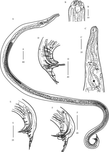
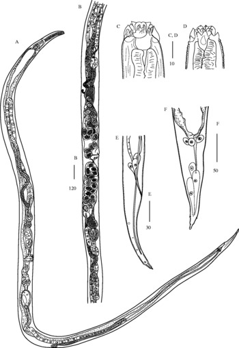
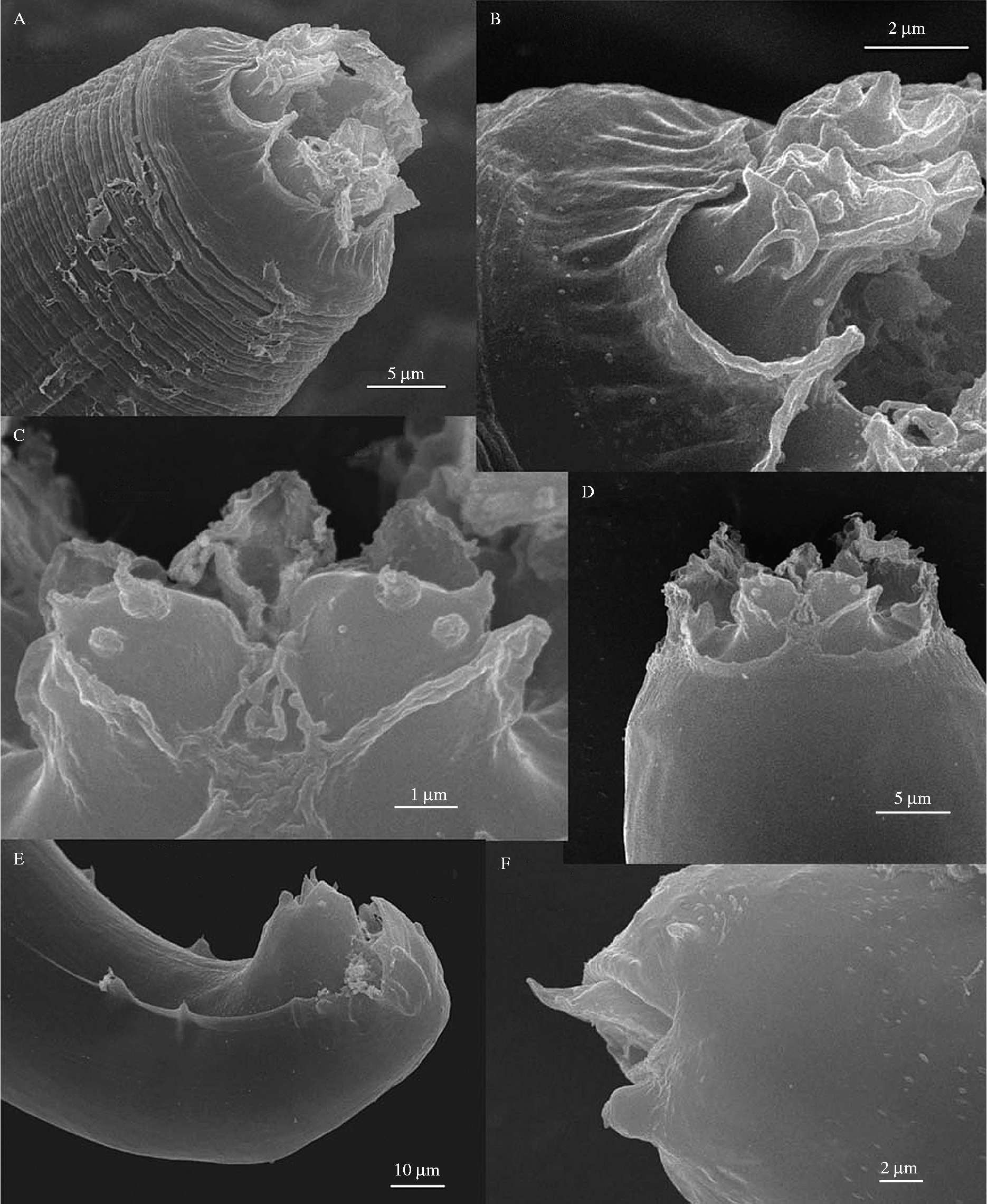
Medium-sized angiostomatids (figs 1–3). No marked sexual dimorphism present. Cylindrical body gradually tapering to both ends, sometimes very slightly expanded at basal bulb level. Body cuticle annulated (annuli c. 1–2 μm wide) and finely striated. Lateral field very slightly protruding at the anterior end and flat in further course, c. 2–4 μm wide at ends and c. 10–12 μm at mid-body. Head with two circlets of six probolae with triangular, forward-pointing lobes: internal probolae just as high as lips (c. 8–10 μm) whereas external are slightly lower. Six isolated, attenuated, rhomboid lips present. Prominent papillae at the outer contour of lips, two on each lip. Papillae situated closer to mouth aperture are higher and more prominent. Small, pore-like amphids with prominent pouch displaced posteriad at the level of lip bottom. Stoma beneath lips, enlarged, slightly anisotopic, with non-refractive walls expanded to 1.5 μm. Glottoid apparatus absent. Pharyngeal collar absent. Pharynx with long, rather thick corpus, distinct isthmus two times shorter than corpus and medium-sized, pear-shaped bulb with very finely cuticularized valves. Nerve ring on posterior part of corpus or on isthmus, thick. Excretory pore opposite isthmus/bulb. Excretory cell small, ampoule-like. Excretory duct short. Cardia and intestine well developed, intestinal walls broad. Rectal glands present. Phasmids at short distance from tail tip.
Angiostoma glandicola sp. n., male. (A) Entire view; (B) head; (C) anterior end; (D–F) tail. A–E in lateral position, F in ventral position. Scale bars in μm.

Angiostoma glandicola sp. n., female. (A) Entire view; (B) reproductive system; (C), (D) head; (E), (F) tail. All in lateral position. Scale bars in μm.

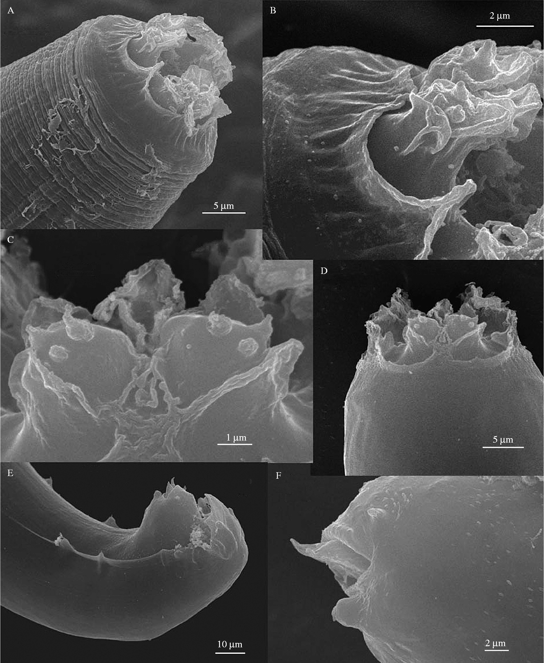
Angiostoma glandicola sp. n., SEM images. (A), (B) Male head; (C), (D) female head; (E) male tail; (F) male cloaca. Scale bars in μm.
Male
Holotype: body length = 2778 μm; max. width = 78 μm; anal width = 50 μm; pharynx length = 227 μm; head to excretory pore = 165 μm; head to nerve ring = 140 μm; tail = 55 μm; spicules measured on arc = 105 μm; spicules measured on chord = 85 μm; gubernaculum length = 45 μm. De Manian indexes: a = 35.6; b = 12.2; c = 50.5.
Paratypes (n = 10): body length = 2950 ± 413 (2583–3718) μm; max. width = 79 ± 14 (62–100) μm; anal width = 46 ± 6 (38–57) μm; pharynx length = 222 ± 15 (205–245) μm; head to excretory pore = 163 ± 24 (124–205) μm; head to nerve ring = 137 ± 16 (113–170) μm; tail = 57 ± 6 (48–65) μm; spicules measured on arc = 105 ± 7 (96–120) μm; spicules measured on chord = 83 ± 6 (75–91) μm; gubernaculum length = 44 ± 7 (32–52) μm. De Manian indexes: a = 37.8 ± 3.3 (34.0–43.1); b = 13.3 ± 1.3 (11.6–15.2); c = 51.9 ± 4.4 (45.1–59.0).
Anterior end with slightly longer pharynx with larger bulb and shorter stoma than in females. Lateral field extends from the level of mid-corpus to cloaca. External probolae 6.3 ± 0.6 (6–7) μm high, internal probolae 8.7 ± 1.2 (8–10) μm high. Lips 8 μm high. Stoma 8.7 ± 1.9 (7–13) μm long and 9.5 ± 1.1 (8–11) μm wide. Pharyngeal corpus 18 ± 2 (15–22) μm wide; isthmus 14 ± 2 (12–17) μm wide; bulb 37 ± 5.2 (30–45) μm wide and 50 ± 6.9 (40–62) μm long. Excretory duct 11 ± 4.1 (6–15) μm long and 1.5 ± 0.5 (1–2) μm wide. Monorchic. Testis reflexes in 599 ± 86 (493–760) μm from anterior. Testis flexure c. 566 ± 53 (475–652) μm long. Growing spermatocytes in two rows. Immature sperm in ejaculatory duct c. 11 μm in diameter. Tail short, wide at cloaca but dramatically narrowing to tip. Tail spike free, not embedded into bursal velum, but barely extends beyond bursa (c. 2–10 μm). Bursa anteriorly open. Bursa supported by nine pairs of longish pedunculate genital papillae: three pairs precloacal and six pairs postcloacal in two groups: GP 4–7 situated closely behind cloaca and GP 8–9, shorter ones, at some distance further posteriad. GP 1–4 and 6, 7, 9 terminate on the ventral surface of the bursa whereas GP 5 and 8 terminate on the dorsal surface. Papillae in pairs 8–9 fused at base. A prominent unpaired ventral precloacal papilla and two smaller subventral postcloacal papillae are present which are not incorporated into the bursa. A precloacal papilla is situated on the anterior cloacal flap close to its edge; c. 4–7 μm long and 2 μm thick with a sensillum protruding from the top. Spicules longer than anal width, with straight shafts and distal tips curved at one-third of spicule body length. Manubria small, rounded, c. 6.2 ± 2 (5–10) μm in diameter; shafts thickened to 9.5 ± 2 (7–14) μm. Tips c. 3 μm thick, pointed. Gubernaculum thin, boat-shaped. Phasmids poorly discernible, c. 10–15 μm from tail tip.
Female
Paratypes (n = 6): body length = 3023 ± 577 (2548–4072) μm; max. width = 91 ± 19 (70–125) μm; anal width = 28 ± 3 (23–32) μm; pharynx length = 213 ± 13 (198–230) μm; head to excretory pore = 162 ± 39 (125–215) μm; head to nerve ring = 125 ± 16 (100–145) μm; percentage of anterior vulva distance to body length (V%) = 45.2 ± 3.2 (41.7–50.4)%. De Manian indexes: a = 33.7 ± 4.0 (27.4–37.5); b = 14.2 ± 2.3 (11.7–17.7); c = 27.5 ± 2.5 (24.1–30.0).
Lateral field extends from the level of mid-corpus to phasmids. External probolae c. 6 μm high, internal probolae 9.5 ± 0.8 (8–9) μm high. Lips 8.5 ± 0.7 (8–10) μm high. Stoma 9.8 ± 1.5 (8–12) μm long and 9.5 ± 1.5 (8–12) μm wide. Pharyngeal corpus 18 ± 2.5 (15–21) μm wide; isthmus 15 ± 2 (12–18) μm wide; bulb 33 ± 2.9 (30–38) μm wide and 46 ± 5.9 (35–52) μm long. Excretory duct 11 ± 1.8 (10–14) μm long and 2.8 ± 0.8 (2–4) μm wide. Didelphic. Ovaries reflexed, each flexure very long and equal to the length of the portion leading to vulva. Ovary length/uterus length ratio c. 3:1. Each ovary starts near distal part of correspondent uterus, then runs on dorsal side of the body toward head (anterior branch) and tail (posterior branch), then reflexes at some distance from both ends. Anterior branch of ovary reflexes in 489 ± 112 (317–660) μm from head end; posterior branch reflexes in 897 ± 466 (597–1712) μm. Oocytes in two, then single row. Oviduct glandular. Uteri divergent, short. Distal parts of uteri morphologically distinct from the rest of the structure and accumulate several dozens of spermatozoa, smaller in size than those in the ejaculatory duct of males. Each uterus contains 12–40 thin-shelled eggs in different stages of development, 59 ± 6 (55–68) μm in length and 43 ± 2 (40–45) μm in width. Vulval lips flat. Vagina straight, slightly longer than half-width at the vulva, c. 38 ± 16 (20–62) μm long. Tail conical with arrowhead-like tip bearing no denticles. Phasmids in 40 ± 3.4 (35–45) μm from tail tip.
Etymology
The species name refers to the typical habitat of the nematode in the digestive gland of a mollusc.
Type host
Megaustenia sp.
Type locality
Cat Tien National Park, province Dong Nai, Vietnam; June 1999 and June 2007, in forest litter.
Type habitat
Digestive gland (hepatopancreas).
Type material
Holotype male (1079), two paratype males and one paratype female mounted on the same slide (1080) are deposited in the Museum for the Helminthological Collections of the Centre of Parasitology of the A.N. Severtsov Institute of Ecology and Evolution in Moscow.
Diagnosis and relationships
There are two genera in the family Angiostomatidae, Angiostoma Dujardin, 1845 and Aulacnema Pham Van Luc et al. Reference Pham Van Luc, Spiridonov and Wilson2005, which are differentiated on the basis of the didelphic vs. monodelphic female reproductive system and the median vs. anterior vulval position.
The new species is characterized by: presence of nearly flat, not wing-like lateral fields; a head decorated with six low triangular probolae in an external circle and six higher ones forming an internal circle; six isolated, high, rhomboid lips; 6+6 prominent cephalic papillae situated on the margin of lips; pore-like amphids with a prominent pouch displaced to the level of lip bottom; an enlarged stoma with weakly sclerotized, non-refractive walls; a club-shaped pharynx with plumpish procorpus, a short thick isthmus and pear-shaped bulb; a thick nerve ring surrounding the isthmus; the excretory pore opposite the isthmus/bulb; a small ampoule-like excretory cell; reflexed ovaries with a long, flexed portion; short, divergent uteri and median vulva in females; in males long arcuate spicules with small manubria; a small gubernaculum; one precloacal and two postcloacal ventral circumcloacal papillae; bursa supported by nine pairs of pedunculate genital papillae: three precloacal and six postcloacal; of the latter, four are situated just behind the cloaca and the last shorter two pairs are distanced and fused at the base. All papillae except GP 5 and 8 terminate on the dorsal edge of the bursa whereas the latter ones terminate on the ventral edge. The male tail is short and broadly conical, spike free but barely extends beyond the bursa; the female tail is long and conical with an arrowhead-like tip.
The striking feature of this species is the structure of the cephalic end, decorated with probolae and isolated, elongated lips. It clearly differentiates the new species from other species of the genus. In the aspect of possessing probolae and elongated lips, it is similar to representatives of the Cephalobinae but is clearly distinguished by the male tail structure.
Apart from the head structure, it is related to A. aspersae by having a similar male tail which is also short with a spike extending a short distance beyond the bursa and with the same arrangement of postcloacal GP. However, in the present species there are three vs. four precloacal GP, no paired papilliform appendages in front of the cloaca but two small ventral papillae just behind it and the tail spike is not bilobated.
The new species can be distinguished from A. coloaense (also found in Vietnam), in having probolae vs. no probolae on the head, fused vs. isolated lips, a much smaller stoma, a shorter male tail and a different set of GP (Pham Van Luc et al., Reference Pham Van Luc, Spiridonov and Wilson2005).
Molecular differentiation and phylogenetic relationships
The D2D3 LSU rDNA sequences obtained were deposited in NCBI GenBank: Angiostoma glandicola sp. n., GQ167724; Angiostoma limacis, GQ167725; Angiostoma dentifera, GQ167726; Angiostoma milacis, FJ949063.
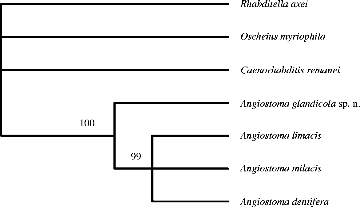
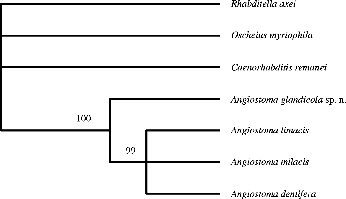
Comparisons of our four obtained Angiostoma sequences and three species of Rhabditidae using ClustalX produced a 631 bp long alignment. Differences between angiostomatids and Rhabditidae species ranged from 100 to 161 bp, whereas intrageneric differences within Angiostoma spp. ranged from 27 to 49 bp. Angiostoma glandicola sp. n. differs from A. limacis (type species), A. dentifera and A. milacis in 33, 49 and 42 nucleotides, respectively (fig. 4). Identical tree topologies were obtained with all three methods of analysis used and the monophyletic status of the Angiostoma clade was strongly supported under all methods. The bootstrap support of the subclade consisting of the three European angiostomatids and the basal position of A. glandicola sp. n. between Angiostoma species was strong under MP and NJ analysis (fig. 4) but lower in the ML tree (data not shown).
Phylogenetic relationships between Angiostoma species and selected Rhabditidae taxa. Optimality criterion = parsimony, of 631 total characters: 343 characters are constant; 131 variable characters are parsimony-uninformative; number of parsimony-informative characters = 157, 1000 bootstrap repeats. Gaps are treated as ‘fifth base’. Identical tree with the same bootstrap values was obtained from neighbour joining (NJ) analysis.
Discussion
The present species is the third member of the family Angiostomatidae found in terrestrial molluscs in Vietnam. Angiostoma coloaense and Aulacnema monodelphis were dissected from the intestines of Cyclophorus sp. and Camaena sp., respectively (Pham Van Luc et al., Reference Pham Van Luc, Spiridonov and Wilson2005).
Angiostoma is the larger genus of the small family Angiostomatidae that contains just two genera, both of which are predominantly associated with terrestrial gastropods, although a few species of Angiostoma have been found in plethodontid salamanders and a sea turtle. The large number of records of these nematodes from various gastropod hosts suggests a long-established association with gastropods. It is much less obvious how well established the relationship is in the case of vertebrate hosts because of the scarcity of recorded findings. In recent years, three new Angiostoma species have been described from vertebrate hosts: A. onychodactyla, A. lamotheargumedoi and A. carettae (Bursey & Goldberg, Reference Bursey and Goldberg2000; Falcón-Ordaz et al., Reference Falcón-Ordaz, Mendoza-Garfias, Windfield-Perez, Parra-Olea and de Leon2008; Bursey & Manire, Reference Bursey and Manire2006).
With a growing number of angiostomatid nematodes recorded in vertebrate hosts more questions arise concerning the biology and taxonomic status of the vertebrate-associated nematodes. Unfortunately, some of the aforementioned descriptions failed to provide some important angiostomatid diagnostic details. The conservative nature of Angiostoma morphology further inhibits the resolution of the taxonomic status of species from vertebrate hosts. Detailed molecular analyses would greatly aid resolution of their taxonomic position.
Acknowledgements
The authors thank Prof. A.A. Schileyko, A.N. Severtsov Institute of Ecology and Evolution RAS, for identification of the mollusc host. This work was supported by the Russian Foundation for Basic Research grants 08-04-00 209 and 09-04-90 300.