Introduction
Sir Harold Gillies, Commander of the Order of the British Empire (‘CBE’) and Fellow of the Royal College of Surgeons (‘FRCS’), has been the subject of many tributes, celebrations and eulogies.Reference Pound1–Reference Bamji7 He started his career as an otolaryngologist and progressed to lay the foundation of plastic surgery in Britain. He is considered one of the greatest otolaryngologists ever.Reference Weir and Seir2 He is responsible for the invention of numerous surgical instruments and operative techniques.
This paper provides a closer inspection of the history surrounding the rhinoplasty,Reference Rana and Arora8, Reference Bennet9 and, in particular, of the inception, dissemination and evolution of the tubed pedicle flap (Gillies Archives (Frognal Centre of Medical Education, Queen Mary's Hospital, Sidcup), unpublished works).
Predecessors
Plastic surgery techniques have been in existence for centuries. The ‘Sushruta Samhita’, a chapter in the Vedas (a collection of Indian religious books), contains detailed descriptions of the teachings and practice of an ancient surgeon Sushruta, credited for the first recorded rhinoplasty.Reference Rana and Arora8 Over the years, Sushruta's method was modified in India to what is now termed a paramedian forehead flap. This procedure reached Europe when an article in the Madras Gazette of 5th August 1794 detailed the restoration of the lost nose of Cowasjee, who was at the time a prisoner of the Sultan.Reference Bennet9 However, the procedure was not attempted in the UK until October 1814, when JC Carpue first performed it in the repair of the nasal defect of a British Army officer without the benefit of anaesthesia.Reference Rana and Arora8 The procedure was subsequently performed at the Edinburgh Royal Infirmary on the 28th of April 1840 by Mr Ferguson, later Sir William Ferguson, Professor of Surgery at King's College Hospital, London.Reference Bennet9
Gillies' early life
Gillies was born in Dunedin, New Zealand on the 17th of June 1882. He was sent to Lindley Lodge, a boarding preparatory school in Nuneaton, UK before returning to New Zealand to complete his secondary education at Wanganui College. He managed to overcome the handicap of a stiff elbow, which he had sustained from a childhood accident when captaining the ‘first 11’ team in cricket.
He went on to study medicine at Cambridge University, where he displayed a vast array of talents. He rowed for Cambridge in the 1904 boat race, played golf for England against Scotland, won the Royal St George Grand Challenge at Sandwich in 1913 and received a chapel choir scholarship.Reference Pound1, Reference Negus3, Reference Millard5–Reference Bamji7
He was known as the best house surgeon during his time at St Bartholomew's Hospital, London. He progressed to ENT surgery and applied for a job in Harley Street, London as assistant to Sir Milsom Rees, the ENT surgeon to King George. He was to have a lucrative career with Sir Rees, who is reported to have been more impressed by Gillies' golfing skills than his clinical prowess.Reference Pound1, Reference Bamji6, Reference Bamji7
World War One
With the outbreak of World War One, Gillies' lucrative career prospects in conventional ENT were altered. In 1915, he volunteered for service with the British Red Cross and was posted to France. Gillies met with Valadier, a French-American dentist who had established a 50-bed facial, dental and jaw unit in Wimereux. The intrinsic nature of trench warfare led to an increased incidence of injuries to the face and head. Gillies decided to devote himself to facial surgery and arranged to observe Europe's most celebrated facial surgeon at the time, the Creole Hippolyte Morestin.Reference Pound1
He subsequently returned to England to commence his new career as a facial plastic surgeon. He persuaded the Army Surgeon-General Alfred Keogh that facial work should be concentrated in one place and initially proceeded to take over some wards at the Cambridge Military Hospital in Aldershot. After the massacre of the Battle of the Somme in 1916, Gillies persuaded Sir William Lane, Director of Surgery to the British Army, that a much larger facility should be established to deal with all facial injuries. This was to be built on the estate of the 1st Viscount Sydney (after whom the city in Australia is named), at Frognal House in Sidcup, which later became Queen Mary's Hospital.Reference Pound1, Reference Bamji6, Reference Bamji7
Queen Mary's Hospital
Queen Mary's Hospital, Sidcup was the UK's first specialist centre for facial reconstruction. With over 1000 beds between 1917 and 1921, it admitted in excess of 5000 service men, and more than 11 000 operations were performed.
There were dozens of surgeons from around Britain, and the various nations in its empire, collected as a team. Gillies introduced the concept of a multidisciplinary team by utilising a diverse array of expertise at this centre. The team included: radiologists; physicians; the dental surgeon (Sir) Kelsey Fry, who was known for his contribution to the development of maxillofacial surgery; (Sir) Ivan Magill, who invented endotracheal anaesthesia at Sidcup; and Henry Tonks, an artist (sculptor) and surgeon, another essential member of Gillies' operative team.
Gillies recognised the importance of post-operative rehabilitation and as such the Queen Mary's Hospital held practical classes where post-operative patients were encouraged to socialise with civilians. Gillies was known as a friend to his patients.Reference Pound1, Reference Negus3, Reference Millard5–Reference Bamji7
Gillies' team, which included Archibald McIndoe, developed cartilage and bone allografts. Gillies is reported to have had a proclivity for nasal reconstruction. This is depicted in the following case series and the accompanying photographs. These depict the invention, evolution and dissemination of what is arguably his greatest technical achievement: the Filatov–Gillies flap.
Case notes
Case one
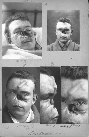
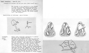
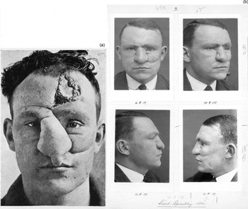
Case one illustrates the use of a cartilage graft and forehead flap. Lieutenant Spreckley was 28 years old when he was admitted on the 30th of January 1917 from a casualty station in Ypres. In his case notes, Gillies describes the patient's condition as ‘the whole of the tissues covering the nose carried away…’ (Gillies Archives, unpublished works). Gillies planned to recreate the skin and the cartilage supporting it. He shaped a piece of cartilage from the soldier's ribcage and transplanted it under the skin of the forehead (Figures 1 and 2). By the time Spreckley was discharged, his face was almost as good as new (Figure 3). He died in old age with 35 grandchildren, having fought again in World War Two.
A collection of clinical photographs from the case notes of Lieutenant Spreckley. The cartilage implant is noted under the skin of his forehead. Reproduced with the permission of the Gillies Archives, Queen Mary's Hospital, Sidcup, UK.

Operation notes from the case notes of Lieutenant Spreckley. Reproduced with the permission of the Gillies Archives, Queen Mary's Hospital, Sidcup, UK.

Clinical photographs from the case notes of Lieutenant Spreckley (a and b). The ones in part b were taken prior to discharge, after Gillies had excised Spreckley's forehead scar. Gillies' final entry reads ‘…results are satisfactory. Forehead scar negligible’. Reproduced with the permission of the Gillies Archives, Queen Mary's Hospital, Sidcup, UK.

Case two
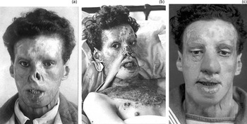
Case two describes the invention of the tubed pedicle flap. Able Seaman Willie Vicarage was 20 years old when he was admitted on the 2nd of August 1917. He had been wounded in the field in May 1916 and was referred to Gillies for treatment of his extensive facial burns. These are described by Gillies in the case notes as ‘the whole tip of the nose and both alae have been burnt away’ (Gillies Archives, unpublished works). Figure 4a depicts the effects his facial injuries have had on his profile. Gillies' plan for the operation is outlined in the notes and the invention of the tubed pedicle flap is noted (Figure 4b). Its tubed design, which reduces the rate of infection, also enables large defects to be filled, achieving a satisfactory cosmetic outcome (Figure 4c).
Clinical photographs from the case notes of Able Seaman Vicarage, showing his appearance before (a) and after (c) surgery. Part b depicts the first tubed pedicle flap, termed a ‘Masonic Collar Flap’ in the notes. Reproduced with the permission of the Gillies Archives, Queen Mary's Hospital, Sidcup, UK.

Case three
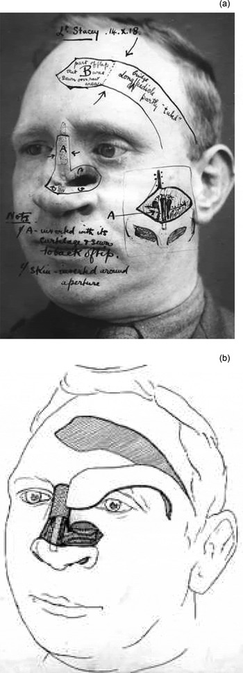
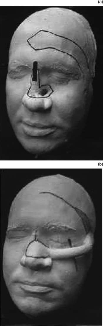
Case three depicts the evolution of the tubed pedicle flap. Second Lieutenant Stacey Anson was admitted in April 1918, 4 days after being wounded in battle. The tubed pedicle flap was developed in the treatment of this case. Gillies planned the operation meticulously in the notes with the assistance of a sculptor (Figures 5 and 6). Gillies recorded Anson's condition as ‘fractured maxilla, perforated left antrum, nasal septum fractured’ (Gillies Archives, unpublished works). There was a large defect to fill and a cartilage implant was needed. Gillies utilised the left temporal artery as a pedicle, as is depicted in Figures 5 and 6. The pedicle was divided in stages every two weeks until Gillies was satisfied with the vascularisation of the pedicle and a good outcome (Figure 7) had been achieved (Gillies Archives, unpublished works).
Clinical photograph (a) and illustration (b) from the case notes of Lieutenant Stacey Anson. Here, Gillies' meticulous design is evidenced as he plans the different operative stages. Reproduced with the permission of the Gillies Archives, Queen Mary's Hospital, Sidcup, UK.

Clinical photographs (a and b) of the sculptures commissioned by Gillies in the planning of the reconstructive rhinoplasty of Lieutenant Stacey Anson. Reproduced with the permission of the Gillies Archives, Queen Mary's Hospital, Sidcup, UK.

Pre-operative (a and b) and post-operative (c and d) clinical photographs from the case notes of Lieutenant Stacey Anson. Reproduced with the permission of the Gillies Archives, Queen Mary's Hospital, Sidcup, UK.

Case four
Case four describes the dissemination of the tubed pedicle flap. Private Tringham was 19 years old on admission in November 1918. His condition is described in the case notes as ‘total loss of upper 4/5th of nose … attempt at rhinoplasty elsewhere…’ (Gillies Archives, unpublished works). He was referred to Queen Mary's Hospital for repair of this rhinoplasty attempt, which had left him severely disfigured (Figure 8a). Henry Pickerill, a fellow New Zealander, was the operator for this case and he performed as taught by Gillies, using the tubed pedicle flap in his reconstruction (Figure 8b) and achieving a very good cosmetic outcome (Figure 8c).
Clinical photographs from the case notes of Private Tringham (a–c), who was operated on by Henry Pickerill as taught by Gillies. Reproduced with the permission of the Gillies Archives, Queen Mary's Hospital, Sidcup, UK.

Gillies' other achievements
Gillies was knighted in the King's Birthday Honours list in 1930. He pioneered the use of the fascia lata graft in facial paralysis and presented this at the Royal Society of Medicine in 1934.Reference Gillies10 Throughout World War Two, Gillies was consultant adviser in plastic surgery to all three services of the British Armed Forces, the Ministry of Health and the Ministry of Pensions. He helped establish plastic surgery units throughout the UK. He continued his work in Basingstoke and trained many doctors from Commonwealth nations.Reference Pound1 Gillies made history again in 1946 by performing the first female to male sex reassignment surgery.Reference Kennedy11
Conclusion
The trench warfare seen in World War One produced a host of craniofacial injuries which potentiated surgical progression. While other countries had sites where surgeons became pioneers, these sites did not offer the same opportunity for teaching or such a concentrated group of disfigured patients as the Queen Mary's Hospital, Sidcup. This enabled Gillies to pioneer and advance himself to be the father of modern plastic surgery.
The 10 principles that follow, as outlined in his book The Principles and Art of Plastic Surgery,Reference Gillies and Millard12 are eternally applicable:
(1) Observation is the basis of surgical diagnosis: there is no better training for a surgeon than to be taught observation by a physician. (2) Diagnose before you treat… (3) Make a plan and a pattern for this plan: use paper or carry out a pretence operation in reverse. Do not rush in… (4) Make a record: start with a diagram in the notes … follow up the case with a camera. (5) A good style will get you through: surgical style is the expression of personality and training exhibited by the movements of fingers; its hallmark dexterity and gentleness. (6) Treat the primary defect first: borrow from Peter to pay Paul only when Peter can afford it. (7) Never let routine methods become your master: routine methods must be mastered but never let them master you. (8) Consult other specialists: the reaction of one man's mind to another's is increased by the stimulus of sharing mutual problems. (9) Speed in surgery consists of not doing the same thing twice: this is the old story of the hare racing back and forth at terrific speed while the tortoise without retracing one step, slowly crosses the finish line. (10) Never do today what can honourably be put off till tomorrow: …when in doubt, don't… It is well to remember that time, although the plastic surgeon's most trenchant critic, is also his greatest ally.
Acknowledgements
The author would like to thank Dr Andrew Bamji, Curator of the Gillies Archives at Queen Mary's Hospital in Sidcup, for granting access to the archives and permission to reproduce the clinical photographs. Thanks also go to Mr Neil Weir, Consultant Otolaryngologist at the Royal Surrey County Hospital in Guildford, for his suggestions for this paper.