Introduction
Acute rheumatic fever (is a systemic autoimmune disease that develops as an abnormal immune response to Group A beta-haemolytic streptococcal pharyngitis). Reference Carapetis, Beaton and Cunningham1 Diagnosis is established using the revised Jones criteria, which include major and minor criteria. Reference Gewitz, Baltimore and Tani2 Acute rheumatic fever can affect the cardiac conduction system and most commonly causes first-degree AV block. Higher-degree AV blocks, atrial and ventricular ectopic beats, supraventricular tachycardia, and junctional rhythm have also been reported. Reference Karacan, Isikay, Olgun and Ceviz3–Reference Olgun and Ceviz5 However, the simultaneous occurrence of multiple AV block types alongside ventricular tachycardia within the same episode of acute rheumatic fever is exceedingly rare and warrants detailed reporting. Reference Yakut, Eybek, Eroglu and Karacan6,Reference Freed, Sacks and Ellman7
Case report
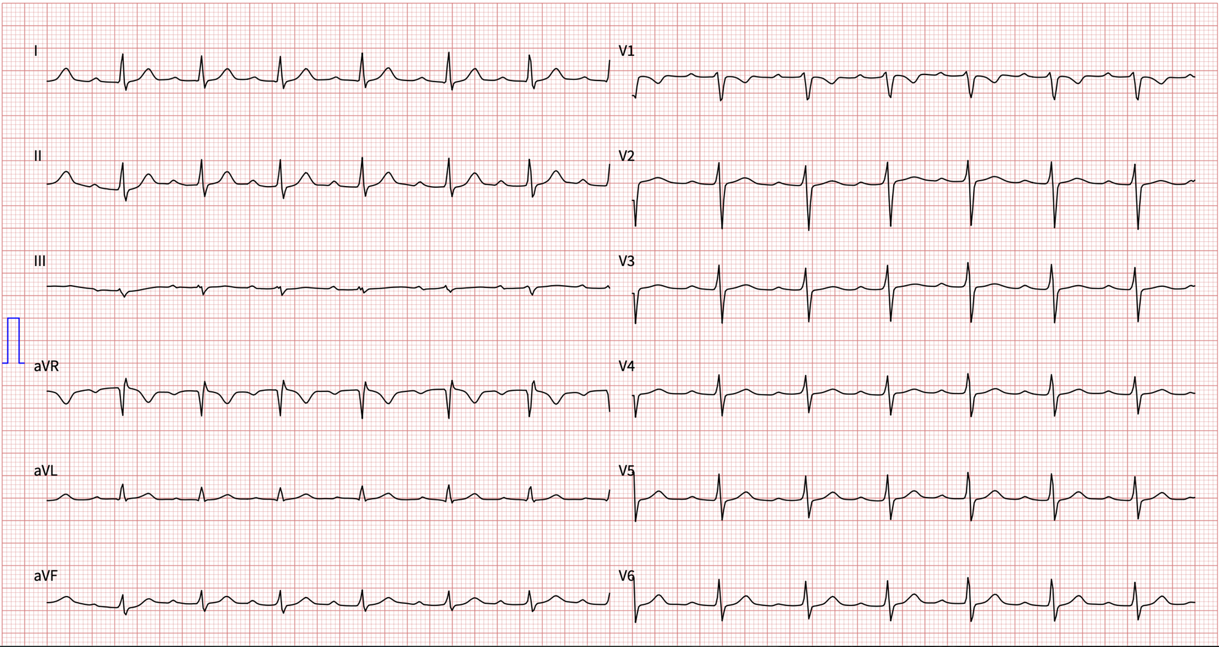
A 15-year-old boy presented to the emergency department with a five-day history of knee arthralgia. His medical history was notable for an upper respiratory tract infection approximately two to three weeks prior to admission. Laboratory investigations revealed a C-reactive protein level of 263 mg/L (reference range: <5 mg/L), an anti-streptolysin O titre of >777 IU/mL (reference range: <150 IU/mL), a cardiac troponin T level within normal limits, and an erythrocyte sedimentation rate of 40 mm/hour. Echocardiography demonstrated mild aortic and mitral regurgitation with a preserved left ventricular ejection fraction. The initial electrocardiography showed first-degree AV block with a prolonged PR interval of 250 msn (Figure 1). A diagnosis of acute rheumatic fever was established in accordance with the 2015 Jones criteria, taking into account the clinical findings, electrocardiography, echocardiography, and elevated inflammato ry markers. Reference Gewitz, Baltimore and Tani2
Initial electrocardiography at presentation to the emergency department demonstrating first-degree AV block.

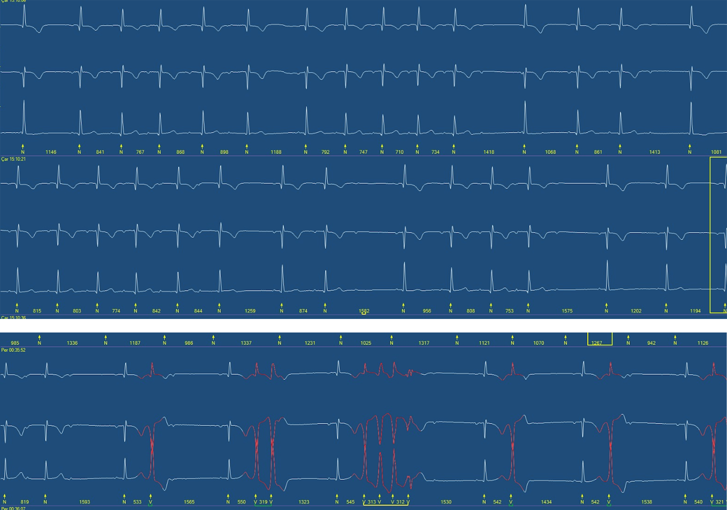
The patient was admitted to the paediatric cardiology ward. Benzathine penicillin G was administered intramuscularly, and a non-steroidal anti-inflammatory drug was initiated. The electrocardiography obtained on the second day of treatment demonstrated second-degree Mobitz type 1 (Wenckebach) AV block. A 24-hour Holter electrocardiography recording showed predominantly second-degree Mobitz type 1 and type 2 AV block, along with moderately frequent (5%) uniform ventricular extrasystoles and episodes of nonsustained ventricular tachycardia at a rate of approximately 210 beats per minute (Figure 2).
Holter electrocardiographies demonstrating ventricular extrasystoles with nonsustained ventricular tachycardia and 2nd degree type 1 AV Block.

Following these findings, anti-inflammatory therapy was escalated to oral prednisolone at a maximum dose of 2 mg/kg/day (60 mg/day), and intravenous amiodarone infusion was commenced. A repeat 24-hour Holter electrocardiography performed on the fifth day of steroid and amiodarone therapy showed resolution of the AV blocks and non-sustained ventricular tachycardia, with only rare multifocal ventricular extrasystoles remaining.
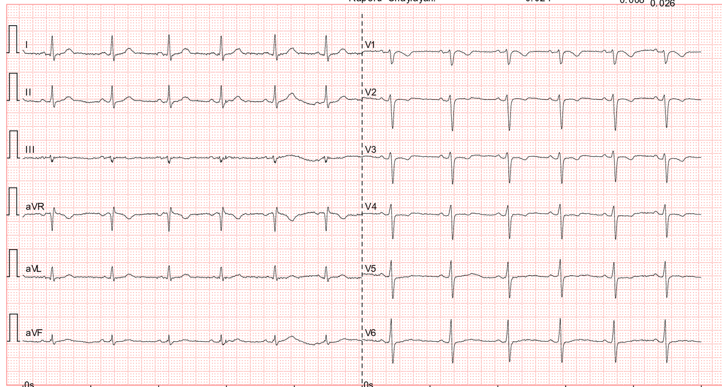
After approximately two weeks of hospitalisation, acute-phase reactants normalised, and clinical improvement was observed. The electrocardiography at that time demonstrated normal sinus rhythm (Figure 3). Prednisolone was tapered over a further two weeks and then discontinued. Naproxen sodium (20 mg/kg/day in two divided doses) was added during the steroid taper and was also discontinued after four weeks. No recurrence of AV block or ventricular tachycardia was observed during follow-up. Secondary prophylaxis with benzathine penicillin G was continued at 21-day intervals.
Follow-up electrocardiography showing normal sinus rhythm.

Discussion
Cardiac involvement in acute rheumatic fever, known as rheumatic carditis, is one of the most serious complications of the disease and represents the leading cause of acquired valvular heart disease in developing countries. Reference Carapetis, Beaton and Cunningham1,Reference Steer and Carapetis8 While first-degree AV block is the most frequently described conduction abnormality in acute rheumatic fever, higher-degree AV blocks are uncommon and typically transient. Reference Karacan, Isikay, Olgun and Ceviz3,Reference Agnew, Neutze, Lowe and Barratt-Boyes9 The mechanisms underlying conduction disturbances in acute rheumatic fever are thought to include inflammation-mediated oedema of the conduction system, immune complex deposition, and myocarditis. Reference Zalzstein, Maor, Zucker and Katz4,Reference Çevlik, Öztürk and Tunca Sahin10
The simultaneous occurrence of Mobitz type 1 and Mobitz type 2 second-degree AV block in a single patient with acute rheumatic fever is particularly unusual, as these two subtypes reflect distinct pathophysiological mechanisms—functional block at the AV node level versus structural infranodal block, respectively. Reference Yakut, Eybek, Eroglu and Karacan6,Reference Venu Reddy, Chun and Yamamoto11 The additional finding of non-sustained ventricular tachycardia further underscores the severity of myocardial inflammation in this case. Reference Freed, Sacks and Ellman7 The prompt response to corticosteroid therapy and intravenous amiodarone, with complete resolution of arrhythmias, supports an inflammatory rather than a degenerative aetiology. Reference Olgun and Ceviz5,Reference Çevlik, Öztürk and Tunca Sahin10
Ventricular tachycardia in the setting of acute rheumatic fever has been reported rarely and is most commonly described in patients with severe valvular involvement. Reference Ramoğlu, Epçaçan and Yeşilbaş13 The occurrence of ventricular tachycardia and a cardiac murmur in our patient, in the absence of significant valvulitis, is therefore particularly uncommon. Myocarditis was considered in the differential diagnosis given the clinical overlap; however, cardiac troponin level was within normal limits, making myocarditis less likely. Reference Kamblock, Payot and Iung14
This case highlights the importance of continuous cardiac monitoring, including 24-hour Holter electrocardiography, in all patients with acute rheumatic fever, even when the initial electrocardiography shows only mild conduction abnormalities. Reference Karacan, Isikay, Olgun and Ceviz3,Reference Seckeler and Hoke12 Early recognition and aggressive anti-inflammatory treatment may prevent progression to complete heart block or haemodynamically significant ventricular arrhythmias. Reference Zalzstein, Maor, Zucker and Katz4,Reference Çevlik, Öztürk and Tunca Sahin10
The use of intravenous amiodarone in this case was prompted by the haemodynamic significance of the ventricular arrhythmia in the context of active myocardial inflammation. Intravenous amiodarone has been reported as an effective antiarrhythmic agent for children with acute life-threatening ventricular tachyarrhythmias, particularly in the setting of impaired or at-risk ventricular function, given its broad-spectrum antiarrhythmic properties. Reference Yildirim, Tokel, Saygili and Varan15 The rapid resolution of arrhythmias following the combination of corticosteroid and amiodarone therapy supports a transient inflammatory mechanism rather than a fixed structural abnormality of the conduction system.
Conclusion
We report a rare case of acute rheumatic fever complicated by concurrent second-degree Mobitz type 1 and Mobitz type 2 AV block and non-sustained ventricular tachycardia in a 15-year-old boy. The arrhythmias resolved fully with corticosteroid and amiodarone therapy. This case emphasises the need for thorough cardiac monitoring in paediatric patients with acute rheumatic fever, as conduction abnormalities may be more complex than the initial electrocardiography suggests.
Financial support
No funding was received for this work.
Competing interests
The authors declare no conflict of interest.
Patient consent
Written informed consent was obtained from the patient and his family for publication of this case report.