Introduction
Cystic echinococcosis is caused by the larval stage of the tapeworm, Echinococcus granulosus (Eckert & Thompson, Reference Eckert and Thompson1997). Secreted and membrane-bound proteins secreted by the larval stage of the parasite play an important role in feeding, reproduction and defence against host immune attacks (Rosenzvit et al., Reference Rosenzvit, Camicia, Kamenetzky, Muzulin and Gutierrez2006). Among these critical proteins, antigen B (AgB) has received much attention, mainly for its immunogenicity in humans (Williams et al., Reference Williams, Pérez Esandi and Oriol1971) and animals (Ibrahem et al., Reference Ibrahem, Craig, McVie, Ersfeld and Rogan1996). AgB is involved in immune evasion because its multimers of 8 kDa subunit are involved in detoxifying mechanisms, sequestering and buffering xenobiotics (Chemale et al., Reference Chemale, Ferreira, Barrett, Brophy and Zaha2005). On the basis of molecular mining five clades of AgB have been characterized so far, namely AgB1 (Shepherd et al., Reference Shepherd, Aitken and McManus1991), AgB2 (Fernández et al., Reference Fernández, Ferreira, Fernández, Zaha and Nieto1996), AgB3 (Chemale et al., Reference Chemale, Haag, Ferreira and Zaha2001), AgB4 (Arend et al., Reference Arend, Zaha, Ayala and Haag2004) and AgB5 (Haag et al., Reference Haag, Alves-Junior, Zaha and Ayala2004). Antigenic variability is critical for establishment of infection by this parasite (Haag et al., Reference Haag, Zanotto, Alves-Junior, Gasser, Zaha and Ayala2006). Down- or up-regulation of the AgB family during different developmental stages of Echinococcus multilocularis has been described earlier in the literature (Mamuti et al., Reference Mamuti, Sako, Xiao, Nakaya, Nakao, Yamasaki, Lightowlers, Craig and Ito2006). However, transcription levels of the AgB family of E. granulosus isolated from naturally infected buffalo have not been reported. Our study sheds light on the expression pattern of the AgB family of bubaline isolates of E. granulosus in active and inactive cysts.
Materials and methods
Collection of cyst samples
A total of 50 samples were collected from buffalo. Eight samples were collected from buffalo with white germinal membrane with protoscoleces and clear cyst fluid (S1). Ten samples were collected from cysts with transparent germinal membrane and clear cyst fluid without protoscoleces (S2). A total of ten samples were collected from cysts with yellowish germinal membrane and turbid cyst fluid along with protoscoleces (S3). Twelve samples were collected from cysts with white germinal membrane and clear cyst fluid without protoscoleces (S4). And finally, ten samples were collected from cysts without any protoscoleces and turbid cyst fluid in which calcification had initiated (S5).
Polymerase chain reaction (PCR) analysis
Total RNA was isolated from fresh protoscoleces and germinal membrane using Tri reagent (Sigma, St Louis, Missouri, USA). Each sample was incubated for 45 min at 37°C with 1 unit of RQ1 RNase-free DNase (Promega, Madison, Wisconsin, USA) and heat denatured at 65°C for 10 min using DNase stop buffer (Promega). The total RNA was quantified by determining the optical density at 260 nm. From each isolate, 3 μg total RNA was used for cDNA preparation. The cDNA was synthesized from each RNA sample using high-capacity cDNA reverse transcription kits (Applied Biosystems, Foster City, California, USA). cDNA samples were stored at − 70°C for future use.
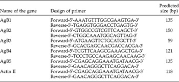
Five sets of primers were designed specifically for amplification of the five E. granulosus AgB genes (AgB1–AgB5) using Primer Premier version (Premier Biosoft International, San Diego, California, USA). The primers used were a forward primer annealing to 5′ region inside the second exon and a reverse primer annealing to 3′ flanking region of EgAgB1 (AY942154), EgAgB2 (EU622922), EgAgB3 (DQ148521), EgAgB4 (AY569350) and EgAgB5 (AY871024). Actin II gene was considered as a housekeeping gene and the primer was designed from available sequence information provided in PubMed public gene database (L07774). The amplicon size was restricted to less than 150 bp. The details of design of high performance liquid chromatography-purified sense and antisense primers have been detailed in table 1.
Design of sense and antisense primers for amplification of AgB and actin II.

Singleplex real-time PCR was carried out with StepOne™ Real-time PCR system (Applied Biosystems). Reactions were set up in a total volume of 20 μl using 5 μl cDNA (diluted 1:4), 10 μl of 2 × Power SYBR® Green PCR Master Mix (Applied Biosystems) and 20 pmol of each gene-specific primer. Composition of the reaction mixture was the same for both target and housekeeping genes. The reaction mixture was placed in MicroAmp™ Fast Optical 48-Well Reaction Plate (Applied Biosystems) and covered with MicroAmp™ 48-Well Optical Adhesive Film (Applied Biosystems). Amplification of cDNA molecules derived from either different AgB genes or housekeeping genes was repeated three times. A control PCR for each sample included isolated total RNA from the aliquot that had not been treated with reverse transcriptase, in order to confirm that no residual genomic DNA contamination persisted. PCR was initiated by a hold of 10 min at 95°C followed by 40 cycles of denaturation (95°C for 15 s), annealing (53°C for 60 s) and extension (72°C for 30 s). Fluorescence acquisition was carried out at 74°C for 1 s. Predicted size of amplicon and absence of non-specific products were confirmed by conventional PCR with the same primers.
The relative quantification was measured with the levels of expression of a target gene (AgB1–AgB5) and compared to levels of an internal control (housekeeping gene; actin II). In order to quantify our results, all samples were expressed as a ratio of the housekeeping gene. The ratios were then compared. Finally, the results were analysed using mathematical models viz. R = 2− ΔΔCt method (Livak 1997 and Reference Livak2000; Livak & Schmittgen, Reference Livak and Schmittgen2001).
Numerical values were analysed by utilizing the GraphPad Prism software (La Jolla, California, USA) to determine significant differences in expression between the five morphotypes of cysts and among the AgB family.
Results
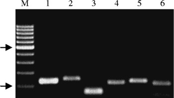
After amplification with respective primers, amplicon sizes of the five isoforms of AgB were determined to be 135 bp (AgB1; FJ897577), 150 bp (AgB2; FJ897578), 59 bp (AgB3), 121 bp (AgB4; FJ897580) and 135 bp (AgB5; FJ897581). The amplicon size of the internal control (actin II) was 181 bp (fig. 1).
Lanes 1–6 indicate the amplified products of AgB1, AgB2, AgB3, AgB4, AgB5 and actin II, respectively. Lane M, 100 bp DNA ladder (arrows indicate 100 and 500 bp fragments from bottom to up).

Cysts with white germinal membrane and protoscoleces and clear cyst fluid (S1) exhibited a uniform expression pattern of the five isoforms of the AgB family (AgB1–AgB5). The highest relative expression of AgB1 was seen in S5 cysts followed by S4, S3 and S2. The highest relative expression of AgB2 was seen in cysts with protoscoleces (S1 and S3) followed by cysts without protoscoleces. The highest expression of AgB3 was seen in S2 cysts followed by S4, S3 and S5 in descending order. The relative expression of AgB4 was the highest in S3 cysts followed by S2, S5 and S4 in descending order. AgB5 expression was higher in S2 cysts followed by S3, S5 and S4 in descending order (table 2).
Relative quantification of the AgB family by the 2−ΔΔC t method.

* Significant difference compared with the standard (P < 0.05).
Discussion
During the present investigation, we classified cysts into five categories depending upon the type of germinal membrane, colour of cyst fluid, presence or absence of protoscoleces and signature of calcification. Cysts with protoscoleces are termed fertile cysts. On the other hand, cysts without protoscoleces are termed sterile cysts. Numerical data with respect to the relative expression could not be represented based on fertility and sterility of cysts because of significant variation in data. Therefore, the cysts were classified based on the WHO–Office International Des Epizooties criteria. Active cysts may be fertile or sterile. On the other hand, inactive cysts may be degenerating or calcified cysts (Pawlowski et al., Reference Pawlowski, Eckert, Vuitton, Ammann, Kern, Craig, Dar, Rosa, Filice, Gottstein, Grimm, Macpherson, Sato, Todordov, Uchino, von Sinner and Wen2001). Therefore, during the present investigation, the cysts categorized as S1, S2 and S4 were classified as active cysts, whereas, cysts categorized as S3 and S5 were deemed inactive cysts.
Once the oncosphere establishes the fully developed cysts contain an inner germinal membrane. The germinal membrane is supported by a host-derived tough, elastic, acellular laminated layer of variable thickness. The laminated layer is surrounded by a host-produced fibrous adventitial layer. The protoscoleces are formed by asexual proliferation from the germinal membrane (Thompson, Reference Thompson, Thompson and Lymbery1995). During the present study, typical unilocular cysts (S1) exhibited a uniform expression pattern of the five isoforms of the AgB family. Therefore, the cysts were considered as a control or reference group during relative quantification measurements when data were compared to expression levels of housekeeping genes.
Active, transitional and inactive cysts have been correlated with genotypes of the parasite (Guarnera et al., Reference Guarnera, Parra, Kamenetzky, García and Gutiérrez2004), but relative expression of AgB isoforms in active and inactive cysts has not been reported in the literature.
AgB of E. granulosus is a major component of hydatid fluid because this antigen is expressed by the metacestode stage of the parasite (Maddison et al., Reference Maddison, Slemenda, Schantz, Fried, Wilson and Tsang1989; Ito et al., Reference Ito, Ma, Schantz, Gottstein, Liu, Chai, Abdel-Hafez, Altintas, Joshi, Lightowlers and Pawlowski1999; Mamuti et al., Reference Mamuti, Yamasaki, Sako, Nakaya, Nakao, Lightowlers and Ito2002). Evidence has indicated that AgB is encoded by a multigene family (Siracusano et al., Reference Siracusano, Margutti, Delunardo, Profumo, Rigano, Buttari, Teggi and Ortona2008). Using phylogenetic tools, the AgB family can be clustered into two groups – AgB1/B3/B5 and AgB2/B4 (Frosch et al., Reference Frosch, Hartmann, Mühlschlegel and Frosch1994; Chemale et al., Reference Chemale, Haag, Ferreira and Zaha2001; Arend et al., Reference Arend, Zaha, Ayala and Haag2004; Haag et al., Reference Haag, Alves-Junior, Zaha and Ayala2004; Kamenetzky et al., Reference Kamenetzky, Muzulin, Gutierrez, Angel, Zaha, Guarnera and Rosenzvit2005). Antigenic variation is well documented and has been described extensively in microparasites such as bacteria, fungi and protozoans. Switching of AgB from one isoform to another aids the parasite evade the host immune system (Haag et al., Reference Haag, Alves-Junior, Zaha and Ayala2004).
In our study, down- and up-regulation of the five isoforms of AgB were seen. We found that expression of AgB1 was the highest in cysts, where calcification has initiated. The most plausible reasoning for this may be that this isoform is essential for survival of metacestodes in intermediate hosts (Mamuti et al., Reference Mamuti, Sako, Xiao, Nakaya, Nakao, Yamasaki, Lightowlers, Craig and Ito2006). Higher expression of AgB2 and AgB4 was seen in cysts with protoscoleces. This observation suggests that AgB2 and AgB4 are more abundantly secreted by fertile cysts. The highest expression of AgB3 was seen in cysts with white germinal membrane and clear cyst fluid without protoscoleces (S2) and the lowest expression in cysts with signs of calcification. AgB3 is expressed constitutively in all parasitic stages of E. multilocularis (Mamuti et al., Reference Mamuti, Sako, Xiao, Nakaya, Nakao, Yamasaki, Lightowlers, Craig and Ito2006). However, data reflecting this observation are not yet available for E. granulosus in its different cystic morphotypes. However, this study reveals decreased expression of AgB3 in metacestodes, where calcification has commenced.
Highest expression of AgB5 was seen in cysts with transparent germinal membranes and clear cyst fluid without protoscoleces (S2) and the lowest expression in cysts with white germinal membranes and clear cyst fluid without protoscoleces (S4). From the pattern of expression of AgB5, conclusions could not be drawn because relative expression of AgB5 has been observed in the metacestode stage of the parasite. Moreover, AgB5 expression has already been documented in the adult stage of the parasite (Mamuti et al., Reference Mamuti, Sako, Xiao, Nakaya, Nakao, Yamasaki, Lightowlers, Craig and Ito2006).