Introduction
Dirofilariosis, caused by Dirofilaria immitis, is found worldwide, but the most endemic areas are those with high temperatures and appropriate mosquito vector populations. Dirofilaria immitis is one of the most pathogenic nematode parasites of dogs. Adult nematodes are normally found in the pulmonary arteries of dogs. Adult heartworms may cause clinical signs ranging from mild cough to congestive heart failure, intravascular haemolysis and pulmonary thromboembolism, which are often fatal if untreated (Soulsby, Reference Soulsby1986; Kassai, Reference Kassai1999). The infective larval stage (L3) can develop in approximately 70 species of mosquitoes. Since these vectors are widespread throughout the world and are commonly found near human habitats, human infection, although rare, is nevertheless also possible, creating concern about potential zoonosis (Fan et al., Reference Fan, Su, Lin, Liao, Du and Chiou2001; Simon et al., Reference Simon, Lopez-Belmonte, Marcos-Atxutegi, Morchon and Martin-Pacho2005). Dirofilaria immitis in dogs can be diagnosed through careful morphological examination of circulating microfilariae, detection of circulating antigens, histochemical or immuno-histochemical staining of circulating microfilariae or, more recently, through molecular approaches. Morphological identification of circulating microfilariae, however, is not always easy and is potentially misleading (Casiraghi et al., Reference Casiraghi, Bazzocchi, Mortarion, Ottina and Genchi2006; Rishniw et al., Reference Rishniw, Barr, Simpson, Frongillo, Franz and Alpizar2006).
Few studies have been published regarding the distribution and prevalence of D. immitis in dogs in Turkey. Dirofilaria immitis was first reported in a dog in Turkey in 1951 (Guralp, Reference Guralp1981). Tasan (Reference Tasan1983) detected microfilaraemia in 53/283 (18.7%) stray dogs in Elazig. Coskun et al. (Reference Coskun, Tinar, Akyol, Aydin and Demir1992) documented infection in 5/168 dogs (2.98%) in Bursa. The prevalence was recorded in Sivas (Atas et al., Reference Atas, Ozcelik and Saygi1997), Van (Agaoglu et al., Reference Agaoglu, Akgul, Ceylan and Akkan2000), Ankara (Oge et al., Reference Oge, Doganay, Oge and Yildirim2003), Istanbul (Oncel & Vural, Reference Oncel and Vural2005) and Kayseri (Yildirim et al., Reference Yildirim, Ica, Atalay, Düzlü and Inci2007) as 6%, 46.2%, 9.3%, 1.52% and 9.6%, respectively. These different prevalence rates may reflect different testing methodologies or true regional differences. The prevalence of D. immitis in dogs has been determined traditionally by postmortem inspection, detection of microfilariae and serological testing. However, dogs with occult heartworm infections are amicrofilaraemic. In addition, some antiparasitic treatments such as macrolides may render an infected dog amicrofilaraemic for 6–9 months (Hoover et al., Reference Hoover, Campbell, Fox, Claypool and Mullins1996). Thus, serological and microfilarial examinations should be applied together for screening D. immitis in dogs. DNA-based diagnostic tests for D. immitis infections have been shown to overcome some deficiencies of parasitological and serological diagnosis, and specific and sensitive polymerase chain reaction (PCR)-based assays have been reported (Cancrini et al., Reference Cancrini, Allende, Favia, Bornay, Anton and Simon2000; Mar et al., Reference Mar, Yong, Chang and Fei2002; Rishniw et al., Reference Rishniw, Barr, Simpson, Frongillo, Franz and Alpizar2006).
The present study was performed to investigate the prevalence of canine D. immitis infection with antigen-detecting enzyme-linked immunosorbent assay (ELISA) and PCR in Turkey and to compare the prevalence in five enviromentally distinct regions of Turkey. Furthermore, this study is the first to use PCR-based detection of microfilaria in peripheral blood of dogs and cats in Turkey.
Materials and methods
Sample collection and study area
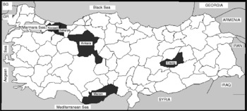
The study was carried out in five environmentally distinct provinces in Turkey (fig. 1). Environmental conditions of these areas, such as climate, altitude, temperature, relative humidity in the summer and throughout 2005 are provided in table 1. Of the 211 dogs used in the current study, 93 (44.1%) were males and 118 (55.9%) were females; 143 (67.7%) were 0.5–2 years old, 57 (27%) were 3–5 years old and 11 (5.2%) more than 6 years old; 125 (59.2%) were strays and 86 (40.7%) were owned dogs. Blood samples were collected between March 2005 and March 2006 from randomly selected dogs in five different provinces: Kocaeli (n = 71), Sakarya (n = 65), Elazig (n = 29), Ankara (n = 27) and Mersin (n = 19). Demographic information for each dog, including age, sex, breed, life status (stray or owned) was recorded. Serum was separated by centrifugation and stored at − 20°C in a freezer for subsequent serological analysis. Whole-blood samples were collected for PCR assay from each dog and stored at − 80°C until analysis. Fifteen cats (14 owned and 1 stray) from Ankara province were examined by PCR for D. immitis infection.
Location of study areas (shaded in black) in Turkey.

Environmental and climatic conditions of five Turkish provinces in which dirofilarial testing was performed.

Control samples
A total of three different D. immitis genomic DNA samples were used as controls in this study. One DNA control sample was extracted from an adult D. immitis parasite (this worm was collected from a dog that had been necropsied at Firat University, Faculty of Veterinary Medicine, Department of Pathology). The second and third DNA control samples were extracted from microfilariae present in the blood of infected dogs (one sample was obtained from another research group (Yildirim et al., Reference Yildirim, Ica, Atalay, Düzlü and Inci2007) and microfilariae were visualized with Giemsa staining; while the other sample was provided by one of the authors, M.R.).
Serological analysis
Circulating D. immitis antigen was detected using a commercial antigen ELISA kit (DiroCHEK, Synbiotics Corp., USA), according to the manufacturer's instructions. DiroCHEK Canine Heartworm Antigen Test kit is an enzyme immunoassay designed to detect the presence of circulating antigen from adult females of D. immitis in serum. All sera were tested with this kit, and sera were re-tested when inconclusive results were obtained.
DNA isolation and PCR
DNA extraction from canine and feline blood was performed according to the method described by Clausen et al. (Reference Clausen, Wiemann, Patzelt, Kalkaire, Potzsch, Peregrine and Mehlitz1999) with slight modifications. Briefly, 125 μl of blood were added to 250 μl of lysis mixture (0.32 m sucrose, 0.01 m Tris, 0.05 m MgCl2, 1% TritonX-100, pH 7.5) and the mixture was centrifuged at 11,600 g for 30 s. The pellet was washed five times with lysis buffer. Following this, 250 μl PCR buffer (50 mm KCl, 10 mm Tris–HCl pH 8, 0.1% TritonX-100 pH 8.3) containing 50 μg ml− 1 proteinase-K (Sigma, St Louis, Missouri, USA) was added to the pellet and incubated at 56°C for 3 h. At the end of this period, samples were boiled at 95°C for 10 min. Five microlitres of the supernatant were used as templates in the PCR. This method was used for both whole blood and adult worm extraction procedures. For DNA extraction from the adult worm, a small section of worm was macerated and this tissue was then used for genomic DNA extraction as described above. The PCR was performed using two sets of primers. Initially, we used one primer pair: 5′-CATCAGGTGATGATGTGATGAT-3′ (forward), and 5′-TTGATTGGATTTTAACGTATCATTT-3′ (reverse), which have been described by Mar et al. (Reference Mar, Yong, Chang and Fei2002) for the amplification of a 302 bp ITS-2 gene segment. Unexpectedly, this primer pair non-specifically amplified a 350 bp product from all samples, including all positive controls. Therefore, a second set of primers was used: 5′-AGTGTAGAGGGTCAGCCTGAGTTA-3′ (forward) and 5′-ACAGGCACTGACAATAC CAAT-3′ (reverse), identical to those described in a previous study (Rishniw et al., Reference Rishniw, Barr, Simpson, Frongillo, Franz and Alpizar2006) for the amplification of a 203 bp cytochrome oxidase subunit 1 (CO1) gene fragment. The PCR was performed in a touchdown thermocycler in a total reaction volume of 50 μl containing 4 μl MgCl2 (25 mm, Promega, Madison, Wisconsin, USA), 4 μl dNTP (10 mm dNTP mixture, Promega), 5 μl 10 × PCR buffer (Promega), 1.2 μl Primer F (68.2 nmol, lyophilized) and 1.2 μl Primer R (64.8 nmol, lyophilized), 0.2 μl Taq DNA polymerase (5 U μl− 1, Promega), 29.4 μl distilled water and 5 μl template DNA. Positive control DNA was used at each PCR and distilled water was used as a negative control. The PCR procedure consisted of a pre-denaturing step at 94°C for 1 min and 32 cycles of denaturing (30 s at 94°C), annealing (30 s at 63°C), extension (30 s at 72°C) and a final extension (7 min at 72°C). The amplified products were separated by gel electrophoresis in 1.5% agarose gel with a Tris–boric acid–EDTA (TBE, pH 8.3) buffer at 90 V for 45 min. Following electrophoresis, the amplified products were visualized with ethidium bromide (0.5 μg ml− 1) staining for 45 min at room temperature. A visible band of 203 bp in the PCR was considered to be a positive result.
Statistical analysis
Pearson's chi-square (χ2) test was used to evaluate the differences between proportions of infected dogs or cats detected by each method. Statistical comparisons were carried out using SPSS 11.0 statistical software (SPSS Inc., Chicago, Illinois, USA). A P < 0.05 was considered significant.
Results
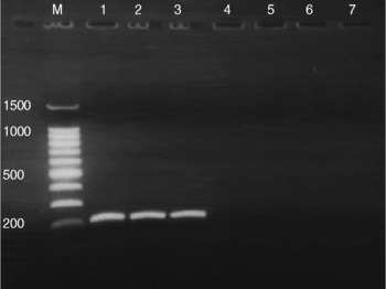
All dogs and cats were negative by PCR analysis. Figure 2 summarizes our findings with a variety of negative and control DNA templates amplified by the D. immitis specific primer pairs.
Species-specific PCR amplification of Dirofilaria immitis CO1 (203 bp). Lanes: M, molecular weight marker; 1, positive control (template DNA isolated from adult Dirofilaria immitis); 2, positive control (template DNA isolated from infected dog blood); 3, positive control, (template DNA isolated from infected dog blood); 4, negative control (distilled water); 5–7, randomly selected test samples.

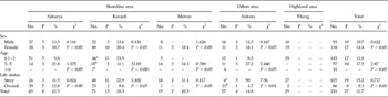
The results of the ELISA examination on the canine dirofilariosis and the status (owned or stray) are shown in table 2. Twenty-seven (12.8%) of 211 samples tested positive with the antigen-detecting ELISA kit. There were no differences in ELISA-positive results between sexes (females, 14.4%; males, 10.7%; P>0.05). The mean overall positivity rates of D. immitis infection were 11.8% in the 0.5–2-year-old group, 17.5% in 3–5-year-old group. The prevalence rates between stray dogs (15.2%) and owned dogs (9.3%) did not differ (P>0.05). Highest seropositivity rate was detected in Kocaeli province (18.3%), followed by Ankara (14.8%), Sakarya (12.3%) and Mersin (10.5%) provinces. No dogs from Elazig province tested positive.
Seroprevalence (%) of ELISA by sex, age, life status and provinces.

a,b,c Values with different letters within the same column are significantly different (P < 0.01).
No., Number of dogs examined; P, number of dogs positive by ELISA; %, positivity rate.
Discussion
Our study further documents the prevalence of D. immitis in dogs in Turkey, complementing and expanding the data of previous studies (Oge et al., Reference Oge, Doganay, Oge and Yildirim2003; Oncel & Vural, Reference Oncel and Vural2005; Yildirim et al., Reference Yildirim, Ica, Atalay, Düzlü and Inci2007). This study demonstrates that the overall prevalence is relatively low (12.7%), varies regionally (18.3%, 14.8%, 12.3%, 10.5% and 0% in Kocaeli, Ankara, Sakarya and Mersin and Elazig provinces, respectively), and may be increasing in some areas – in the one province we examined where previous serological data were available (Ankara), antigen positivity increased from 9.3 to 14.8% over the past 4 years (Oge et al., Reference Oge, Doganay, Oge and Yildirim2003). Whether this apparent increase is due to random sampling differences, differences in methodology, or an actual increase in prevalence, is unknown. It is not surprising that regional differences in prevalence exist. A previous study of dirofilariasis in Korea demonstrated that prevalence was significantly higher in coastal areas (69.5%) than in urban and mountain areas (Song et al., Reference Song, Lee, Hayasaki, Shiramizu, Kim and Cho2003). Sakarya, Kocaeli and Mersin are coastal areas and have warmer climates than Ankara and Elazig. We expected the highest prevalence to be in Mersin, due to the warmest climate; however, Mersin had the lowest prevalence of any region in which dirofilaria were detected. Thus, high ambient temperatures are necessary but not sufficient to predict prevalence rates for canine dirofilariasis. Other factors influence prevalence, most important of which is a suitable mosquito population, which may depend on non-thermal factors, such as humidity (Ahid et al., Reference Ahid, Lourenco-de-Oliveira and Saraiva1999). We could find no difference in seropositivity between stray and pet dogs. Previous studies have examined seropositivity in indoor and outdoor dogs with and without chemoprophylaxis, and suggested that outdoor dogs have a tenfold increased prevalence due to increased exposure to mosquitoes (Byeon et al., Reference Byeon, Kim, Kim, Yu, Jeong and Ock2007). In our study population, all pet dogs received monthly prophylaxis and stray dogs in Kocaeli, Sakarya and Ankara provinces received monthly ivermectin injections. This would eventually render dogs amicrofilaraemic and may explain why we could not detect any microfilarial DNA in peripheral blood samples by PCR.
Prevalence showed an initial increase with age, followed by a decline: prevalence was 11.8% in the 0.5–2-year age group, 17.5% in 3–5-year age group and 0% in dogs >6 years old. This is similar to previous findings of an increase in prevalence until middle age (about 4–8 years), with a subsequent decline occurring in later years (Glickman et al., Reference Glickman, Grieve, Breitschwerdt, Mika-Grieve, Patronek, Domanski, Root and Malone1984; Capelli et al., Reference Capelli, Poglayen, Bertotti, Giupponi and Martini1996; Byeon et al., Reference Byeon, Kim, Kim, Yu, Jeong and Ock2007), and is suspected to be the consequence of prophylaxis in older dogs (Grieve et al., Reference Grieve, Frank, Stewart, Parsons, Belasco and Hepler1991; Byeon et al., Reference Byeon, Kim, Kim, Yu, Jeong and Ock2007). Additionally, we included some dogs that may have been too young to seroconvert, although all dogs were more than 6 months old. We did not find any differences in prevalence between genders. This is similar to the results of some previous studies (Oge et al., Reference Oge, Doganay, Oge and Yildirim2003; Song et al., Reference Song, Lee, Hayasaki, Shiramizu, Kim and Cho2003), but different from those of others (Montoya et al., Reference Montoya, Morales, Ferrer, Molina and Corbera1998; Byeon et al., Reference Byeon, Kim, Kim, Yu, Jeong and Ock2007; Yildirim et al., Reference Yildirim, Ica, Atalay, Düzlü and Inci2007) where prevalence rates in males were significantly higher than in females.
The lack of any PCR-based positivity in our study is a little perplexing. This method requires sufficient DNA to be present in the sample to act as a template and is usually provided by circulating microfilariae. Several possibilities for our results exist. First, it is possible that the seropositive dogs in this study had occult infections. As stated earlier, pet dogs were all receiving chemoprophylaxis, and stray dogs were being given monthly ivermectin injections. Alternatively, the blood collection method may have affected our study – blood for PCR was collected into EDTA tubes and DNA was subsequently extracted using the procedure described above. A critical component of PCR reactions, Mg2+, which acts as a cofactor for Taq polymerase, can be chelated by EDTA. It is possible that the Mg2+ was chelated by residual EDTA from the blood sample. However, the DNA extraction procedure should have diluted and removed the majority of the EDTA, and the amount of DNA added to a PCR reaction is on the order of 5% of the total reaction volume, so this effect of EDTA is extremely unlikely. Additionally, dirofilarial PCR from blood samples collected into EDTA has been performed routinely by one of the authors without complications.
Feline dirofilariosis is now recognized as a potential cause of serious disease and can present both a clinical and diagnostic challenge to practising veterinarians. Feline heartworm infection can be symptomatic in some cats. However, it is possible that the presence of only one or two adult worms in the lungs and/or heart may cause severe illness (Borgarelli et al., Reference Borgarelli, Venco, Piga, Bonino and Ryan1997). We could not find any studies of prevalence of feline dirofilariosis in Turkey. We examined 15 cats by PCR in this study but had no positive results. This is not surprising, since the PCR reactions require infected animals to be microfilaraemic – a state that cats rarely achieve for any length of time. Further studies of the prevalence of feline dirofilariosis may require serological testing; however, given the prevalence of canine dirofilariosis in Turkey, the predicted prevalence in cats would be extremely low.
In conclusion, D. immitis is prevalent at relatively modest rates throughout several Turkish provinces, but appears to be absent in others. Therefore, heartworm prophylaxis is advisable for dogs in certain areas (which should be determined by further regional prevalence studies) or for dogs travelling to endemic areas.
Acknowledgements
The authors would like to thank all veterinarians and technicians for their kind help during sample collection and also to Dr A. Yildirim for control samples. This work was supported financially by a grant (TOVAG-106 O 014) from The Scientific and Technical Research Council of Turkey (TUBITAK).