INTRODUCTION
Atz (Reference Atz, Armstrong and Marshall1964) has defined hermaphroditism as ‘the existence of both the male and female sex in a single individual’. Although hermaphroditism is rare in elasmobranchs, it is evident that it can be expressed in a variety of forms in this group. Thus, it can range, for example, from individuals that are essentially female except for their possession of claspers (e.g. Yano & Tanaka, Reference Yano and Tanaka1989; Iglésias et al., Reference Iglésias, Sellos and Nakaya2005), to those with a full complement of male and female reproductive organs (Pratt, Reference Pratt1979; Jones et al., Reference Jones, White and Potter2005) to those in which a testis is combined with an ovarian component to produce an ovotestis (e.g. King, Reference King1966; Yano & Tanaka, Reference Yano and Tanaka1989; Iglésias et al., Reference Iglésias, Sellos and Nakaya2005). Accounts of hermaphroditism in elasmobranchs have only occasionally included detailed quantitative comparisons between their reproductive structures and those of normal individuals (eg. Iglésias et al., Reference Iglésias, Sellos and Nakaya2005; Jones et al., Reference Jones, White and Potter2005; Scenna et al., Reference Scenna, Díaz de Astarloa and Cousseau2007) and have apparently only once provided photomicrographs of their gonad histology (Capapé et al., Reference Capapé, Chadli and Baouendi1979).
The Port Jackson shark Heterodontus portusjacksoni comprises a substantial proportion of the elasmobranch bycatch of the commercial fisheries in southern Australian waters (McAuley & Simpfendorfer, Reference McAuley and Simpfendorfer2003; Walker et al., Reference Walker, Hudson and Gason2005; Jones et al., Reference Jones, Hall and Potter2008). During an initial study of the bycatch of demersal fisheries in south-western Australia, one of 353 individuals of the Port Jackson shark collected was a hermaphrodite (Jones et al., Reference Jones, White and Potter2005). This individual was unusual for a hermaphroditic elasmobranch in that it possessed claspers and a full complement of both male and female reproductive tracts. However, the female component was better developed than the male component and its right ovary contained large vitellogenic follicles that were indistinguishable from those of normal mature females.
Subsequent sampling yielded a second but different form of hermaphroditic H. portusjacksoni. In contrast to the first hermaphrodite, the internal male reproductive organs of this individual were better developed than its female organs, and its right testis was combined with an ovary to form a single ovotestis. This short paper quantitatively compares the reproductive tracts of this hermaphrodite with those of gonochoristic males and females of H. portusjacksoni. Emphasis was also placed on using histological sections of the hermaphrodite's gonads to determine whether the characteristics of the ovotestis were analogous to those of the discrete testis and whether its germ cells had progressed as far through spermatogenesis.
MATERIALS AND METHODS
This second hermaphrodite was one of a further 203 Heterodontus portusjacksoni obtained, between November 2003 and April 2007, through regular sampling of the commercial catches of demersal gillnet, longline and trawl-net fishers operating between 31°S on the west coast and 118°E on the south coast of Western Australia. All individuals collected, were stored frozen and later dissected in the laboratory.
The total length (TL) of the hermaphrodite and the lengths of its claspers, the widths of its oviducal glands and uteri and the diameter of its largest ovarian follicle were measured to the nearest 1 mm. The weight of this individual, and of its single left testis and single right ovotestis, were recorded to the nearest 1 g and all ovarian follicles with a diameter ≥5 mm, were counted. The testis and both the testicular and ovarian components of the ovotestis were each assigned a gonadal stage following the criteria of White et al. (Reference White, Platell and Potter2001). Corresponding measurements, weights, counts and gonadal stages were recorded for five normal males and five normal females closest in size to the hermaphrodite. The normal males were all mature, while the normal females, which reach maturity at a far larger size than males (Jones et al., Reference Jones, Hall and Potter2008), were immature. Thus, for further comparison, measurements, weights, follicle counts and gonadal stages were also recorded for normal mature females with lengths at the lower end of those of normal mature females.
The testis and ovotestis were cut transversely into three approximately equal segments. Each segment was fixed in a 10% neutral buffered formalin solution for 2 days, dehydrated in an ascending series of alcohols, embedded in paraffin wax and cut into 6 µm sections. Alternate sections were stained with Ehrlich's haematoxylin–eosin and Mallory's trichrome and photographed using an Olympus DP70 camera attached to an Olympus BX51 compound microscope. Although freeze-storage without cryopreservatives prevented the optimal fixation of tissues, histological sections were still of sufficient quality to enable the spermatogenic stages in the testis and ovotestis to be determined.
RESULTS
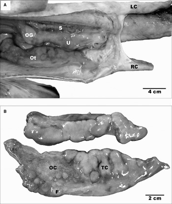
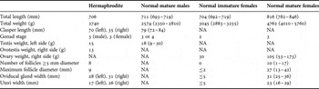
The hermaphrodite measured 706 mm total length, weighed 2740 g and had two fully calcified claspers (Figure 1A). The left clasper approached the length of normal mature males (Table 1) and possessed a typical rhipidion groove and clasper spur. The right clasper was far shorter, i.e. less than half that of normal mature males (Table 1), had a less well developed rhipidion groove and lacked a spur.
(A) Body cavity of the hermaphroditic Heterodontus portusjacksoni, with the testis removed to reveal the left oviducal gland, uterus and sperm duct; (B) testis above and ovotestis below, removed from the left and right sides of the body cavity, respectively. F, follicle; LC, left clasper; OC, ovarian component of ovotestis; Ot, ovotestis; OG, oviducal gland; RC, right clasper; S, sperm duct; TC, testicular component of ovotestis; U, uterus.

Comparisons between measurements and counts recorded for the hermaphrodite of Heterodontus portusjacksoni and those of five normal mature males, five normal immature females with lengths similar to that of the hermaphrodite, and five normal mature females. Values for normal individuals are given as mean and range (in parentheses). The measurements of the right clasper of the males, and the right reproductive components of females, which are normally symmetrical, are shown. NA, not applicable.

The left side of the body cavity contained a lobed testis of comparable appearance and weight to that of normal mature males (Table 1), whereas the right side possessed an ovotestis in the same position as the right functional ovary of normal mature females (Figure 1A, B). However, both sides contained a coiled sperm duct, oviducal gland and uterus (Figure 1A), with the diameters of the latter two structures far exceeding those of normal immature female H. portusjacksoni (Table 1).
Within the ovotestis, the testicular component is located posteriorly and the ovarian component anteriorly (Figure 1B). As with the testis, the testicular component of the ovotestis was lobed and associated with a coiled sperm duct and thus likewise meets the criterion for maturity stage 3, which is considered mature (Jones et al., Reference Jones, Hall and Potter2008). The ovarian component of the ovotestis contained numerous small non-vitellogenic follicles ≤2 mm in diameter and 14 follicles ≥5 mm in diameter (Figure 1B) eight of which were pale yellow and thus apparently undergoing vitellogenesis, while six were orange and flaccid and thus appeared to be undergoing atresia. The possession of yolked follicles and enlarged uteri fulfils the criteria for maturity stage 3, albeit on the basis of the far smaller follicle size than those of normal mature females (Table 1) at an early phase of that stage. In contrast to the hermaphrodite, all follicles in the ovaries of normal mature females closest in size to the hermaphrodite, were unyolked and ≤2 mm in diameter (Table 1), which, together with the presence of thin uteri, show that these normal females had only reached maturity stage 2 and were thus immature.
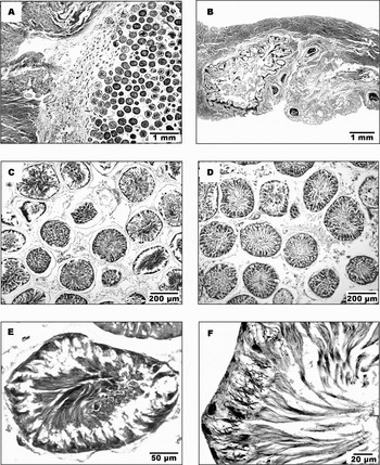
In the ovotestis, the ovarian stroma closest to the testicular component did not contain follicles (Figure 2A), whereas the main part of the ovarian component contained numerous small follicles and a few large follicles, some of which were atretic (Figure 2B). The testicular component of the ovotestis was histologically indistinguishable from the discrete left testis (Figure 2C, D). Although the germ cells in any given spermatocyst were at the same stage in spermatogenesis, different spermatocyst maturation stages were found across the testis and the testicular component of the ovotestis (Figure 2C, D). These stages included late stage spermatids, with their elongated flagella directed inwards towards the lumen of the spermatocyst (Figure 2E, F). No spermatozoa were observed in the sperm ducts.
(A) Section of ovotestis showing ovarian component on the left and testicular component (showing numerous spermatocysts) on the right, separated by connective tissue; (B) section through ovarian component of ovotestis, with a large atretic follicle on the left and five small follicles on the right; sections through testicular component of ovotestis (C) and testis (D), both of which contain numerous spermatocysts; (E) section showing spermatids in a spermatocyst of the ovotestis; (F) section showing elongated tails of late stage spermatids directed towards the lumen of a spermatocyst of the ovotestis. Section (A) was stained with Ehrlich's haemotoxylin–eosin, while those in (B) to (F) were stained with Mallory's trichrome.

The weight of the right ovary of normal immature females is far less than that of normal mature females (Table 1). Although the hermaphrodite did not possess the very large follicles found in normal mature females, the widths of its oviducal glands and uteri were similar to those of normal mature females and thus many times greater than those of normal immature females (Table 1). However, the uteri of the hermaphrodite exhibited marked asymmetry, with the right being wider.
DISCUSSION
The two hermaphrodites caught during the present and previous studies (Jones et al., Reference Jones, White and Potter2005) were among 556 Heterodontus portusjacksoni obtained from the bycatch of commercial fisheries in south-western Australia. This very low prevalence (0.36%), together with the absence of reports of this phenomenon in previous detailed studies of the reproductive biology of H. portusjacksoni elsewhere (Jones & Jones Reference Jones and Jones1982; Tovar-Avíla et al., Reference Tovar-Avíla, Walker and Day2007), emphasizes that hermaphroditism is rare in this species. Hermaphroditism is also uncommon in other elasmobranchs, apart from, for example, in certain hexanchiforms (Atz, Reference Atz, Armstrong and Marshall1964) and the catshark Apristurus longicephalus (Iglésias et al., Reference Iglésias, Sellos and Nakaya2005).
The reproductive tracts of the present hermaphrodite differ from those of the individual described earlier (Jones et al., Reference Jones, White and Potter2005) in that the male reproductive components are better developed than those of the female. Thus, this new hermaphrodite possessed a discrete testis on the left and an ovotestis on the right, the latter being located in the same position as the functional ovary in normal mature female H. portusjacksoni. Although the testicular tissue on the right constituted part of an ovotestis, it still had the same internal structure and associated sperm duct as the discrete left testis. Furthermore, as in the left testis, various stages in spermatogenesis, including late spermatids sensu Bonner Engel & Callard (Reference Bonner Engel, Callard and Hamlett2005), were present. However, only the left clasper, i.e. that on the same side as the discrete testis, was as well developed as that of normal mature males.
Although the testis and ovotestis both produced late stage spermatids, the ovarian component of the ovotestis had not produced the large follicles found in normal mature females. While the testis and the testicular component of the ovotestis were more developed than the ovarian component of the ovotestis, the follicles, oviducal glands and uteri were better developed than those of normal females of similar size, and which were immature. Indeed, the oviducal glands and uteri were of similar widths to those of mature females, even though the latter individuals were far larger than the hermaphrodite. As testosterone influences follicle growth in female elasmobranchs (e.g. Koob & Callard, Reference Koob and Callard1999, Awruch et al., Reference Awruch, Pankhurst, Frusher and Stevens2008), the presence of larger follicles in the hermaphrodite, may reflect an influence of the production of higher levels of testosterone by its testicular tissues than would be expected in normal maturing females. However, as some of the large follicles were undergoing atresia, which in our previous work (Jones et al., Reference Jones, White and Potter2005) had not been observed in females in early stage 3, an early stimulatory effect may have subsequently been replaced by an inhibitory effect. Furthermore, the far larger sizes of the uteri and oviducal glands in the hermaphrodite than those of normal immature females, demonstrate that the stimuli responsible for their development, must have been strong and occurred early in life.
ACKNOWLEDGEMENTS
Gratitude is expressed to commercial fishers, particularly Hugh Gilbert, Jamie Thornton and Alan Butler, and Rory McAuley and Justin Chidlow (Department of Fisheries Western Australian), the crew of the ‘Maritime Image’ and friends and colleagues for helping to obtain H. portusjacksoni. Thanks also to Megan Storrie and Jim Cummins for their advice, to the referees for providing very helpful suggestions for improving our paper and especially to Gordon Thompson for his histological expertise. Financial support was provided by Murdoch University and Fishcare WA.