Introduction
Interrupted aortic arch is an uncommon cardiac anomaly characterised by a lack of continuity between the ascending and descending aorta. Reference Talner1 Interrupted aortic arch rarely occurs in isolation, with almost all cases being associated with patent ductus arteriosus and ventricular septal defect, collectively known as the interrupted aortic arch triad. The presence of interrupted aortic arch in adults is extremely rare, and there is limited documentation of such cases in the literature. In this article, we present a unique case of interrupted aortic arch in an adult diagnosed through angiography. This case falls under the anatomical classification of type B interruption, although the blood supply to the left subclavian artery originates from the ascending aorta. To our understanding, no previous literature has reported cases of this particular type of interrupted aortic arch.
Case report
A 29-year-old female was preliminarily diagnosed with congenital heart disease during childhood but did not receive any interventions. She experiences limited physical activity and cannot tolerate strenuous exercise. In May 2022, her exercise tolerance progressively declined, manifested as dyspnoea and chest tightness during brisk walking or climbing two flights of stairs, leading her to seek medical consultation at our department in September 2022. Physical examination revealed a height of 165 cm and weight of 67 kg. Blood pressure in the right upper limb was measured at 98/46 mmHg, while in the left upper limb it was 92/40 mmHg. Arterial oxygen saturation levels were as follows: left upper limb – 93%, right upper limb - 96%, left lower limb – 85%, right lower limb – 85%. P2 heart sound intensified, presence of auscultatory noise in the precordial region, and broad conduction. The patient had clubbing in both lower limbs. The electrocardiogram showed signs of right ventricular overload (Figure 1). The complete blood count revealed a red blood cell count of 6.52*10^12/L and haemoglobin level of 197 g/L. A 6-minute walk test demonstrated a distance of 320 m with oxygen desaturation to 85%. The echocardiogram demonstrated overall cardiac enlargement with a left ventricular ejection fraction of 67%. The pulmonary artery was dilated with a diameter of 33 mm, and estimated pulmonary artery systolic pressure was 65 mmHg. There was a continuous interruption in the interventricular septum (membranous portion) with a defect diameter of approximately 15 mm, resulting in a left-to-right shunt. The ascending aorta did not connect to the descending aorta, and the left subclavian artery originated from the ascending aorta, suggesting a diagnosis of type A interruption. An abnormal channel measuring approximately 11 mm was observed between the descending aorta and the left pulmonary artery, indicating a possibility of patent ductus arteriosus.
Electrocardiogram: sinus rhythm, prolonged P-R interval, complete right bundle-branch block, right ventricular overload, and clockwise transposition.

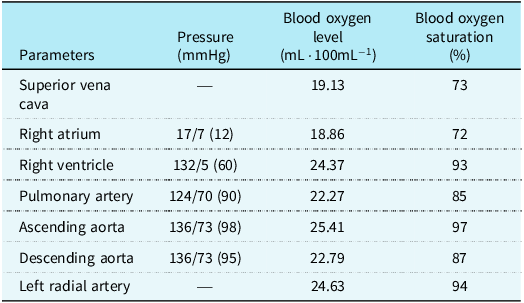
If the patient is diagnosed with type A interruption, the arterial oxygen saturation levels in both upper limbs should be the same. However, there is a certain difference in the measured arterial oxygen saturation levels. Is this difference due to measurement errors or unidentified anatomical structures? Given the patient’s deteriorating haemodynamic status (pulmonary artery systolic pressure 65 mmHg) and ambiguous echocardiographic findings, invasive angiography was prioritised over non-contrast CT angiography to expedite definitive diagnosis and guide urgent surgical planning. Therefore, we performed a vascular angiography, and the results are as follows: The 6F Pigtail catheter was advanced through the right radial artery to the ascending aorta for pressure and oxygen saturation measurements. Angiography of the ascending aorta did not reveal visualisation of the descending aorta. The right subclavian artery, right common carotid artery, and left common carotid artery originated from the ascending aorta, while the left subclavian artery was also visualised, similar to the findings in type A interruption angiography (Supplement Video 1). However, the left subclavian artery did not connect with the aortic arch. A 6F Pigtail catheter was introduced through the right femoral vein into the pulmonary artery to measure pressure and oxygen saturation. The angiography of the pulmonary artery revealed visualisation of the descending aorta, which exhibited a course on the right side of the spine, and no visualisation of the left subclavian artery. Multiple sites of angiography involving the descending aorta and pulmonary artery showed no visualisation of the left subclavian artery. Subsequent angiography of the ascending aorta demonstrated a branch originating from the ascending aorta, which curved upwards and then downwards to supply blood to the left subclavian artery. A puncture was made in the left radial artery, and a guidewire was successfully advanced from the left subclavian artery to the pulmonary artery. Angiography revealed a connection between the left subclavian artery and the descending aorta (Figure 2). The haemodynamic parameters of each vessel are presented in Table 1, and the calculated pulmonary vascular resistance is 11.19 Wood units.
Angiography results. ( a ) The ascending aorta is not connected with the descending aorta, and the right subclavian artery, right common carotid artery, left common carotid artery, and left subclavian artery can be seen. ( b ) The branches of the aortic arch first bend upwards and then bend downwards to supply the left subclavian artery(white box). ( c ), ( d ) Pulmonary angiography: the left and right pulmonary arteries are thickened, the descending aorta is visualised and located on the right side of the spine, and the left subclavian artery is not visualised. ( e ) The catheter can enter the descending aorta from the pulmonary artery. ( f ) The catheter can pass through the left radial artery to the left subclavian artery, then descending aorta to pulmonary artery. RSA = right subclavian artery; RCA = right common carotid artery; LSA = left subclavian artery; LCA = left common carotid artery; DAO = descending aorta; AAO = ascending aorta.

The application of a pigtail catheter facilitated the precise measurement of blood pressure, oxygen content, and oxygen saturation in different segments

The final diagnosis was made as congenital interrupted aortic arch (type B), congenital ventricular septal defect, patent ductus arteriosus, severe pulmonary arterial hypertension, and secondary polycythaemia. Treatment plan includes the administration of sildenafil (10 mg twice daily) and ambrisentan (5 mg once daily), with surgical intervention planned once the pulmonary artery pressure decreases. Following three months of pharmacotherapy, repeat 6-minute walk test in December 2022 showed improved performance (380 m) and oxygen saturation maintained above 90%.
Discussion
Interrupted aortic arch represents a rare and intricate congenital cardiovascular ailment characterised by an anatomical discontinuity between the ascending and descending aortas, accounting for approximately 1% of CHDs. Reference Talner1 Its manifestation primarily occurs during childhood, and patients reaching adulthood without surgical intervention is exceedingly uncommon. Reference Dahiya, Thamilarasan and Arruda2 Interrupted aortic arch is classified into three distinct types based on the location of aortic interruption Reference Celoria and Patton3 : Type A, prevailing at the distal end of the left subclavian artery, most prevalent among adults; Type B, existing between the left subclavian artery and the left carotid artery, commonly found in infants but remarkably rare in adults; and Type C, involving the interruption between the innominate artery and the left carotid artery. In the retrospective study conducted by Gordon et al. concerning 38 adult cases of interrupted aortic arch, 79% of the patients exhibited type A, followed by type B and type C, constituting 16 and 3%, respectively. Reference Gordon, Person and Kavarana4 In this instance, the angiography confirmed the origin of the left subclavian artery from the descending aorta. According to the aforementioned anatomical classification, it should be classified as type B interruption. However, the blood flow to the left subclavian artery is supplied through collateral circulation from the ascending aorta, which contrasts entirely with the haemodynamics of classical type B interruption. Instead, it bears similarities to the haemodynamics of type A interruption, albeit not entirely identical. Given that the oxygen saturation in the right upper limb is slightly higher than that in the left upper limb, we hypothesise the existence of a small collateral circulation between the descending aorta and the left subclavian artery. From a functional classification perspective, this presents a unique subtype of Type B interruption, hitherto unobserved in previous case reports.
Interrupted aortic arch can be classified into isolated and complex types. Reference Oosterhof, Azakie and Freedom5 The former is rare, while the latter is more prevalent and often accompanied by cardiac malformations such as ventricular septal defects and patent ductus arteriosus, making it the most common congenital cardiovascular condition associated with interrupted aortic arch. Reference Vriend, Lam and Mulder6 In patients with interrupted aortic arch, survival relies on the arterial blood flowing from the left ventricle into the right ventricle through the ventricular septum, which then supplies the descending aorta via the ductus arteriosus. In a review by Sharratt et al., among 9 cases that survived into childhood, only 2 cases had a patent ductus arteriosus. Reference Sharratt, Carson and Sanderson7 As the ductus arteriosus gradually closes, collateral circulation usually develops in the descending aorta to ensure a certain degree of blood flow. However, in our case, collateral circulation is limited due to the persistent presence of the ductus arteriosus, providing sufficient blood flow to the descending aorta, allowing the patient to survive into adulthood. As a result, a significant disparity in oxygen saturation between the lower limbs and upper limbs is observed, leading to the manifestation of clubbing in the lower extremities, a long-term consequence of chronic hypoxia. Additionally, another manifestation of chronic hypoxia in this patient is secondary polycythaemia.
The assessment of interrupted aortic arch primarily relies on various imaging techniques used for detection. Transthoracic echocardiography is the preferred diagnostic modality, applicable for both paediatric and adult cardiac abnormalities. However, echocardiogram has limitations in visualising the detailed anatomy of the aortic arch, posing certain constraints. Reference Davis, Gilani and Al-najjar8 In addition, magnetic resonance angiography and CT angiography are reliable non-invasive examinations with multiplanar capabilities and three-dimensional imaging. Under these imaging presentations, the morphological features of interrupted aortic arch can be clearly depicted, albeit the haemodynamic conditions of individual components may be challenging to ascertain. Although invasive, angiography remains the gold standard for diagnosing interrupted aortic arch. Apart from identifying common interrupted aortic arch cases, angiography excels in discerning the anatomical relationships of more complex cardiac malformations. In our patient’s case, the left subclavian artery originates from the descending aorta, but it does not receive blood supply from the descending aorta; instead, it is supplied by the ascending aorta. This can be inferred from the comparison of oxygen saturation between the descending aorta and the left radial artery, as well as between the ascending aorta and the left radial artery. Through analysis of pressures and oxygen saturations across various components, we have clarified their haemodynamic relationships and subsequently determined the overall pulmonary resistance, all of which contribute to the diagnosis and treatment process.
Conclusion
In conclusion, interrupted aortic arch is a rare and severe congenital vascular anomaly that is exceptionally uncommon in adults. Through analysis of echocardiography, magnetic resonance angiography, CT angiography, and angiographic findings, a definitive diagnosis of interrupted aortic arch can be established, facilitating the development of subsequent pharmacological or surgical treatment strategies.
Supplementary material
The supplementary material for this article can be found at https://doi.org/10.1017/S1047951125001805.
Financial support
This work was supported by the grant from the National Natural Science Foundation of China (No. 82260056).
Competing interests
None.
Declaration of Patient Consent
The authors certify that they have obtained all appropriate patient consent forms. In the form, the patient has given her consent for her images and other clinical information to be reported in the journal. The patient understands that her name and initials will not be published and due efforts will be made to conceal her identity, but anonymity cannot be guaranteed.