Introduction
Many of the technological applications envisioned for organic electronic materials, ranging from photovoltaics to sensors to light-emitting diodes, involve the absorption and emission of light. The low-lying bound electronic states (excitons) that determine the absorption and emission properties of these materials can be probed using optical spectroscopy, but the characterization of excitons in organic materials such as polymers is often complicated by the disordered nature of the sample. In principle, single crystals provide the best opportunity for drawing predictive structure–function relationships thanks to their high degree of structural order and chemical purity. Furthermore, the outstanding performance of small molecule, crystalline materials in applications such as light-emitting diodes, solar cells, and field-effect transistors suggests that this class of materials will play an important role in practical applications of organic electronic materials. Advances in sample preparation and characterization, spectroscopy, and theory are leading to new insights that will provide guidance for the design of new generations of crystalline molecular electronic materials. The purpose of this review is to provide a basic overview of exciton structure and dynamics using the molecule tetracene as a prototype system to demonstrate some of the relevant phenomena.
Overview of electronic states in molecular crystals
The term “exciton” has often been used as a generic label for an excited state in an organic material, and several reviews discuss their structure.Reference Davydov1–Reference Scholes and Rumbles7 Here we present a very basic picture based on concepts developed almost 50 years ago.Reference Murrell and Tanaka8, Reference Kasha, Rawls and El-Bayoumi9Figure 1 provides a schematic outline of how the electronic eigenstates evolve as individual molecules self-assemble into a crystal. At large separations, when two molecules, A and B, have no measurable Coulomb interaction, absorption of a photon leads to either molecule A or molecule B being in its excited state (denoted with an asterisk), with the two possible product wave functions ( $\Psi _A^ * \Psi _B $ and
 $\Psi _A^{} \Psi _B^ * $) of the system given in the figure. Similarly, if we consider ionized states that are usually higher in energy than excited neutral states, we also have two possible system wave functions that are degenerate. As molecules A and B are brought closer to each other, the intermolecular interaction becomes significant, and the “localized” excited states
 $\Psi _A^ * \Psi _B $ and
 $\Psi _A^{} \Psi _B^ * $ give way to their symmetric and anti-symmetric linear combinations, where the excitation now resides on both A and B simultaneously. A similar change occurs for the ionized states. As additional molecules are added, they interact and create N different states that are now delocalized over N molecules. The collection of N states forms a “band.” If the band is composed of neutral excited states, it is termed a Frenkel exciton band, while if it is composed purely of ionized states, it is termed a charge-transfer (CT) exciton band.Reference Bounds, Petelenz and Siebrand10, Reference Pope and Swenberg11 Note that a single material can have both Frenkel and CT bands, and this situation is more complex than the simple valence-conduction band picture commonly described in introductory treatments of excitons in solid-state systems.Reference Scholes and Rumbles7 The CT exciton can be thought of as a localized, structured version of the Wannier–Mott exciton, where a high dielectric constant permits the radius of the Coulombically bound electron-hole pair to extend over many sites and the excitonic wave function resembles that of a hydrogen atom.Reference Knox12 Wannier–Mott excitons tend to play a dominant role in the optical properties of inorganic semiconductors. It is interesting that a quantum confinement effect, analogous to that seen in inorganic quantum wells, has been observed for CT excitons in ultrathin films of perylene tetracarboxylic-dianhydride.Reference Shen and Forrest13 Ideally, in a macroscopic crystal N→∞, and the new states form a continuous band. Although this limit can be approached in inorganic semiconductors, the delocalization lengths for excitons in ordered organic systems generally lie in the range N = 10–1000. In the limit of N = 2, a CT exciton is more commonly termed an excimer. For such small delocalization lengths, it is possible to regard the exciton as the excited state of a “super-molecule” consisting of several molecules arranged together, rather than as an exciton in the traditional sense.Reference Wagersreiter and Mukamel14–Reference Clark, Qin and Li16
A schematic picture of exciton formation, starting with two separate molecules, A and B. For molecule A,  $\Psi _A $,
 $\Psi _A^* $,
 $\Psi _A^ + $, and
 $\Psi _A^ - $ denote the ground state, neutral excited state, and positively and negatively charged ionic states, respectively. For molecule B,
 $\Psi _B $,
 $\Psi _B^* $,
 $\Psi _B^ + $, and
 $\Psi _B^ - $ denote the ground state, neutral excited state, and positively and negatively charged ionic states, respectively. These wave functions are eigenstates of the individual molecular Hamiltonians. At large separations, there is no interaction between molecules, and the total wave function of an excited state is the product state of the individual molecular wave functions. As the two molecules approach each other, Coulombic interactions change the Hamiltonian and mix the product states to form new dimer superposition states where the excitation is delocalized over both A and B. Similarly, the ionic product states, Ψ–AΨ+B and Ψ+AΨ–B, mix to form delocalized charge resonance states. As the crystal grows to include N molecules, the neutral states grow into a Frenkel exciton band, with N excitonic states. The k th excitonic wave function (Ψ(k)Frenkel) is given by a sum over N product states
 $\Psi _1 ..\Psi _i^ * ..\Psi _N $, where the subscript i denotes the excited molecule, and
 $c_i^{(k)} $ is the weighting of this term in the overall wave function. Similarly, the k th state in the charge-transfer (CT) exciton band (Ψ(k)CT) is given by a weighted sum over product states with the negatively charged electron on molecule i and the positively charged hole on molecule j.

Given the picture in Figure 1, we can make two observations about exciton states in molecular crystals. First, both Frenkel and CT excitons can have wave functions that are delocalized over many sites.Reference Spano17, Reference Yamagata and Spano18 It is the type of molecular states that are mixed, and thus the degree of charge separation, that differentiates these two types of excitons. Second, although we have shown the case where the CT and Frenkel exciton bands are well-separated, in reality, the two bands can interact and overlap, and recent work has shown that Frenkel-CT mixing can have a profound impact on the optical properties of the low-lying excited states in polyacene crystals, which were originally presumed to be purely Frenkel excitons.Reference Schuster, Knupfer and Berger19, Reference Yamagata, Norton, Hontz, Olivier, Beljonne, Brédas, Silbey and Spano20
The lack of charge separation for Frenkel excitons means that they must be considered a full shell species (i.e., the exciton state possesses two electrons in close contact that can communicate with each other). When the spin angular momentum operator is diagonalized for these two-electron states, the solutions include one symmetric spin state (the singlet) and three antisymmetric states (the triplets).Reference Levine21 Since the ground state is singlet in character, spin selection rules make the three triplet states effectively dark, with very weak absorption and phosphorescence. But these states are always present at energies below that of the singlet, and it is the 3:1 triplet:singlet ratio that limits the efficiency of organic light-emitting diodes to 25% if only singlet states are emissive.Reference Yersin, Rausch, Czerwieniec, Hofbeck and Fischer22, Reference Baldo, O’Brien, You, Shoustikov, Sibley, Thompson and Forrest23 Transitions between singlet and triplet states, referred to as intersystem crossing, require that an electron undergo a spin flip, and they are typically quite slow. But as we will show later, multiple exciton interactions can provide spin-allowed ways to connect the singlet and triplet manifolds.
Preparation of molecular crystalline samples for spectroscopic study
Crystal growth is still somewhat of an art, and contradictory experimental results exist from different groups on what are nominally identical samples. There is a range of methods for both the purification of the molecule of interest and the growth of single crystals, many of which have been developed for the fabrication of field-effect transistors, where the charge mobility depends critically on sample purity.Reference d. Boer, Gershenson, Morpurgo and Podzorov24 The study of excitons requires the consideration of solid-state optical effects that can distort the spectroscopic response in both the time and frequency domains. For example, most single crystals are optically thick, absorbing so much light that simple transmission measurements become difficult,Reference Tavazzi, Campione, Laicini, Raimondo, Borghesi and Spearman25 and reflection measurements must be used.Reference Philpott26 For fluorescence measurements, the complications from self-absorption of the high-energy portion of the luminescence, wave guiding from total internal reflection in the high index crystal, and depolarization due to the crystal birefringence can all lead to distortions of the spectroscopic response.
Many of these complications can be reduced or eliminated by using optically thin (optical density less than one) samples. For molecular crystals, this typically means a physical sample thickness on the order of 100 nm or less. Thin single crystals can be grown by sublimation or solvent evaporation, but their isolation and characterization typically require microspectroscopy techniques. Another way to generate crystals smaller than optical wavelengths is to grow polycrystalline thin films by evaporationReference Baldo, Deutsch, Burrows, Gossenberger, Gerstenberg, Ban and Forrest27 or to make nanocrystalline colloidal suspensions.Reference Nalwa, Kasai, Okada, Oikawa, Matsuda, Kakuta, Mukoh and Nakanishi28–Reference Kostler, Rudorfer, Haase, Satzinger, Jakopic and Ribitsch31 A final challenge for photoluminescence measurements is distinguishing between emission from excitons existing in the intrinsic crystal structure and that from structural defects (whose type and concentration will depend on sample preparation conditions). Time-resolving the emission can help distinguish between intrinsic species (which are directly excited and should dominate the early-time emission) and defect states (which should appear later, after exciton diffusion and trapping).
Absorption and fluorescence spectroscopy of excitons
The absorption and emission spectra of crystals can provide important clues as to the type of exciton generated by photoexcitation. Both the coupling of the intermolecular exciton to the intramolecular vibrations and the oscillator strength of the emitting state can be quite different from those of an isolated molecule. Both effects depend sensitively on the details of the intermolecular interaction and thus on the crystal packing. CT or excimer interactions typically lead to shifted, broadened absorption and emission spectra with lower oscillator strengthsReference Hochstrasser32 and also different signatures in their transient absorption signals.Reference Lanzani, Frolov, Lane, Vardeny, Nisoli and Silvestri33 In some cases, the initially excited state has Frenkel character but rapidly relaxes into a lower energy state with more CT character, a process often referred to as “self-trapping.”Reference Matsui34, Reference Yago, Tamaki, Furube and Katoh35 In fact, the energies of the CT and Frenkel states can be reversed depending on how the molecules pack in the crystal, as seen for the α (face-to-face packing) and β (edge-to-face herringbone) crystal polymorphs of perylene.Reference Tanaka36 However, it should also be noted that CT states can support well-structured line shapes as well, as recently demonstrated for crystalline polydiacetylene.Reference Yamagata and Spano18
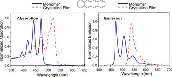
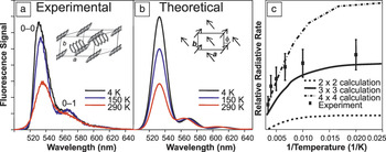
The absorption and luminescence spectra of neutral Frenkel excitons often bear a superficial resemblance to those of the neutral molecular states, with small shifts, narrow line widths, and vibronic progressions, as seen for tetracene in Figure 2. Despite this similarity, however, there are important differences between the optical properties of a localized excited state on a single molecule and those of a delocalized exciton in a molecular crystal. First, depending on the crystal structure, the absorption may exhibit Davydov splitting, where two distinct peaks appear, corresponding to the two energy levels outlined in Figure 1.Reference Muccini, Schneider, Taliani, Sokolowski, Umbach, Beljonne, Cornil and Bredas37 Depending on the orientation of the molecular transition dipole moments, Frenkel excitons can be further classified as J-type (after the first report by JelleyReference Jelley38) or H-type (denoting the hypsochromic shift of the absorption to shorter wavelengthsReference Eisfeld and Briggs39). In J-type systems, which include the polyacene crystals, the lowest lying exciton state has enhanced oscillator strength that gives rise to an enhanced radiative decay rate commonly referred to as “super-radiance.” Similar to the phenomenon of Dicke super-radiance observed in atomic systems,Reference Dicke40–Reference Spano and Mukamel42 the transition dipoles encompassed by the delocalized excitonic wave function radiate in phase, enhancing the emission of the sample. In H-aggregates, the opposite situation holds—the lowest lying exciton state has weakened or nonexistent oscillator strength. These two types of states also have different vibronic line shapes, with the J-type dominated by the 0–0 peak, which is suppressed in the H-type. Note that at room temperature, the emission can originate from multiple closely spaced exciton states, each with a different vibronic coupling and radiative rate. One way to gauge if excitonic effects are important and to determine whether the exciton is J-type or H-type is to lower the temperature and observe the changes in the emission line shape and radiative rate as the Boltzmann distribution condenses into the lowest exciton state. An example of this type of experimental data for the J-type exciton in tetracene is given in Figure 3,Reference Camposeo, Polo, Tavazzi, Silvestri, Spearman, Cingolani and Pisignano43–Reference Voigt, Langner, Schouwink, Lupton, Mahrt and Sokolowski45 and the theory underlying these effects is detailed in review articles by Spano and co-workers.Reference Spano17, Reference Spano46 This approach has clarified the role of excitonic effects in anthraceneReference Ahn, Muller, Al-Kaysi, Spano, Norton, Beljonne, Brédas and Bardeen47 as well as the technologically important oligothiophenes,Reference Meinardi, Cerminara, Sassella, Borghesi, Spearman, Bongiovanni, Mura and Tubino48, Reference Sun, Zhao, Spano, Beljonne, Cornil, Shuai and Brédas49 and signatures of intermolecular electronic delocalization have been found even in nanocrystalline polythiophene films.Reference Osterbacka, An, Jiang and Vardeny50, Reference Clark, Silva, Friend and Spano51
The absorption and emission line shapes for tetracene are plotted for isolated molecules in liquid solution and aggregated molecules in a polycrystalline film. The redshift of the absorption and emission line shapes results from the higher dielectric constant of the crystal environment. In addition, one can see a shoulder appear in the first absorption peak at around 500 nm, corresponding to the Davydov splitting that arises from the creation of the dimer states shown in Figure 1. One effect of the delocalization shown in Figure 1 is that the intensity of the intramolecular vibronic side peaks in the solid declines relative to the main peak, with this change being most pronounced in emission.Reference Lim, Bjorklund, Spano and Bardeen44

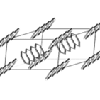
(a) The temperature-dependent emission line shapes of tetracene, integrated over the first 200 ps of emission in order to avoid contamination from trap and defect state emission, are plotted. The inset shows the herringbone crystal structure of tetracene, with a and b crystal axes labeled. (b) Theoretical simulations of the emission line shape at different temperatures assuming an exciton domain size of nine molecules. The inset shows how the transition dipoles of the tetracene molecules are aligned relative to the crystal axes, making an angle φ with respect to the b-axis. The growth of the 0–0 peak at lower temperatures reflects the increasing coherence length of the J-type singlet exciton. (c) Temperature-dependent enhancement in the relative radiative rate (defined as the radiative rate of the crystal exciton divided by the radiative rate of a single tetracene), as measured by the amplitude of the initial fluorescence signal. As the J-type exciton becomes dominated by the lowest state in the band, its radiative rate becomes proportional to the number of molecules that participate in the electronic wave function.Reference Lim, Bjorklund, Spano and Bardeen44

Dynamics of excitons
Figure 4 shows the most common processes that can occur after a photon is absorbed and an exciton is created. We have already touched on the emission and trapping of excitons, and in the remainder of this article, we concentrate on diffusion, ionization, and exciton–exciton interactions.
Schematic illustration of the dynamic processes that can occur after an exciton is created by the absorption of a photon.

Diffusion
After photoexcitation, the newly created exciton can move through the solid-state matrix, carrying its energy with it. For organic photovoltaic cells, also referred to as “excitonic cells,” diffusion of the exciton allows it to encounter an interface where it can be ionized.Reference Brédas, Norton, Cornil and Coropceanu52, Reference Gregg53 The small exciton diffusion lengths seen in most amorphous organic semiconductors (10–20 nm) limit the effective thickness of the films, leading to what one researcher has termed the “exciton bottleneck.”Reference Peumans, Uchida and Forrest54 Both singlet and triplet excitons can diffuse, and the highly ordered crystalline matrix prevents the formation of low energy trap sites that hamper diffusion in systems with more energetic disorder, like amorphous polymers.Reference Lunt, Benziger and Forrest55 Another factor that can enhance exciton diffusion is the formation of delocalized singlet states. Such states can facilitate energy transfer between sites through enhanced long-range dipole–dipole interactions,Reference Jang, Newton and Silbey56, Reference Emelianova, Athanasopoulos, Silbey and Beljonne57 and coherent electronic energy transfer has been implicated in the efficient functioning of biological systems.Reference Engel, Calhoun, Read, Ahn, Mancal, Cheng, Blankenship and Fleming58, Reference Scholes59 The high degree of order and possible existence of delocalized states both provide mechanisms to enhance exciton motion in molecular crystals, and measurements of exciton diffusion have suggested that both band-like “coherent” transport and diffusion lengths on the order of a micron or more are possible for both singlet and triplet excitons.Reference Rose, Righini and Fayer60, Reference Najafov, Lee, Zhou, Feldman and Podzorov61 In the past, the experimental challenges described in the previous section (“Preparation of molecular crystalline samples for spectroscopic study”) have made it difficult for different measurements made on the same material to converge on a single value for the diffusion length.Reference Powell and Soos62 Recent work on directly imaging the spatial extent of the exciton luminescence shows promise for the measurement of longer exciton diffusion lengths.Reference Adams, Kerimo, O’Connor and Barbara63–Reference Irkhin and Biaggio65
Ionization
The scheme shown in Figure 4 assumes that the creation of a bound CT or Frenkel exciton precedes the creation of free charge carriers. In this scenario, most excitons have large (0.5 eV or higher) binding energies and are stable species. If it has enough energy, the exciton can undergo an autoionization process that competes with internal energy relaxationReference Silinsh and Inokuchi66, Reference Petelenz and Mucha67 or diffuse until it encounters a second component (e.g., a fullerene) that dissociates it. This second case is relevant to organic photovoltaic functioning, and understanding how exciton dissociation occurs at molecular interfaces is currently a subject of great interest.Reference Yi, Coropceanu and Brédas68 This picture is consistent with most theoretical calculations and with early work on anthracene and other molecular crystals.Reference Pope and Swenberg11, Reference Kleinerman, Azarraga and McGlynn69–Reference Sai, Taigo, Chelikowski and Reboredo71 More recently, time-resolved photoconductivity and terahertz experiments have been interpreted to show the direct creation of high mobility free carriers.Reference Moses, Soci, Chi and Ramirez72–Reference Thorsmolle, Averitt, Chi, Hilton, Smith, Ramirez and Taylor74 If this is the case, then organic crystals like pentacene may be more analogous to inorganic semiconductors than Figure 4 would suggest. However, most optical measurements, as well as some photoconductivity experiments,Reference Horowitz, Kouki, Valat, Delannoy and Roussel75, Reference Najafov, Biaggio, Podzorov, Calhoun and Gershenson76 tend to point to bound, emissive excitons as being the majority species formed after photoexcitation. To date, there has not been a consistent effort to perform optical measurements and transient conductivity experiments under the same experimental conditions in order to reconcile these two scenarios. It is possible that once variables such as exciton-exciton interactions,Reference Kearns77, Reference Silver, Olness, Swicord and Jarnagin78 the polarizability of delocalized excitons,Reference Gelinck, Piet, Wegewijs, Mullen, Wildeman, Hadziioannou and Warman79 and sample preparation are taken into account, an accurate estimate of the relative yields of free carriers versus excitons created by photoexcitation can be obtained.
Exciton–exciton interactions
The high density of sites and facile exciton motion make it possible for excitons to encounter each other when they are created within the same crystal. When two singlet excitons encounter each other, they can fuse into a single high-energy exciton that rapidly relaxes back down into its lowest singlet state.Reference Greene and Millard80 The net result is the transformation of two excitons into one. This type of exciton–exciton annihilation can induce a rapid decay in the excited state singlet population, and care must be taken to account for this effect, especially in laser experiments where high densities of singlet excitons are generated. But two excitons can also combine to form a new, higher energy state. This nonlinear excitonic process, termed “energy pooling” in covalent dendrimer macromolecules,Reference Andrews and Bradshaw81 is usually referred to as exciton fusion in molecular crystals.Reference Pope and Swenberg11 While the fusion of two singlet excitons always produces another singlet, fusion of two triplet excitons encompasses spin-allowed pathways for the creation of high-energy excitons with different spin states, including the singlet. The conversion of two low-energy, optically dark triplets into a bright singlet state results in “delayed fluorescence”Reference Parker, Hatchard and Joyce82, Reference Ryansnyanskiy and Biaggio83 that survives much longer than the intrinsic singlet lifetime. There is much interest in harnessing this 2→1 fusion process in order to “upconvert” two low-energy photons into a high-energy photon that can be absorbed by a high bandgap solar cell.Reference Insangulov, Kozlov and Castellano84, Reference Baluschev, Miteva, Yakutkin, Nelles, Yasuda and Wegner85
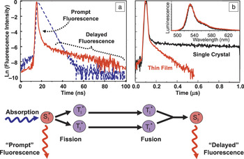
If exciton fusion is an allowed process, then its inverse, exciton fission, must also be allowed. Indeed, the 1→2 reaction in which a singlet exciton (S1) spontaneously splits into a pair of triplet excitons (T1) is known to occur in a variety of solid-state organic materials,Reference Smith and Michl86 including molecular crystals such as the polyacenes (anthracene,Reference Singh, Jones, Siebrand, Stoicheff and Schneider87 tetracene,Reference Swenberg and Stacy88–Reference Chan, Ligges and Zhu91 pentaceneReference Rao, Wilson, Hodgkiss, Albert-Seifried, Bassler and Friend92–Reference Chan, Ligges, Jailaubekov, Kaake, Miaja-Avila and Zhu94), isobenzofuran,Reference Johnson, Nozik and Michl95 and polyenes,Reference Wang and Tauber96 as well as in ordered polymers such as polydiacetylene.Reference Lanzani, Cerullo, Zavelani-Rossi and Silvestri97 In order to conserve energy, the singlet and triplet energies (E) of a molecule used for this application must fulfill the condition 2E (T1)  $ \le $E (S1). Fortunately, recent theoretical and experimental work has identified several classes of conjugated organic molecules that fulfill this requirement.Reference Paci, Johnson, Chen, Rana, Popovic, David, Nozik, Ratner and Michl98 From a practical standpoint, singlet fission is of interest as a way to enhance the efficiency of photovoltaic cells, and devices based on this phenomenon have shown promise.Reference Jadhav, Mohanty, Sussman, Lee and Baldo99, Reference Ehrler, Wilson, Rao, Friend and Greenham100 For a photon whose energy is well above the bandgap, most of its excess energy is lost as heat as it decays to the band edge. This waste of excess photon energy is one factor that led Shockley and Queisser to calculate an upper limit of ∼32% for the efficiency of a single junction solar cell.Reference Shockley and Queisser101 If the high-energy exciton could be split into a pair of low-energy excitons close to the band edge, then the excess photon energy would generate an extra electron-hole pair rather than dissipating into the phonon bath. The more complete utilization of the solar spectrum through the transformation of both above-bandgap photons (through exciton fission) and below-bandgap photons (through exciton fusion) can boost the overall efficiency of a solar cell by as much as 40%.Reference Hanna and Nozik102, Reference Shpaisman, Niitsoo, Lubomirsky and Cahen103 Both singlet fission and triplet fusion can be observed in crystalline tetracene, as illustrated in Figure 5a.Reference Burdett, Gosztola and Bardeen89 Note that although the short-lived singlet fluorescence is not very sensitive to sample preparation, the delayed fluorescence due to the long-lived triplets is, as shown in Figure 5b. This result nicely illustrates how longer-lived species that diffuse over larger distances are more sensitive to sample morphology.
(a) The single-exponential fluorescence decay of an isolated tetracene molecule in solution (blue dashed) is compared with that observed in a polycrystalline film (red solid). The rapid initial component of the fluorescence decay in the crystalline film corresponds to prompt fluorescence from the initially excited singlet state that decays predominantly through exciton fission. The delayed fluorescence arises when the singlet state (S1*) is regenerated by the fusion of a pair of triplet (T1*) excitons. These two processes are outlined in the schematic diagram below the data. (b) The delayed fluorescence decay in tetracene reflects the lifetime of the triplet excitons. Although the prompt decay is the same, the lifetime of the triplets is significantly longer in a single crystal (black) than in a polycrystalline film (red). The longer diffusion length of the triplet excitons makes them more sensitive to chemical impurities and structural defects than the short-lived singlet excitons. The inset shows that the emission spectra at 1 ns (black) and 1 μs (red) are identical.Reference Burdett, Gosztola and Bardeen89

Conclusion
Molecular crystals provide a structurally well-defined medium where exciton dynamics can be studied and understood at a fundamental level. Moreover, it is becoming clear that excitons in molecular crystals have unique properties, such as delocalization, enhanced diffusion, and fission/fusion reactions, which make them interesting for practical applications as well. Advances in the growth and shape control of molecular crystal nanostructuresReference Liu, Wang, Briseno, Mannsfeld and Bao104 may lead to photoactive materials that possess the superior electronic properties of single crystals while retaining the cost advantages associated with polymeric materials.
Acknowledgment
This work was supported by the National Science Foundation under Grant CHE-1152677.