Introduction
The use of osseointegrated implants for bone-anchored hearing aid (BAHA) retention is now a well established practice, following commercial availability of the device in 1987.Reference Tietze and Papsin1 There are a great many reports acknowledging the benefits of the BAHA, both in terms of audiological competence and patient well being and (in the paediatric population) development. The BAHA has also proven to be enormously useful in cases of congenital aural atresia, and provides an alternative to canal and middle-ear reconstructive surgery.Reference Mylanus, Snik and Cremers2–Reference Gillett, Fairley, Chandrashake, Bean and Gonzalez5
It is estimated that more than 30 000 BAHA fixtures have been implanted worldwideReference Granstrom6 to date.
Trauma to the Baha is a well documented problem especially in the paediatric population. Such a significant injury/complication as in this case is very rare. The literature describes four other such significant cases.
Clinicians should remember that unusual and unexpected complications of BAHA implantation do occur, albeit rarely. It is wise to remember that the care of the patient continues long after BAHA surgery is complete.
Case report
A nine-year-old girl had previously been fitted with a BAHA to treat moderate bilateral conductive hearing loss.
The patient had been diagnosed with Dubowitz syndrome at birth, a rare autosomal recessive condition characterised by low birth weight, growth retardation and delayed bone maturation, short stature, high sloping forehead with a broad nasal bridge, and sometimes eczema.Reference Gorlin, Cohen and Hennekan7 She had behavioural and learning difficulties, which have also been reported to be associated with this syndrome.Reference Gibson and Potparic8
A left-sided BAHA had been fitted when the patient was four years of age. This procedure had been performed in the usual, well described fashion,Reference Tjellstrom, Lindstrom, Hallen, Albrektsson and Branemark9 employing two stages, with a healing time of 16 weeks for osseointegration.
There had been some initial problems with wound infection, but these had settled with conservative management.
Over a five-year period, the patient had made excellent progress and had managed her BAHA well, without complications.
However, prior to the current presentation, the patient fell and sustained a blow to the side of her head. The injury was initially thought to be minor, and the patient experienced no immediate problems. However, later that day, the BAHA abutment was noticed to be embedded deep in the scalp.
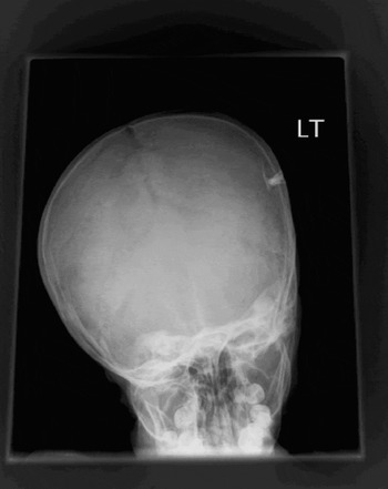
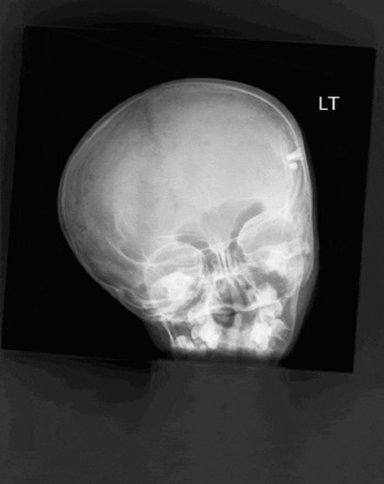
Examination revealed an intracranial intrusion of both the fixture and the entire abutment. Plain radiographs confirmed the intracranial position of the abutment (Figures 1 and 2).
Surgical removal of the implant was undertaken. At the time of surgery, all bony fragments were removed and the dura was found to be intact.
Plain radiograph demonstrating intrusion of the bone-anchored hearing aid abutment and associated skull fracture.

Plain radiograph demonstrating a left-sided, complete intracranial intrusion of both the bone-anchored hearing aid fixture and abutment. The ‘sleeper’ fixture is also noted.

The patient made an uneventful recovery. However, at the time of writing she was having difficulty with conventional hearing aids, and was keen to have another BAHA.
Discussion
A considerable amount of data have been collected on patients with implanted BAHAs. A significant number of patients have sustained traumatic injuries to their BAHA site, although this phenomenon is almost unique to the paediatric population. Institutions with a BAHA programme involving children require a good clinical team in order to support these patients long after the surgical procedure has been completed. In many cases, trauma results in repeated damage to the BAHA sound processor rather than to the abutment and/or fixture. It is not uncommon for families to request a spare BAHA sound processor in case of such an injury.
The vast majority of studies reporting paediatric BAHA outcomes describe fixture loss as being a direct result of trauma.Reference Granstrom6, Reference Lloyd, Almedya, Sirimanna, Albert and Bailey10–Reference Seeman, Liu and Di Toppa12
Fortunately, serious and unusual complications of BAHA implantation are very rare. We report one such serious complication. This was the only such injury experienced in both the Birmingham adult and paediatric BAHA programmes since their commencement in the late 1980s.
On review of the literature, four previous cases of unusual and serious BAHA complications were identified. Only one involved a traumatic injury in a child.
Deitmer et al. Reference Deitmer, Krassort and Hartmann13 reported the first case of BAHA intrusion injury. This case involved a young girl who had a fall and sustained a blow to the head on the same side as her BAHA. As in our case, there was no haematoma or intracranial complications. The fixture and abutment were surgically removed immediately, and the child made a full recovery and was re-implanted at a later date. This child was not reported to have any significant underlying medical condition.
Our patient had Dubowitz syndrome, which is known to be associated with growth retardation, with reports of delayed bone maturation. This may have increased her risk of an intrusion-type injury following trauma.
In November 1993, the first report of an intracerebral abscess after a BAHA abutment change was published by Deitmer et al. Reference Deitmer, Krassort and Hartmann13 The reported patient had a significant soft tissue reaction around the abutment and the abutment was therefore removed, leaving the well osseointegrated fixture in place. The wound was reported to be almost completely healed when the patient developed an intracranial abscess.
Scholz et al. Reference Scholz, Eufinger, Anders, Illerhaus, Matthias and Schmieder14 reported the second case of an intracranial abscess developing after an abutment change. In this case, the abscess was diagnosed three months following the procedure.
The interesting points from this second case were, firstly, the fact that change of the abutment was reported to be ‘long lasting’ and ‘complicated’. Secondly, the abutment did not actually appear to have been replaced at the end of the procedure. As discussed by Tjellstrom and Niparko,Reference Tjellstrom and Niparko15 abutment change is usually a simple procedure lasting just a few minutes and is often performed in the out-patient setting.
Scholz et al. Reference Scholz, Eufinger, Anders, Illerhaus, Matthias and Schmieder14 described successful drainage of the abscess via the screw hole once the fixture had been removed. Unfortunately, the patient reported by Deitmer et al. Reference Deitmer, Krassort and Hartmann13 required formal neurosurgical drainage.
Both these patients were adults and both made a full recovery after appropriate drainage of the abscess and antibiotic therapy.
It has been reported that 8.5 per centReference Tjellstrom and Granstrom16 of fixtures in adults and 21 per cent Reference Granstrom, Bergstrom, Odersjo and Tjellstrom17 of fixtures in children are placed in contact with the dura. It is therefore surprising that there are not more reports of intrusion injury and intracranial infection, especially as many centres are now implanting younger children.
In Birmingham, more than 3000 BAHA fixtures have been implanted (in the adult plus paediatric programmes) over the past 15 years, with only one significant complication, as reported above. This rarity is reflected in the world literature. It has been estimated that more than 30 000 BAHA fixtures have been implanted worldwide to date,Reference Granstrom6 with only four reports of unusual and/or serious complications.
• A nine-year-old girl with Dubowitz syndrome sustained an intrusion injury of her bone-anchored hearing aid fixture and abutment following a fall
• The patient underwent immediate removal of the implant and subsequently made a full recovery
We have previously described a case of metastasis of a bronchogenic neoplasm to the soft tissue surrounding the BAHA abutment in a 68-year-old woman.Reference Hadjihannas, McDermott, Warfield, Proops and Reid18 Although this was not a BAHA complication per se, the occurrence is sufficiently rare to warrant a mention when discussing unusual BAHA complications.
Conclusion
The BAHA is used worldwide, with very few serious complications. This case report serves to remind all clinicians involved with BAHA provision that they must monitor their patients carefully. Clinicians should remember that unusual and unexpected complications of BAHA implantation, although rare, do occur.