I. INTRODUCTION
Chitosan, which can be extracted from crustaceans shellsReference Abdullin, Shipovskaya, Fomina, Artemenko, Ovchinnikova and Pchelintseva1 or microbial cell walls such as fungi,Reference Wang, Du and Wang2 is a linear polysaccharide composed of glucosamine and N-acetyl glucosamine linked through β-1,4 glucosidic bonds. It has become one of the most widely used biomaterials due to its excellent biocompatibility, biodegradability, nutritional function and pharmacological efficacy.Reference Tsujikawa, Kanauchi, Andoh, Saotome, Sasaki, Fujiyama and Bamba3, Reference Seol, Lee, Park, Lee, Young-Ku, Rhyu, Lee, Han and Chung4 As a promising biomaterial in tissue engineering application, the degradation behavior of chitosan plays a crucial role on the long-term performance of the tissue-engineered cell/material constructs. The degradation kinetics may affect many cellular processes, including cell growth, tissue regeneration, and host response.Reference Ren, Yi, Wang and Ma5 But the disadvantage of using chitosan alone as a scaffold is that chitosan itself has a very slow degradation rate, and its molecules degrade in an uncontrollable manner.Reference Lu, Kong, Sheng, Wang, Gong and Zhang6 Many investigations have been published on the degradation of chitosan scaffold. Most of them showed that the molecular weight and degree of N-deacetylation (DD) are the key factors controlling the degradation of chitosan.Reference Freier, Koh, Kazazian and Shoichet7–Reference Li, Du and Liang9 The problem of effectively controlling the chitosan scaffold degradation needs to be resolved when the molecular weight and DD are certain parameters. A few publications have focused on the influence of porosity and fiber diameterReference Wan, Yu, Wu, Wang and Wen10, Reference Cunha-Reis, TuzlaKoglu, Baas, Yang, EI Haj and Reis11 or different ratios of chitosan blendsReference Ren, Yi, Zhang, Xie, Wang and Ma12, Reference She, Zhang, Jin, Feng and Xu13 on the degradation of chitosan scaffolds. Others were mainly about adjusting the extent of crosslinking to control the degradation rate.Reference Prashanth, Lakshman, Shamala and Tharanathan14–Reference Hong, Song, Gong, Mao, Gao and Shen16 In our opinion, a biomimetic approach may be attempted to solve this problem.
Lysozyme is widely studied as an antibacterial proteinReference Kuijpers, Wachem, Luyn, Engbers, Krijgsveld, Zaat, Dankert and Feijen17 or model protein because of its size similar to many therapeutically effective cytokines such as interferons and interleukinsReference Kang, Jiang, Hinderliter, DeLuca and Singh18 in controlled release of drugs and also in tissue engineering. Some researchers have reported that lysozyme could induce amyloid toxicity in vivo,Reference Morozova-Roche19, Reference Mossuto, Dhulesia, Devlin, Frare, Kumita, Laureto, Dumoulin, Fontana, Dobson and Salvatella20 but this might be decelerated by intact-protein binding and conversion.Reference Mishra, Sörgjerd, Nyström, Nordigården, Yu and Hammarström21 Cerven et al. also studied the 28-day repeated dose oral toxicity of recombinant human lysozyme in rats, the results demonstrated a lack of toxicity and there were no treatment-related, toxicologically relevant changes in clinical signs, growth, food consumption, hematology, clinical chemistry, organ weight and pathology.Reference Cerven, DeGeorge and Bethell22 So, lysozyme may be a safe material for in vivo uses. In addition, since chitosan itself has a very slow degradation rate and its molecules degrade in an uncontrollable manner,Reference Lu, Kong, Sheng, Wang, Gong and Zhang6 lysozyme has been regarded as the main contributing agent to chitosan degradation in vivo, which is commonly present in various human body fluids and tissues. But due to the presence of inhomogeneous distribution of lysozyme that causes the uneven degradation of chitosan scaffold, it is very hard to control the degradation rate of chitosan to match the building rate of new tissue regeneration. Considering the self-existence and degradation property of lysozyme, a new strategy needs to be used by controlling the lysozyme release and further influencing the scaffold degradation.
In this study, we prepared poly(D,L-lactic-co-glycolic acid) (PLGA) microspheres by water-in-oil-in-water (w/o/w) method and studied the degradation-controlling efficacy. PLGA is approved by the US Food and Drug Administration (FDA) and most importantly the degradation products are acidic, which can be neutralized by chitosan and its degradation products. These acid-alkali reactions may alleviate the potent inflammatory reactions caused by PLGA or chitosan degradation products in vivo.Reference Jiang, Nukavarapu, Deng, Jabbarzadeh, Kofron, Doty, Abdel-Fattah and Laurencin23–Reference Ganji and Abdekhodaie25 Some authors have studied incorporation of PLGA microspheresReference Tan, Wu, Lao and Gao26 or nanoparticlesReference Nandagiri, Gentile, Chiono, Tonda-Turo, Matsiko, Ramtoola, Montevecchi and Ciardelli27 into chitosan blend porous scaffolds to improve the compressive strength of the natural polymer scaffolds without affecting the biocompatibility. Interestingly, Martins also described an innovative self-regulated degradation system in a biomimetic route.Reference Martins, Pereira, Leonor, Azevedo and Reis28 This biomimetic view is different from what we proposed in this article, and the main differences are listed as below: (i) In Martins’ work, lysozyme was incorporated into calcium phosphate (CaP) coatings which meant that lysozyme was at the surface of the scaffold, whereas in our work, lysozyme was incorporated into the PLGA microspheres and then dispersed in the whole matrix of the scaffold. (ii) In Martins’ view, the biomimetic route utilized the antibacterial properties of lysozyme to prevent infection upon implantation, whereas in our view, the biomimetic strategy utilized the degradation behavior of lysozyme toward β-1,4 glucosidic bonds of chitosan. Therefore, incorporation of lysozyme-loaded microspheres into chitosan porous scaffold may result in degradation of the chitosan scaffold in a controlled manner.
Bearing all these considerations in mind, the aim of the present study was to develop a new strategy for controllable degradation of chitosan scaffolds. Here, we describe the preparation of PLGA microspheres and the combined scaffolds. The chitosan scaffold degradations in vitro and in vivo as well as the primary biocompatibility tests were also investigated in this article.
II. MATERIALS AND METHODS
A. Materials
Poly(D,L-lactic-co-glycolic acid) (PLGA, 50:50, Mw 6,218) was purchased from Chengdu Hangli Fine Chemicals Co. Ltd. Chitosan (Mη 1.3 × 10⁶, DD 85%; Sichuan Province, China) was obtained from Jinan Haidebei Marine Bioengineering Co. Ltd. (Shangdong Province, China) The lysozyme was kindly provided by Sigma Co. (distributed in Sino-American Biotechnology Co., Guangzhou, Guangdong Province, China) Poly (vinyl alcohol) (PVA-124, viscosity range 50–60 mPa s, hydrolysis degree is above 97%) was obtained from Guangzhou Tianma Fine Chemical Plant (Guangzhou, Guangdong Province, China). Bicinchoninic acid protein assay (BCA) kit and Micrococcus lysodeikticus were purchased from Pierce (USA) and Sigma respectively. All other compounds were of analytical purity and were used as received.
Spraugue Dawley (SD) rats (1∼2 d) and BALB/C mice (female, SPF Grade, 9–10 wk, 20–26 g) were kindly provided by Experimental Animal Center of Sun Yat-sen University.
B. Microsphere preparation
Lysozyme was encapsulated into PLGA using a double emulsion solvent extraction/evaporation method, which has been reported elsewhere.Reference Wong, Wang and Wang29, Reference Jiang, Woo, Kang, Singh and DeLuca30 Briefly, a primary water-in-oil emulsion (w/o) of the protein solution (6% w/v, 200 μL) in phosphate-buffered saline (PBS, pH 7.4) and polymer solution (20% w/v, 4 mL) in dichloromethane was prepared by homogenization (Ultra Turrax T18 basic, IKA, Staufen, Germany). The resulting w/o emulsion was then added to an aqueous solution of PVA (2.5% w/v, 200 mL) to produce a secondary water-in-oil-in-water (w/o/w) emulsion. The secondary emulsion was stirred for 5 h at 600 rpm using a mechanical stirrer (Eurostar Pcv, IKA, Staufen, Germany), under ambient temperature and pressure, to completely remove the solvent and solidify the microspheres. The microspheres were collected by centrifugation, washed three times with deionized water and vacuum freeze-dried. Microsphere shape and morphology were analyzed by Scanning Electron Microscope (SEM, PHILIPS XL-30 ESEM series, Amsterdam, Netherlands) and Atomic Force Microscope (AFM, AutoProbe CP Research, Thermomicroscopes, Sunnyvale, CA). Microsphere size and distribution were determined by Laser Particle Sizer (LPS, LS230, BECKMAN-COULTER, Brea, CA).
C. Protein loading of microspheres
Lysozyme loading within the microspheres was determined by dissolving accurately weighed 10 mg of freeze-dried microspheres, in duplicate, in 3 mL of 0.1 M sodium hydroxide solution containing 5% (w/v) sodium dodecyl sulfate (SDS). After incubation at 30 °C for 15 h, the solution was centrifuged and the protein content in the supernatant was analyzed by the well-known bicinchoninic acid protein assay (BCA) method. Protein loading and encapsulation efficiency were calculated according to the results.Reference Sharif and O’Hagan31, Reference Blanco and Alonso32
D. Protein release studies
Microspheres (10 mg) were suspended in 2 mL of fresh PBS at pH 7.4 at 37 °C under 100 rpm. At a predetermined time interval, the samples were subjected to centrifugation at 2000 rpm for 3 min. The protein content in the supernatant was analyzed by the BCA method and the buffer was replaced with fresh PBS. The cumulative release profiles were performed in triplicate.Reference Wong, Wang and Wang29, Reference Blanco and Alonso32
E. Enzymatic activity assay
The activity of lysozyme was measured at 450 nm with a microplate reader (MK3, Labsystem, Helsinki, Finland) by a Micrococcus Lysodeikticus assay as described elsewhere.Reference Kang, Jiang, Hinderliter, DeLuca and Singh18, Reference Bezemer, Radersma, Grijpma, Dijkstra, Feijen and Blitterswijk33, Reference Krishnamurthy, Lumpkin and Sridhar34 Relative activity (RA) of each sample was calculated from the equation RA = (As/Af) × 100. Here, As refers to activity of sample and Af refers to activity of fresh lysozyme standards of which weight were the same as the samples.Reference Deng, Zhou and Luo35
F. Combined microsphere-chitosan scaffold preparation
Chitosan was dissolved in 2% (v/v) aqueous acetic acid to prepare a 2% (w/v) chitosan solution. The solution was filtered and centrifuged to remove the undissolved impurities and air bubbles. The microspheres were added (ultrasonic dispersion before adding) into the solution and stirred for homogenization. Then the microsphere-chitosan blend was slowly poured into a tetrafluoroethylene petri dish mold (diameter: 38 mm, depth: 7 mm). The mold was quickly frozen at −80 °C for 1 d and then lyophilized in a vacuum freeze-dryer (MC2L, UNICRYO, Martinsried, Germany) for another 1 d. The obtained scaffold was washed successively with a 2.5% (v/v) aqueous ammonia solution and a large amount of water. The wet material was again frozen and lyophilized to obtain a porous combined microsphere-chitosan scaffold. The combined scaffold was characterized by SEM.
G. In vitro and in vivo degradation tests
Different mass ratios of microsphere to chitosan (blank, 1:50, 1:20, 1:10, 1:5, 1:2) were used to study the degradation efficiency. In vitro degradation test was carried out by incubating the scaffold in pH 7.4 of PBS at 37 °C. In vivo degradation test was carried out by implanting the scaffold underneath the skin of the back of clinical trial mouse (BALB/C, female, SPF Grade, 9–10 wk, 20–26 g). At a predetermined time interval, the scaffolds were taken from the medium or mouse, washed with distilled water and freeze-dried. The degradation rate D was examined by weight loss from the formula below, which was widely used as the index of in vitro degradation.Reference Lu, Kong, Sheng, Wang, Gong and Zhang6, Reference Freier, Koh, Kazazian and Shoichet7, Reference Cunha-Reis, TuzlaKoglu, Baas, Yang, EI Haj and Reis11, Reference Ren, Yi, Zhang, Xie, Wang and Ma12
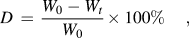
where W 0 denotes the original weight, while Wt is the weight at time t. Each degradation experiment was repeated three times and the average value was taken as the percentage degradation.
H. Cytotoxicity assay
Osteoblasts were isolated from SD rat skull and identified by alkaline phosphatase (ALP) assay and Von Kossa staining in our lab. The osteoblasts were added directly to each combined scaffold in 100 μl medium suspension (1 × 10⁵ cells/5mm × 5 mm × h scaffold). After 3 h, 1 mL medium was slowly added to each well. The plate was then incubated at 37 °C and 5% CO2. For cell viability assessment, 3-(4,5-dimethylthiazol-2-yl)-2,5-diphenyltetrazolium bromide (MTT) test was performed to quantify cytotoxicity.Reference Lu, Kong, Sheng, Wang, Gong and Zhang6, Reference Hong, Song, Gong, Mao, Gao and Shen16 Briefly, after injection of 50 μl MTT solution (5 mg/mL) into the cells/scaffold construct, the construct was cultured at 37 °C for 4 h. Then the MTT solution was removed and insoluble formazan crystals were dissolved in 300 μl dimethylsulfoxide (DMSO). Solution (200 μl) from each well was transferred into a 96-well plate for absorbance measurement at 492 nm under a microplate reader (MK3, Labsystem, Finland). The absorbance was directly proportional to cytotoxicity.
I. Cell attachment study
The osteoblasts were added directly to each combined scaffold in 100 μl medium suspension (1 × 10⁵ cells/5mm × 5 mm × h scaffold). After 3 h, 1 mL medium was slowly added to each well. The plate was then incubated at 37 °C and 5% CO2. After 7 d, seeded scaffolds were prepared for SEM analysis by first rinsing three times in PBS. Then, the samples were placed in 2.5% glutaraldehyde in phosphate buffer for 2 h, after which, the samples were rinsed in PBS and then dehydrated in a series of ethanol washes (50, 70, 90 and 100%) and 100% isoamyl acetate. Finally, the seeded scaffolds were dried by supercritical fluid carbon dioxide (SCF CO2) and placed on a SEM stub and coated for imaging.Reference Jaklenec, Wan, Murray and Mathiowitz36
J. Histological evaluation
After implantation at different periods, the scaffold, the skin tissue and the subcutaneous tissue around the scaffold were carefully taken off. These specimens were fixed in 4% paraformaldehyde for 24 h, embedded in paraffin, and sectioned (5 μm in thickness), followed by staining with hematoxylin and eosin (HE).Reference Malafaya, Santos, Griensven and Reis37 The stained microscope slides were observed using a computer program of Leica Qwin Plus (DMRA2 fluorescence microscopy, Leica, Germany).
III. RESULTS AND DISCUSSION
A. Microsphere characterization
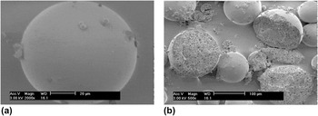
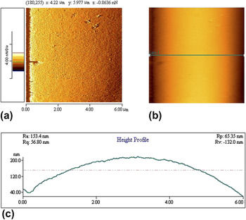
PLGA was characterized by gel permeation chromatography (GPC) (Mn 3849, Mw 6218, Mp 5928, Mz 8922, Mz/Mw 1.43) before use. The microsphere morphology was observed from SEM and AFM (Fig. 1 and Fig. 2). In SEM experiment, all samples were previously attached to copper mounts and coated with gold. In AFM experiment, the surface of the microsphere was imaged in air in tapping mode. Microfabricated silicon cantilevers (Park Scientific Instruments) of sharp tips with a tip radius of curvature 10 nm and a force constant of approximately 2.8 N/m were used. The scan speed of the tip was 1 Hz. The images were planar-leveled using the software (Thermomicroscopes Proscan Image Processing Software Version 2.1) provided with the instrument. We can see from these pictures that microspheres have smooth surface [Fig. 1(a) and Fig. 2] and porous structure inside [Fig. 1(b)]. LPS results indicate that the mean diameter of the microspheres is 54.6 ± 30.8 μm.
SEM pictures of microsphere surface and inner morphologies (left: surface; right: inner).

AFM pictures of microsphere surface morphology (left: error signal; middle & right: topology).

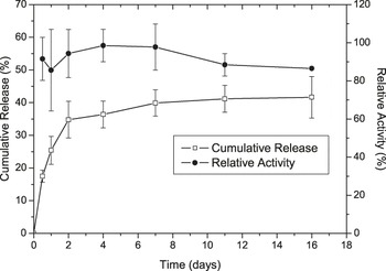
Protein release profiles are listed in Fig. 3. The protein loading was 1.8 ± 0.1% (g/g) and the encapsulation efficiency was 75.0 ± 3.7%. From Fig. 3 we can see that microspheres present excellent drug property of controlled release and enzymatic activity protection property. From 12 h to 2 d, lysozyme cumulative release increased from 17.5 ± 1.9% to 34.8 ± 5.6%, after that period the cumulative release increased slowly, the value was only 41.6 ± 6.4% after 16 d. For assessing the lysozyme stability, we examined the enzymatic activity. We found that the RA was all above 85% during the whole period of microsphere release process. Results indicate that W/O/W method is an appropriate way to prepare lysozyme-loaded PLGA microspheres with high protein activity as other publications reported.Reference Kang, Jiang, Hinderliter, DeLuca and Singh18, Reference Nam, Song, Choi and Park38–Reference Aubert-Pouëssel, Bibby, Venier-Julienne, Hindré and Benoît40
Cumulative release and RA of microspheres.

B. Combined microsphere-chitosan scaffold characterization
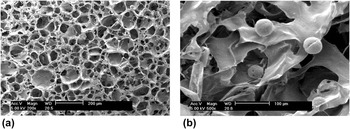
Researches on multifunctional scaffolds have attracted extensive attention in recent years. Some experts focus on the microsphere-integrated scaffolds because microspheres will supply some additional functions.Reference Hiraoka, Yamashiro, Yasuda, Kimura, Inamoto and Tabata41–Reference Niu, Feng, Wang, Guo and Zheng43 In this article, we attempted to prepare biomimetic lysozyme-loaded PLGA microspheres that might control the degradation of chitosan scaffold by combining the microspheres and the scaffold together. The morphologies of the combined scaffolds are shown in Fig. 4. The mean pore size was 88 ± 23 μm and the porosity of all batches was higher than 90% with an interconnected pore network (data not shown).
SEM micrographs of the combined scaffold (left: surface; right: inner).

C. In vitro degradation
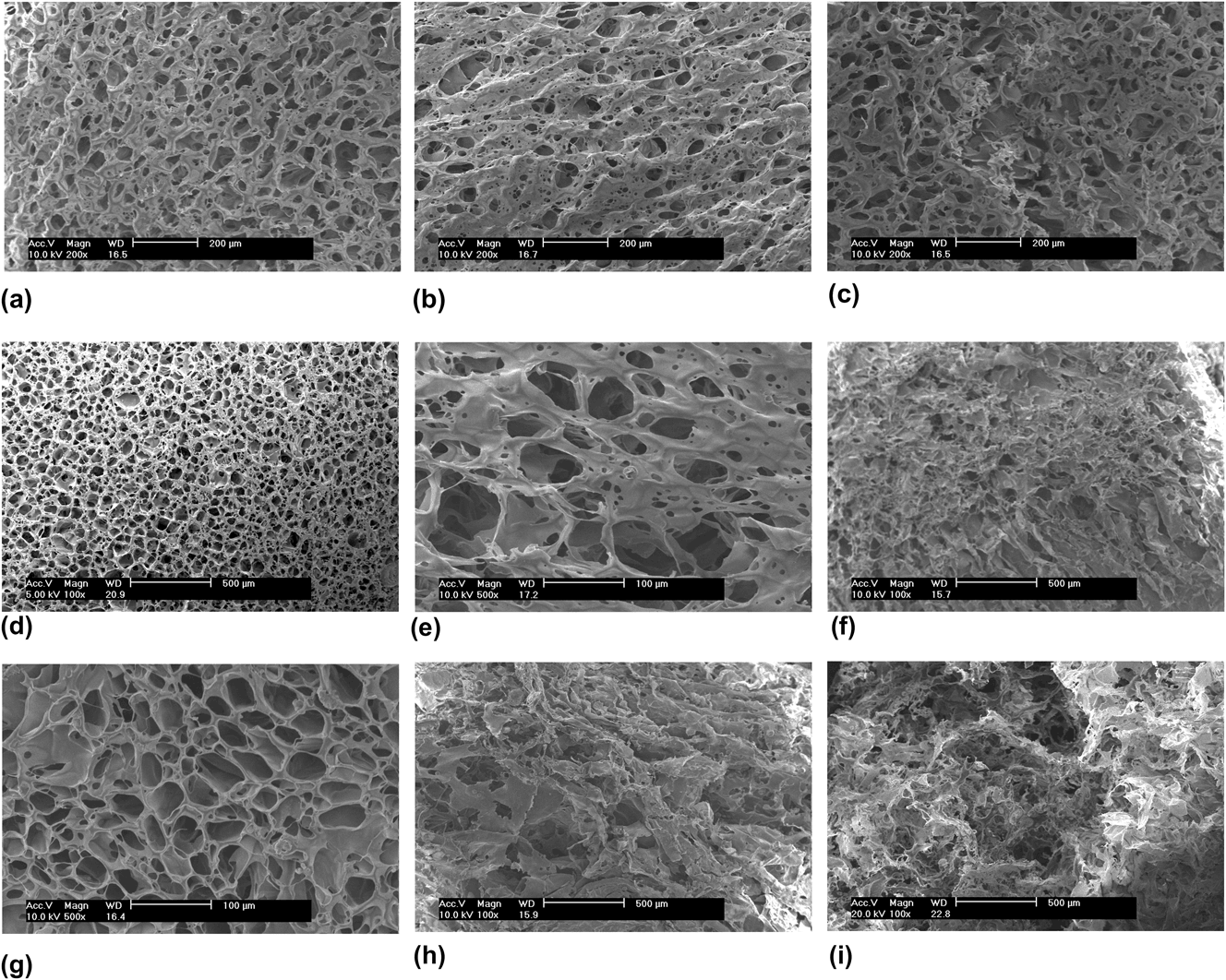
In the study, the mass/volume ratio between the specimens and the test solutions were 1:30, and the specimens were fully immersed in the test solutions according to ISO 10993-13. When the specimens were immersed in PBS at 37 °C, the combined microspheres were gradually surrounded with water in the process of solvent molecules diffusing into the scaffolds. The lysozyme was released from the microspheres in controlled release mode and permitted to react with chitosan, which made the chitosan scaffolds degrade gradually, and the scaffold surface morphology changed (Fig. 5).
Morphology changes of degraded scaffolds with different microsphere/chitosan mass ratio by SEM.
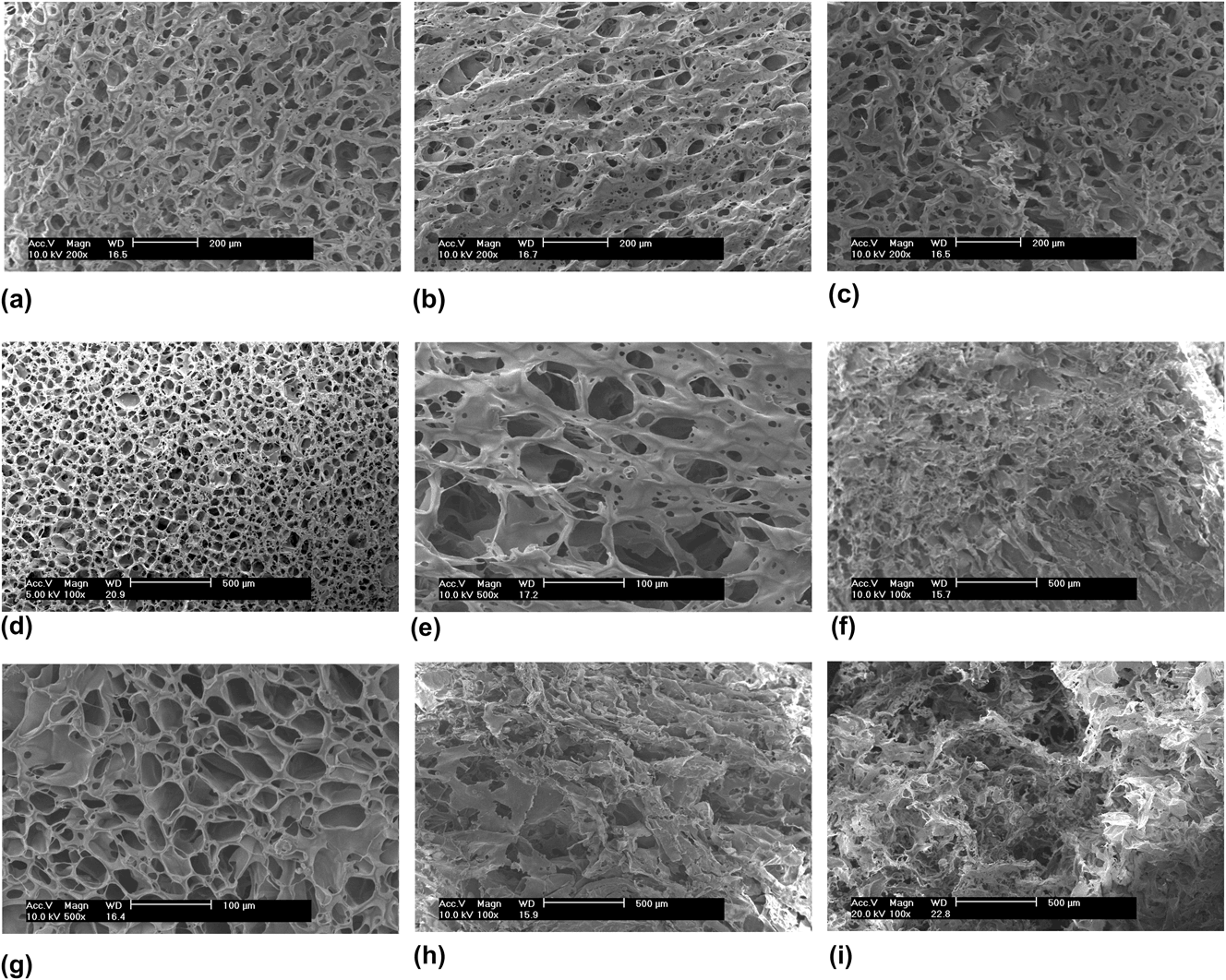
In Fig. 5, blank, 1:20 and 1:2 samples were chosen as examples. As can be seen from these pictures, at 7 d, the surface morphology of all samples did not undergo changes in a visible manner, the pores were still clear. With progress of time, the degradation effect could be observed. At 28 d (1 mo), changes to different extents appeared. The blank sample presented a slightly accidented topography of uneven surfaces and with clear pores whereas the pores of sample 1:20 were irregular and the surface of sample 1:2 had shrunk acutely. At 91 d (3 mo), partial area of sample blank cracked, sample 1:20 became irregular but with discernable ragged edges, and sample 1:2 got blurry. These changes indicate that blank samples degrade the slowest and the degradation effect seems more obvious with increase in the microsphere/chitosan mass ratio. The degradation rate curves support these phenomena (Fig. 6).
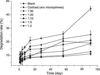
In vitro degradation rate of combined scaffolds with different microsphere/chitosan mass ratios.

In Fig. 6, we added a contrast group, which was chitosan scaffold without microspheres, and we added 0.5 mg/mL lysozyme in the PBS medium in respect of this group. The calculation of the degradation rate was examined by weight loss, which was widely used as the index of in vitro degradation.Reference Lu, Kong, Sheng, Wang, Gong and Zhang6, Reference Freier, Koh, Kazazian and Shoichet7, Reference Cunha-Reis, TuzlaKoglu, Baas, Yang, EI Haj and Reis11, Reference Ren, Yi, Zhang, Xie, Wang and Ma12 Results show that the degradation rate increases when the mass ratio of microspheres-to-chitosan increases. The blank sample was the lowest with degradation rate 11.1% after 3 mo. The numerical value increased from 12.7 to 44.6% after 3 mo when microsphere/chitosan mass ratio increased from 1:50 to 1:2. We also found that the curve of sample 1:2 exhibited an obvious rush point at 28 d, the value increased rapidly from 25.9% (28 d) to 44.6% (91 d). The contrast group showed a different situation. There was a visible turning point at 28 d, the value decreased from 20.5% (28 d) to 19.0% (91 d).
D. In vivo degradation
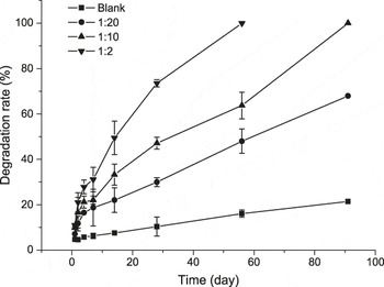
After implantation of the specimens underneath the skin of the mice, the body fluid surrounds and saturates the specimens. The scaffold is degraded under the combined action of body fluid and lysozyme in the microspheres. Figure 7 gives details of degradation rate in vivo. Four groups, including blank, 1:20, 1:10 and 1:2 samples were chosen in this experiment. It must be mentioned that all the animal tests have complied with institutional ethical use protocols of Huaqiao University for care and use of laboratory animals.
In vivo degradation rate of combined scaffolds with different microsphere/chitosan mass ratios.

We can see that the degradation rate in vivo was much faster than that in vitro (Fig. 6). The degradation rate of the blank sample was 21.4% after 3 mo, whereas the 1:2 sample had degraded completely in 2 mo. The 1:10 sample also degraded completely after 3 mo. This is because the body fluid takes part in the degradation process. The other reason is that the PBS may cause amidogen crosslink between chitosan molecules. The existence of resultant insoluble crosslinking may slow down the degradation process in vitro. Besides, the mouse body fluid is in a dynamic mode; the degraded products can be absorbed as glycoproteins or be exhaled as CO2. In respect of in vitro tests, the soluble degraded products still exist in the medium solution; they may cause inhibitory effect and decelerate the degradation when they accumulate to a sufficient concentration. Comparing with the degradation rate in vitro, the degradation rate value of blank, 1:20, and 1:10 improved 0.93 times, 2.63 times and 3.72 times, respectively after 3 mo, and 2.86 times for sample 1:2 after 2 mo. We hypothesize that the improved degradation rate multiples mainly are attribute to the cracking inside the scaffolds. When we increased the microsphere percentage, the degradation inside the scaffolds got faster; finally the inner structure of scaffolds cracked and further accelerated the whole degradation process.
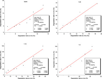
To understand the relationship between in vitro and in vivo degradation, we simulated the correlation by using LineMod model of software Origin Pro 7.0 (Fig. 8). Results showed: blank, y = 2.65093 × (x-3.93055), R2 = 0.85563, r = 0.9250; sample 1:20, y = 5.10307 × (x-5.11406), R2 = 0.96644, r = 0.9831; sample 1:10, y = 6.76294 × (x-7.92769), R2 = 0.94026, r = 0.9697; sample 1:2, y = 5.30067 × (x-9.02569), R2 = 0.93235, r = 0.9656. We checked the critical correlation coefficient, when α = 0.01, r0.01 = 0.8343 (f = n-2 = 8-2 = 6, to blank, 1:20 and 1:10 samples) or 0.8745 (f = n-2 = 7-2 = 5, to 1:2 sample). The relationship between in vitro and in vivo degradation is correlative because r > rα to each group.
Correlation between in vitro and in vivo degradation (microsphere/chitosan mass ratio: blank, 1:20, 1:10 and 1:2).

E. Primary biocompatibility tests
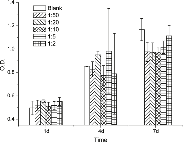
As stated above, we described the preparation of microspheres-loaded chitosan scaffolds and their degradation in vitro and in vivo. But as an appropriate biomaterial, it should have good biocompatibility. So we studied the primary biocompatibility of the combined scaffolds. First, we investigated the material cytotoxicity that could be seen in Fig. 9.
MTT assay on cytotoxicity of osteoblasts on scaffolds with different microsphere/chitosan mass ratios.
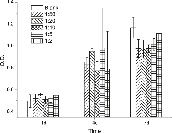
MTT assay is an approach to determine cell viability. The living cells can act with MTT solution and generate insoluble formazan crystals whereas dead cells do not have this function. The absorbance value reflects the number of living cells and is directly proportional to cytotoxicity.Reference Lu, Kong, Sheng, Wang, Gong and Zhang6
Chitosan is a biocompatible biomaterial that has been studied in many publications. Here, we chose blank chitosan scaffold as control. We can see from Fig. 9 that all groups showed improved viability along with prolongation of the culture time. There was no significant difference between the combined scaffold groups and the blank group. These results demonstrate that the chitosan scaffold combined with microspheres has no influence on cell viability. We also studied the cell attachment to the combined scaffolds (Fig. 10).
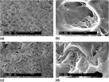
SEM images of combined scaffolds seeded with osteoblasts after 1 wk (a and b) and 2 wk (c and d).

Figure 10 shows the cell proliferation on sample 1:10 combined scaffolds after 1 wk and 2 wk. We can see that the scaffold surface covered with cells and extracellular matrix. Cells were attached across the entire scaffold surface and some had formed bridges across the pores. In addition, cells also penetrated and attached well inside the pores. These images show that the combined scaffolds are favorable to cell attachment and are compatible with the osteoblasts used for this experiment.
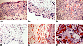
Histology evaluation was performed after the different implantation periods and the obtained results are disclosed in Fig. 11. After 1 wk of implantation of the combined scaffolds, some inflammatory cells, such as lymphocytes and macrophages could be observed in the surrounding skin and subcutaneous tissues. And it was obvious that it had a more extent to subcutaneous tissue than skin tissue in response to the implanted materials. After 4 wk of implantation, the inflammatory phenomena alleviated or disappeared in respect both of skin and subcutaneous tissues. Furthermore, blood vessels [Fig. 11(e)] and fat cells [Fig. 11(f)] were also observed in the space of scaffolds. These features demonstrate that the combined scaffolds were well tolerated by the host and the ingrowth of host blood vessels and fat cells into the scaffolds structure is an evidence of good integration.
HE staining sections of combined scaffolds and surrounding tissues (original magnification, ×200).

IV. CONCLUSION
In this study, we managed to control chitosan scaffold degradation by combination with lysozyme-loaded microspheres. In vitro and in vivo degradation tests indicate that the degradation rate of chitosan scaffolds can be controlled by adjusting the mass ratios of microspheres-to-chitosan. These results show a new strategy to control the chitosan scaffold degradation when the molecular weight and DD are certain parameters. The correlative relationship between in vitro and in vivo data means that the degradation rate in vitro may be predictive of that in vivo. The primary biocompatibility tests show that the scaffolds combined with PLGA microspheres do have little effect on cell attachment and viability. The combined scaffolds have good integration and tolerance to the host. Consequently, the lysozyme-loaded PLGA microspheres are suitable candidates for use in controlling chitosan scaffold degradation in tissue engineering applications.
ACKNOWLEDGMENTS
Financial supports from the Natural Science Foundation of China (31000441, 81171459) are gratefully acknowledged. The authors also thank Dr. Sun Y for the collaboration in the in vivo studies, as well as Dr. Liao W for the initial assistance with the AFM equipment.