Introduction
Worry is a mental process that is widely considered a key component of anxiety (Borkovec et al. Reference Borkovec, Ray and Stober1998) and it is the central feature of generalized anxiety disorder (GAD) as defined by the DSM-IV (APA, 1994; Brown et al. Reference Brown, O'Leary, Barlow and Barlow2001; Fresco et al. Reference Fresco, Mennin, Heimberg and Turk2003). Worry is also frequently associated with depressive rumination (Borkovec, Reference Borkovec, Davey and Tallis1994; Segerstrom et al. Reference Segerstrom, Tsao, Alden and Craske2000; Fresco et al. Reference Fresco, Frankel, Mennin, Turk and Heimberg2002, Reference Fresco, Mennin, Heimberg and Turk2003; Watkins et al. Reference Watkins, Moulds and Mackintosh2005), which is characterized by the predominance of chronic, excessive and uncontrollable negative thoughts (Borkovec, Reference Borkovec, Davey and Tallis1994).
People tend to use worry as a strategy for gaining emotional self-control. However, excessive worry is a fallacious strategy to solve objective and subjective difficulties. In fact, worrying people do not plan complex responses to overwhelming events, rather they tend to repeat to themselves that things will get worse. In figuring out strategies to overcome negative events, worriers use both verbal thinking and visual imagery (Schönpflug, Reference Schönpflug, Spielberger and Sarason1989).
Worry is a prolonged phenomenon, not easy to stop because worrying individuals have tightly organized clusters of worry-related information stored in long-term memory (Pratt et al. Reference Pratt, Tallis and Eysenck1997). This is an interesting feature of worry, in that once the process is triggered, it tends to continue, particularly in GAD patients.
The neural correlates of normal and pathological worry are not fully understood. Functional imaging offers the opportunity to study such neural correlates. To date, there is only one published specific functional magnetic resonance imaging (fMRI) experiment (Hoehn-Saric et al. Reference Hoehn-Saric, Schlund and Wong2004); the authors reported that, before treatment with citalopram, anxiety-inducing sentences elicited stronger brain activations in the frontal cortex and in the cingulate region. However, the relatively small number of subjects and the absence of a placebo-control group and/or normal controls make it difficult to generalize the findings of this study and to evaluate whether the brain regions involved in pathological worry are similar to those involved in worry-like mental activity in normal subjects. In addition, it remains to be established whether brain areas associated with worry induced by verbal stimuli are comparable with those associated with mood induction for other stimuli, for example pictures or faces.
In this paper we report a study aimed at clarifying these issues. Normal and GAD subjects underwent two mood-induction experiments during fMRI scanning: in one experiment we used worry-inducing sentences or neutral sentences; in the second experiment, subjects were asked to empathize with the mood, sad or neutral, represented by faces. Comparisons between groups were made to assess commonalities and/or differences between subjects with GAD and normal controls.
Method
Subjects
One hundred and thirty-five Italian university students (mean age 18–34 years) were recruited for an initial behavioral assessment that involved collection of a general medical, neurological and psychiatric history, and an evaluation of the presence of a abnormal worry using the Penn State Worry Questionnaire (PSWQ; Meyer et al. Reference Meyer, Miller, Metzger and Borkovec1990). The same subjects also completed the Generalized Anxiety Disorder Questionnaire-IV (GAD-Q-IV; Newman et al. Reference Newman, Zuellig, Kachin, Constantino, Przeworski, Erickson and Cashman-McGrath2002). On the basis of the PSWQ (Behar et al. Reference Behar, Alcaine, Zuellig and Borkovec2003) and the GAD-Q-IV scores, subjects were identified as ‘low worriers’ (PSWQ score <45 and negative GAD-Q-IV, score <5.7) or as ‘high-worriers’ (PSWQ score >62 and positive GAD-Q-IV, score >5.7; Newman et al. Reference Newman, Zuellig, Kachin, Constantino, Przeworski, Erickson and Cashman-McGrath2002).
Twenty right-handed subjects, taken from the above sample (12 low worriers and eight high worriers), agreed to take part in the fMRI experiment. For all eight high worriers, the diagnosis of GAD was further confirmed by a clinical psychiatric interview carried out by an experienced psychiatrist (G.M.R.). The purpose of this interview was to confirm, by direct interaction with the patient, beyond the collection of answers to questionnaires, the severity of anxiety and its clinical relevance (DSM-IV criterion E), and the uncontrollable nature of their worries, and to exclude cases with severe anxiety due to different disorders.
All GAD subjects were neither under pharmacological nor psychotherapeutic treatment at the time of testing.
A summary of the characteristics of the subjects who took part in the fMRI study is presented in Table 1.
Demographic and behavioral characteristics of subjects

PSWQ, Penn State Worry Questionnaire; GAD-Q-IV, Generalized Anxiety Disorder Questionnaire-IV; F, female; M, male; s.d., standard deviation.
fMRI experimental design
The fMRI protocol involved two mood-induction tasks, one based on auditory verbal stimuli and another based on faces. In the mood-induction task for sentences, subjects were presented with 12 worry-inducing (WI) sentences (experimental condition) and 12 neutral (N) sentences (control condition), delivered aurally through earphones. The sentences for the two conditions were identified after a pilot study with 24 subjects who rated 72 sentences for their semantic content; the 12 sentences that were unanimously rated as good worry inducers (e.g. think about the subject that most worries you at present) and the 12 sentences that were rated as ‘neutral’ (e.g. think about how the energetic problem could be solved) were adopted. During fMRI scanning, WI sentences were alternated with N sentences. Each sentence represented a 30-s event that was further split into smaller events in an event-related fMRI analysis. A list of representative stimuli is reported in the Appendix. During those 30 s, subjects were asked to listen carefully to the sentences (for 6 s) and to generate thoughts in their inner speech according to the theme of the sentence for the following 15 s. At the end of this time slot, subjects heard a sound and pressed a key on a key-pad to indicate whether they had succeeded in focusing and in generating thoughts on the topic introduced by the sentence. The following 9 s were a resting-state time during which subjects were asked to stop thinking and to relax.
Standardized instructions were: ‘In this task you will be listening to sentences through the earphones. Please try to generate thoughts according to the theme brought up by the sentence. After a few seconds, you will hear a sound to mean that you should stop thinking. Press a key on the key-pad to indicate that you succeeded in thinking according to the instructions. After this, please relax and stop thinking. Another sentence will come up in a short while.’
In the ‘mood-induction’ task with faces, subjects stared at alternating blocks of ‘sad’ or ‘neutral’ faces. Subjects were instructed to empathize with the affective state of the faces. Standardized instructions were: ‘During this task you will see some faces with either a sad or a neutral facial expression. Please try to share the feelings of the faces.’ Each block of stimuli lasted 30 s. There were 12 alternating blocks of stimuli. Half of the subjects performed the task starting with neutral faces, and half started with the sad faces. As this task was completed by only 12 subjects (two high worriers), the fMRI data for this second experiment are presented with within-group analyses only.
fMRI data acquisition and analysis
For each subject we collected 250 complete brain volumes for the ‘sentences task’ and 130 for the ‘faces task’ with a blood oxygen level-dependent (BOLD)/fMRI echo planar imaging (EPI) sequence [repetition time (TR)=3050 ms, echo time (TE)=60 ms; flip angle=90°) using a 1.5-T Marconi Picker Infinion Scanner. The first 10 volumes of each sequence were discarded from the analyses.
After standard image pre-processing (image realignment, stereotactic normalization and smoothing with a 10×10×10 mm Gaussian filter), high-pass filtering and proportional scaling, the experimental conditions were modeled according to an event-related design and condition-specific effects were estimated using SPM2, and the general linear model (Friston et al. Reference Friston, Frith, Turner and Frackowiak1995) by convolving the BOLD response with a canonical hemodynamic response function (HRF).
Mood-induction experiment with sentences
First-level analyses
The following events were modeled: (1) listening to WI sentences; (2) generating thoughts following WI sentences; (3) post-worry resting state; (4) listening to N sentences; (5) generating thoughts following N sentences; and (6) resting state following N sentences. These first-level analyses generated for each subject contrast images containing the information about the fMRI signal change observed for the statistical comparison of interest.
Second-level analyses
To obtain results that can be generalized to a population level, the individual first-level data were then submitted to a second-level random effect analysis (Frison & Pocock, Reference Frison and Pocock1992; Friston et al. Reference Friston, Holmes and Worsley1999). This included a one-way ANOVA for which we entered the contrast images of the effects of the following comparisons:
(1) Simple effects of listening to WI sentences compared with N sentences
(2) Simple effect of worrying compared with neutral thinking
(3) Post-worry resting state compared with resting state following N sentences.
To characterize brain areas of shared activation in the worry-sentences events (listening and generating), a conjunction analysis was performed between the two conditions. These analyses were first performed on the sample of 20 subjects as a whole. We then compared the BOLD response of the two groups of subjects, high worriers and low worriers. The post-scanning debriefing led to an interesting observation, that high worriers systematically reported having had problems in stopping generating ‘worried thoughts’ during the resting state that followed the WI sentences. This observation set the rationale for further statistical fMRI analyses in which we compared the two groups during the post-worry resting state as well. In addition, using a linear regression analysis, we correlated the PSWQ scores, collected outside the scanner during the screening assessment for GAD, with the BOLD response differences derived from the comparisons of the post-worry resting scans versus the resting scans collected after ‘neutral’ thinking.
Mood-induction experiment with faces
This experiment was analyzed according to a standard box-car block design, after convolving the BOLD signal with a canonical HRF as modeled in SPM2. The individual first-level analyses of the comparisons of sad faces minus neutral faces were used for a random effect analysis at a second level in an ANOVA. In the same ANOVA we also entered the data of the sentence-based mood-induction experiment. This permitted the calculation of significant activations associated with mood induction for faces and the conjunctions with the worry-related BOLD response for sentences.
Statistical thresholds
The statistical threshold for all analyses was set at p<0.001. In the tables we indicate which of the foci of activation would survive one of the corrections for multiple comparisons offered by SPM2 [family-wise error (FWE) or false discovery rate (FDR) corrections] or which activation peak belonged to a region of activation significant for spatial extent.
Results
Behavioral results during fMRI
All subjects complied with the instructions during fMRI scanning. The motor response collected after the active thinking phases led us to exclude from the analyses the few trials for which subjects were not able to generate thoughts. In addition, all eight high worriers reported having had problems in stopping their worrying during the resting state that followed the WI sentences.
fMRI results
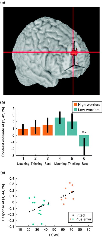
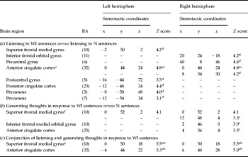
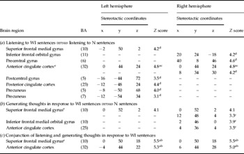
The brain activations associated with the mood-induction experiments are described in Tables 2 and 3 and in Fig. 1.
(a) Illustration of the brain area activated during worry in both high and low worriers while listening to worry-inducing sentences, during active worrying. (b) The same area remained significantly active during the post-worry resting state in the high worriers only. ** Comparison between the two groups for the post-worry resting state scans was significant. (c) The linear regression plot illustrates the blood oxygen level-dependent (BOLD) response in relation to scores on the Penn State Worry Questionnaire (PSWQ).

Functional magnetic resonance imaging (fMRI) results (1)
BA, Brodmann area; WI, worry-inducing; N, neutral.
a Family-wise error (FWE) corrected.
b False discovery rate (FDR) corrected.
c Cluster level corrected.
d Cluster level uncorrected.
e Activation peaks with x stereotactic coordinate 0 are assigned to both sides.
Functional magnetic resonance imaging (fMRI) results (2)
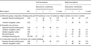
BA, Brodmann area; WI, worry-inducing; N, neutral.
a Family-wise error (FWE) corrected.
b False discovery rate (FDR) corrected.
c Activation peaks with x stereotactic coordinate 0 are assigned to both sides.
Mood-induction experiment with sentences
The within-group analyses over the whole sample showed a significant and enduring activation in the medial regions of the frontal lobe – in the anterior cingulate and in the medial superior frontal gyrus – for the stimuli that induced worry; these regions were active during the presentation of the sentences and during the generation of worry-related thoughts (Table 2).
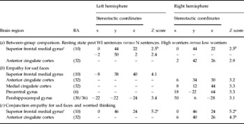
No significant difference emerged between high worriers and low worriers when considering the BOLD response during listening to the WI sentences or when generating worry-related thoughts. However, a difference was found when comparing the post-worry resting state in the two groups. The medial frontal regions associated with worrying in both groups were significantly more active in the post-worry resting state for the GAD high worriers (see Table 3a).
Figure 1 shows a linear regression plot of this BOLD response to illustrate its relationship with the PSWQ scores in the medial frontal region (stereotactic coordinates, Z scores and p values of the two local maxima: x=4, y=44, z=26, Z score=3.0, p=0.001; x=−8, y=48, z=22, Z score=2.9, p=0.002); the plot shows a clear separation of the two groups.
Mood-induction experiment with faces
Analysis of the BOLD response associated with empathy for a sad mood state conveyed by faces showed a very similar activation pattern to that seen with sentences. For faces, however, there was also a significant additional activation in the parahippocampal/amygdala regions (Table 3b). A conjunction analysis of the ‘faces data’ with the ‘sentences data’ confirmed the existence of a shared region for worry-oriented thinking and for empathizing, in the medial frontal and anterior cingulate regions (Table 3c).
Discussion
This simple experiment shows that it is possible to identify specific neural correlates for subtle introspective mental events such as those involved in worry. The brain activations were specific in that they stood over and above activations involved in a similar propositional mental activity such as thinking in response to affectively neutral verbal triggers. Comparison of the brain response in small populations of normal controls and high worriers with GAD allowed us to localize the brain areas involved in ‘normal’ and pathological worry.
The area identified in this study primarily belongs to the mesial frontal cortical region, in the first mesial/dorsal frontal gyrus (area 10) and in the anterior cingulate region. These areas have been implicated in a large repertoire of functions; for example, Ramnani & Owen (Reference Ramnani and Owen2004) discussed the involvement of rostral medial area 10 in ‘processing of internal states’, ‘memory retrieval models’, ‘prospective memory’, ‘branching and re-allocation of attention’, ‘relational integration’ and ‘integration of the outcomes of two cognitive operations’.
Another class of tasks associated with BA 10 activation is particularly interesting in the light of the present study; these tasks may involve ‘reflection on one's own mental states and on mental states of others’ (Frith & Frith, Reference Frith and Frith1999). Because of its involvement with the ‘generation’ or perception of mental states, by analogy with the ventral premotor cortex involvement in motor control and motor observation (Gallese et al. Reference Gallese, Fadiga, Fogassi and Rizzolatti1996), this region has been proposed to be a ‘mirror’ region for mental states (Frith & Frith, Reference Frith and Frith2006). Of note, this medial frontal cortex was active in our subjects when they generated worry-like thoughts introspectively and also when they empathized with the affective state represented by faces. Therefore, our finding reinforces the notion of the medial prefrontal/cingulated region as a ‘mirror’ region for mental states (Frith & Frith, Reference Frith and Frith2006) and suggests that its activation is not dependent on the nature of the stimuli used to induce mental activity.
Of course, much as for the fMRI studies on ‘mirror regions’ in the premotor cortex (Iacoboni et al. Reference Iacoboni, Woods, Brass, Bekkering, Mazziotta and Rizzolatti1999), our statement has a macroscopic validity at a collective level of large populations of neurons. In other words, although it would be possible in principle, we would not want to imply necessarily that the same individual neurons were firing in all conditions (worrying and empathizing), a possibility that could be demonstrated only with invasive single-unit recordings (for a discussion, see Dinstein et al. Reference Dinstein, Thomas, Behrmann and Heeger2008).
According to our results and in the light of the previous functional anatomical literature on the medial frontal cortex in mentalizing, it is plausible to suggest that worry is a dysfunctional way to use and to implement reflective mental functions. These, of course, are not always negative in nature; currently, we do not have sufficient information to distinguish at the neural level between positive and negative mentalizing.
One important question addressed in our study was whether the brain areas involved in pathological worry are distributed similarly in pathological worriers affected by GAD and normal subjects involved in worry-like mental activity. Comparison of the BOLD response during worry-like mental activity of the two groups of subjects did not lead to the detection of any regional difference. This suggests a shared neural system between low and high worriers.
However, when the comparison was made on the BOLD response measured during the post-worry resting state, a clear difference was found between high and low worriers in the same medial prefrontal areas associated with worry-like mental activity for both groups. This correlated with the report made by the subjects of their mental experience during the post-worry resting state scans; low worriers reported having been able to stop their mental activity when asked to do so during the scans, whereas high worriers claimed to have had difficulty in stopping their worrying.
This finding is in keeping with previous psychological investigations. Subjects with GAD are found to be unable to recognize the moment in which it would be useful to stop worrying and believe that it is more appropriate and beneficial to maintain an anxious state of continuous semi-alarm (Borkovec & Inz, Reference Borkovec and Inz1990). This has been associated with the catastrophic beliefs and conceptions of individuals with GAD about the world and life (Beck et al. Reference Beck, Emery and Greenberg1985).
The brain response of the medial prefrontal region during the post-worry resting state, once the post-'neutral-thinking' rest scans had been subtracted, correlated with the PSWQ scores collected during the clinical screening phase of the study days before the fMRI study. This may add some face validity to the scale and further document the relevance of the fMRI finding. Of course, caution should be exercised in making assumptions about a causal relationship between the activity in the medial prefrontal region and a psychiatric condition such as GAD because, when dealing with correlations of this kind, drawing a distinction between causes and effects is difficult, if not impossible. Indeed, we regard this correlation as a neural signature of the ongoing worry activity without implying a causal effect for the presence of the GAD disorder.
It is important to emphasize, once again, that the present study does not lead to a topographical differentiation of the brain regions involved in worry by normal controls and GAD patients, rather it suggest that, at a macroscopic level, the brain regions involved are the same in both classes of subjects.
The absence of a topographical differentiation of the brain regions involved in worry parallels the idea that worry is not qualitatively different in GAD and non-GAD subjects (Borkovec et al. Reference Borkovec, Ray and Stober1998). Pathological worry is in fact a persistent state of cognitive anxiety. Thus, the difference between pathological worry and normal preoccupation is quantitative rather than qualitative. Worry is a disproportionately prolonged preoccupation and not qualitatively different from anxiety (Borkovec et al. Reference Borkovec, Ray and Stober1998).
However, given the spatial resolution of fMRI and the nature of the signal sampled with the technique, it remains plausible that pathological worry is somehow associated with some ‘microscopic’ neural disorder, such as, for example, a neurotransmitter-based dysregulation of the same neural circuitry. The specific neurotransmitter system whose dysregulation may contribute to pathological worry remains to be identified; given current pharmacological approaches used in GAD treatment, one possible candidate may be the serotonergic (5-HT) system (Hoehn-Saric et al. Reference Hoehn-Saric, McLeod and Zimmerli1988; Pollack et al. Reference Pollack, Zaninelli, Goddard, McCafferty, Bellew, Burnham and Iyengar2001). Evidence in this direction has been provided by Hoehn-Saric et al. (Reference Hoehn-Saric, Schlund and Wong2004) in a group of six GAD patients tested before and after 7 weeks of treatment with a selective serotonin-reuptake inhibitor, citalopram.
Given the distribution of the worry-related cortical network, we can also speculate on a possible role for the ventro-tegmental subdivision of the dopaminergic (DA) pathway, which projects primarily to the medial dorsal prefrontal cortex (Williams & Goldman-Rakic, Reference Williams and Goldman-Rakic1998; Frankle et al. Reference Frankle, Laruelle and Haber2006). Several pharmacological studies also suggest the possibility of a pathological interaction between the 5-HT and the DA systems in depression and anxiety (e.g. Alex & Pehek, Reference Alex and Pehek2007). Psychopharmacological fMRI studies and positron emission tomography (PET) studies with specific ligands may help to clarify the role of these systems in the dysregulation of the medial prefrontal cortex in pathological anxiety.
Appendix. Examples of stimuli used for the worry experiment based on sentences
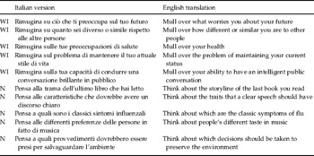
WI, Worry-inducing sentence; N, neutral sentence.
Declaration of Interest
None.