Introduction
Nearly all fish are dependent on vision, to varying degrees, for performing fundamental tasks such as foraging, avoiding predation and mate selection (Guthrie, Reference Guthrie and Pitcher1986). The acquisition, transfer and processing of visual information is part of a complex network of neuronal interactions essential for perception, decision-making and response (Wagner, Reference Wagner2011). When internal or external influences interfere with this process, the ability of a fish to observe and react to environmental cues can be degraded drastically. For example, exposure to certain toxins can result in neuronal stress in the visual processing region of the brain, resulting in fundamental changes to the fish's response to stimuli (Kohler et al., Reference Kohler, Zrenner and Weiler1995). Vision impairment through events such as increased turbidity or prolonged darkness can severely hinder the foraging ability and predator avoidance behaviours of affected individuals (Reid et al., Reference Reid, Fox and Whillans1999; Rowe et al., Reference Rowe, Dean, Williams and Smith2003; Knudsen et al., Reference Knudsen, Klemetsen, Amundsen and Hermansen2006).
Neuronal activity, and thus visual information transfer, can be measured by looking at the expression of genes associated with neuron activity, such as c-Fos. The c-Fos gene is conserved across all vertebrates and expressed as a result of neuronal activation, thus measurement of the resulting upregulated mRNA or Fos proteins is an indirect marker of recent neuronal activity (Dragunow and Faull, Reference Dragunow and Faull1989; VanElzakker et al., Reference VanElzakker, Fevurly, Breindel and Spencer2008; Wiedmann et al., Reference Wiedmann, Rosahl, Brinker, Samii and Nakamura2013; Pacheco et al., Reference Pacheco, Tilden, Grutzner, Lane, Wu, Hengen, Gjorgjieva and Turrigiano2019). In fish model systems, c-Fos expression has been used to determine the effect of a variety of stimuli, from toxins/pollutants to aggression displays, on the central nervous system (CNS) of exposed individuals (Salierno et al., Reference Salierno, Snyder, Murphy, Poli, Hall, Baden and Kane2006; Wai et al., Reference Wai, Lorke, Webb and Yew2006; Topal et al., Reference Topal, Atamanalp, Oruç, Halıcı, Şişecioğlu, Erol, Gergit and Yılmaz2015; Almeida et al., Reference Almeida, Felix, Oliveira, Lopes and Oliveira2019). Rainbow trout Oncorhynchus mykiss, for example, show increased c-Fos expression in the cerebral and cerebellar regions of the brain when exposed to nickel chloride (Topal et al., Reference Topal, Atamanalp, Oruç, Halıcı, Şişecioğlu, Erol, Gergit and Yılmaz2015). The expression of the c-Fos protein expression is altered in the telencephalon and the periaqueductal grey region of the optic tectum of the killifish Fundulus heteroclitus when they are exposed to various neurotoxins associated with harmful algal blooms (Salierno et al., Reference Salierno, Snyder, Murphy, Poli, Hall, Baden and Kane2006).
Measurement of c-Fos expression has revealed that parasitic organisms can make alterations to the CNS in non-fish hosts. By measuring c-Fos expression, Morales-Montor et al. (Reference Morales-Montor, Arrieta, Del Castillo, Rodríguez-Dorantes, Cerbón and Larralde2004) observed changes in hypothalamic and hippocampal activity in Taenia solium-infected mice, which they argued may alter hormonal regulation. The measurement of c-Fos expression can also be used to investigate the underlying mechanisms of parasite-mediated behaviour. House et al. (Reference House, Vyas and Sapolsky2011) used c-Fos expression to reveal altered neural activity in the limbic region's sexual arousal pathways of Toxoplasma gondii-infected rats exposed to cat urine, the parasite's definitive host. Though it had previously been shown that T. gondii-infected rats can be attracted to the odour of cats (Berdoy et al., Reference Berdoy, Webster and Macdonald2000), this study provided insight into the mechanism of the altered behaviour.
Diplostomid trematodes often reside in the eyes of fish as metacercariae where they can inflict variable levels of visual impairment, at times due to direct damage to the host's tissues (Paperna, Reference Paperna1991; Williams and Whitaker, Reference Williams and Whitaker1997; Grobbelaar et al., Reference Grobbelaar, van As, van As and Butler2015; Blasco-Costa and Locke, Reference Blasco-Costa and Locke2017; Locke et al., Reference Locke, Van Dam, Caffara, Pinto, Lopez-Hernandez and Blanar2018). For example, Diplostomum spp. often reside in the lens of their host resulting in the formation of cataracts, with their opacity increasing with intensity of infection, therefore further reducing vision (Chappell, Reference Chappell1995; Wall, Reference Wall1998; Karvonen et al., Reference Karvonen, Seppälä and Valtonen2004; Fig. 1B). Considering the important role vision plays in the environmental perception of many fish species, it is not surprising that parasite-impaired vision has been shown to hinder the host in a variety of ways (Guthrie, Reference Guthrie and Pitcher1986; Owen et al., Reference Owen, Barber and Hart1993; Karvonen et al., Reference Karvonen, Seppälä and Valtonen2004; Seppälä et al., Reference Seppälä, Karvonen and Valtonen2004, Reference Seppälä, Karvonen and Valtonen2005, Reference Seppälä, Karvonen and Valtonen2008; Voutilainen et al., Reference Voutilainen, Figueiredo and Huuskonen2008; Ubels et al., Reference Ubels, DeLong, Hoolsema, Wurzberger, Nguyen, Blankespoor and Blankespoor2018). It has been demonstrated that rainbow trout O. mykiss infected by Diplostomum spp. are more susceptible to predation and form less cohesive shoals compared to uninfected fish (Seppälä et al., Reference Seppälä, Karvonen and Valtonen2004, Reference Seppälä, Karvonen and Valtonen2008). A further example of this is the reduced foraging success of Arctic charr Salvelinus alpinus infected with Diplostomum spathaceum, as evidenced by reduced prey consumption and delayed reaction time to prey in individuals with greater cataract opacity (Voutilainen et al., Reference Voutilainen, Figueiredo and Huuskonen2008). Closely related diplostomids in the genus Tylodelphys reside in the humours of the eye rather than the lens and as such do not cause obvious pathology in the form of cataract formation (Stumbo and Poulin, Reference Stumbo and Poulin2016; Muñoz et al., Reference Muñoz, Staaks and Knopf2017, Reference Muñoz, Bierbach and Knopf2019). However, it has been shown recently that Tylodelphys clavata impairs the reaction distance of infected fish to prey and even reduces their ability to compete with uninfected conspecifics (Muñoz et al., Reference Muñoz, Staaks and Knopf2017, Reference Muñoz, Bierbach and Knopf2019). Tylodelphys spp. when present in the humours move about freely (Blasco-Costa et al., Reference Blasco-Costa, Poulin and Presswell2017), and as such if there is physical pathology associated with infection it is likely to be more subtle than Diplostomum spp. causing cataracts in the lens.
(A) Light refraction through spherical lens in healthy fish eye. (B) Hypothetical light refraction in fish eye with cataracts in the lens. (C) Hypothetical light disruption by metacercariae moving freely within humours of a fish eye. (D) Hypothetical light disruption by semi-transparent metacercariae layered between the lens and the retina of a fish eye.

In New Zealand, the only described diplostomid, Tylodelphys darbyi, infects the eyes of its second intermediate fish host, the common bully Gobiomorphus cotidianus (Blasco-Costa et al., Reference Blasco-Costa, Poulin and Presswell2017; Presswell and Blasco-Costa, Reference Presswell and Blasco-Costa2020). Tylodelphys darbyi is relatively large (>1 mm) in comparison with other species of the genus and freely move about the vitreous chamber of the eye between the lens and the retina (Blasco-Costa et al., Reference Blasco-Costa, Poulin and Presswell2017). There does not appear to be any physical pathology associated with T. darbyi infection, but the parasite does appear to obscure vision by occupying the open region of the vitreous chamber (Fig. 1; Stumbo and Poulin, Reference Stumbo and Poulin2016). During the day, the parasite obstructs ~75% of the observed retinal surface while at night T. darbyi settles to the lower portion of the eye, reducing obstruction to ~30% (Stumbo and Poulin, Reference Stumbo and Poulin2016). It was suggested by Stumbo and Poulin (Reference Stumbo and Poulin2016) that this diel behaviour could cause fish to be predated at higher rates due to increased visual obstruction. However, it has recently been demonstrated that while fish spend more time actively moving about in the open with higher infection levels there does not appear to be an impact of the parasite on anti-predator behaviour (Ruehle and Poulin, Reference Ruehle and Poulin2019; Ruehle and Poulin, Reference Ruehle and Poulin2020). Furthermore, Stumbo and Poulin (Reference Stumbo and Poulin2016) postulated that the metacercariae were layering within the chamber, i.e. lying superimposed instead of adjacent to each other, as the amount of retinal surface area obstruction was independent of parasite intensity (Fig. 1D). Thus, the amount of light reaching the retina may be determined by the number of T. darbyi metacercariae within the eye of an infected fish due to layering and not overall retinal obstruction. If this is the case, then it is possible that impairment of visual information transfer would increase at higher infection levels. Therefore, we hypothesize that the number of T. darbyi metacercariae not only affects the amount of light reaching the retina, due to layering of individuals, but also influences neuronal activity induced by this light stimulus, measured as c-Fos expression, in the visual processing region in the brain of infected bullies. Here, we test the prediction that with decreased light and visual information reaching the retina, regions of the brain involved in visual processing would be less active and thus show lower c-Fos levels.
Materials and methods
Infected bullies were collected with minnow traps from Lake Hayes (44°58′08.0″S, 168°48′44.6″E) in Central Otago, New Zealand, in February of 2016, and transported to the University of Otago in aerated coolers. The fish were housed together in 30-L aquaria for 2 weeks, at 11 ± 2°C and with a 12/12 L/D light cycle, and fed commercial pellets (Ridley AquaFeeds Pty Ltd., Narangba, Qld., Australia) ad libitum. The 2-week holding period should have ensured that most metacercariae had reached infectivity to their avian definitive host. Tanks were provided with PVC pipe shelters (4 cm diameter × 10 cm length) to reduce stress and aggression, and a 75% water change was performed on alternate days. The fish ranged between 45 and 94 mm in length (mean ± s.d. = 63 ± 13 mm), and were selected at random for the following tests.
Light exposures
Fish (n = 22) were placed individually into 1 L beakers with 500 mL of water. The exterior of each beaker was wrapped in opaque tape to prevent fish from observing external information. Black plastic sheeting was placed on all sides of the shelving unit on which the beakers were maintained, limiting outside light, which itself was kept a low enough level to minimize visual stimulation while still allowing the observer to perform the experiment. After a 2 h acclimation period, a flashing light (135 lumens, 10 Hz) positioned 20 cm above the containers was activated from outside the black plastic sheeting for 15 min. Control fish (n = 8) were treated in the same way except for no light stimulus was provided.
Tissue preparation and parasite recovery
Following the experiments, fish were euthanized with MS-222 (tricaine mesylate, 1 g L−1) 45-min after trials, allowing time for the upregulation of the c-Fos protein, and then dissected for parasite load and removal of the brain. Tylodelphys darbyi metacercariae were recovered from the humours in the eyes during dissection under a microscope. The cranium was separated from the body and the dorsal portion was dissected away, exposing the brain, which was then prepared following methods modified from Bosch et al. (Reference Bosch, Maslam and Roberts2001) and Topal et al. (Reference Topal, Atamanalp, Oruç, Halıcı, Şişecioğlu, Erol, Gergit and Yılmaz2015) for later observation of c-Fos activity. Once removed from the cranium, brains were fully submerged in 10% neutral buffered formalin containing 2.5% acrolein for 12 h. After this period, the brains were fully removed and placed into a fresh solution of 2.5% acrolein for 2 h. Brains were then transferred to paraformaldehyde for 2 h, followed by a series of four 15 min washes in potassium phosphate-buffered saline (KPBS). Following this, brains were placed into a 30% sucrose solution until embedding.
Once brains had sunk to the bottom of the solution, indicating full sucrose saturation, they were placed individually into histology moulds containing Cryomatrix™ freezing medium, and frozen using liquid nitrogen-cooled isopropyl alcohol and stored at −80°C. Sectioning was performed using a cryomicrotome (Thermo Scientific, Isceon 89). The sections were cut to 8 μm thickness along the dorsal plane, then mounted onto slides (4/slide) and stored at −80°C.
Immunohistochemistry
Tissue sections of the optic tectum mounted on slides were randomly selected for each fish. Initially, slides were placed in distilled water and incubated at 30°C for 60 min to remove Cryomatrix™, and then rinsed in KPBS for 2 min with mild agitation. A hydrophobic circle was drawn around the tissue sample with a PAP pen before the remaining steps were performed on the slides. A 3-fold, 2 min rinse with KPBS was performed between the application of each solution unless otherwise noted. In order to reduce non-specific background staining, endogenous peroxidases were blocked by incubation with 0.3% H2O2 in 10% methanol in KPBS for 30 min. Incubation with Tween™ (polyoxyethylene (20) sorbitan monolaurate) was then carried out for 5 min to increase the permeability of the cells. A 20 min incubation in 2% normal horse serum in KPBS (Sigma-Aldrich, MO, USA) was used to further reduce non-specific binding. The primary antibody (polyclonal goat anti-c-Fos, 1:200 in 2% normal horse serum, Santa Cruz Biotechnology, TX, USA) was then immediately applied to the slides, which were left to incubate at room temperature for 60 min, and then at 8°C for 48 h.
Following incubation with the primary antibody, the secondary antibody (biotinylated horse anti-goat, 1:500 in KPBS, Vector Laboratories, CA, USA) was applied to the tissue and left to incubate at room temperature for 60 min. An avidin–biotin complex (Vectastain ABC standard kit, Vector Laboratories, CA, USA) was then applied for 60 min. Finally, nickel-DAB chromogen was applied for 20 min to allow for visualization of c-Fos expression and then rinsed with distilled water. Slides were left to dry for 12 h in the dark, after which mounting solution (Shandon IMMU-MOUNT, REF 990402) was applied with a cover slip and sealed with a clear lacquer.
Data analysis
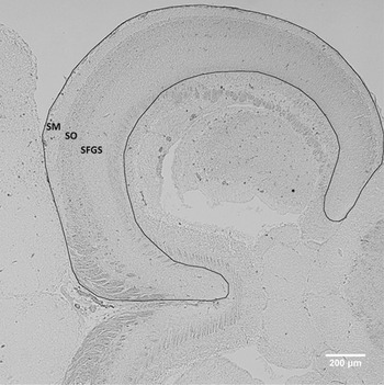
Images of the optic tectum sections were acquired using a compound microscope (Olympus BX51) at 10× magnification, with a mounted camera (Olympus U-TV0.5XC-3). Analysis focused on the stratum marginale (SM), stratum opticum (SO) and stratum fibrosum et griseum superficiale (SFGS) (Fig. 2), as these regions are predominantly associated with vision and directly linked to the retinal ganglion cells (RGCs), which are the neurons responsible for transmitting visual information from the eye to the brain (Northmore, Reference Northmore2011). The periaqueductal grey region was disregarded as it was prone to tearing during histology and is largely a motor area. ImageJ (1.48v, U.S. National Institutes of Health, Bethesda, Maryland, USA) was used to determine the area of the analysed tissue (Fig. 2). c-Fos is expressed in cell nuclei, and these nuclei are revealed as black dots in stained tissue (Fig. 3). The number of nuclei with increased expression of c-Fos was counted manually. A generalized linear model with the quasi-Poisson distribution was performed with R 3.2.3 (R Core Team, 2016) with the number of metacercariae (mean of both eyes) per fish and treatment (control or light exposed) used as predictors, and the number of c-Fos + nuclei per mm2 of tissue treated as the response. Fish length was treated as a proxy for age and used as a covariate.
Histological section of the brain of a common bully Gobiomorphus cotidianus. The outline indicates the visual processing region investigated for neural activation via c-Fos protein staining, comprising the SM, SO and SFGS as defined by Northmore (Reference Northmore2011).

Histological section of common bully G. cotidianus brain after immunohistochemistry staining. Each black dot represents the expression of the c-Fos protein in a cell nucleus, indicating recent neuron activity.

Ethical statement
We used naturally infected fish for our experiments, because the complete life-cycle of T. darbyi is not known. For this reason, we aimed to minimize our sample sizes while keeping them large enough to achieve statistical requirements. Gobiomorphus cotidianus is the most abundant and widespread endemic, freshwater fish in New Zealand (McDowall, Reference McDowall1990) and >100 can be recovered in a single trap night, so the numbers we used in this experiment (n < 40) are not likely to impact the population. In addition, the Lake Hayes population has a T. darbyi prevalence of near 100% (Stumbo and Poulin, Reference Stumbo and Poulin2016), allowing us to collect smaller numbers of fish and still guarantee infection. Fish were kept in holding tanks at stocking densities of <2 L per fish and provided with enough shelter (i.e. 10 cm PVC pipes) that each individual had a territory to itself. Common bullies take longer to acclimate (e.g. pale colouration) to holding conditions when not in a group and without adequate shelter (author's per. observation), so these conditions were intended to reduce stress. Fish were held in captivity for no more than 4 months and at the end of the experiment all individuals were humanely killed via overdose of MS-222 (1 g L−1). These methods were approved by the University of Otago Animal Ethics Committee (no. 52/15).
Results
The mean number of T. darbyi metacercariae per fish among bullies exposed to the flashing light stimulus ranged from 0 to 17.5, with an average ± s.d. of 2.7 ± 4.1 metacercariae. One individual possessed an average of 55 metacercariae per eye, and therefore was treated as an outlier and removed from further analysis. Metacercariae numbers in control fish ranged from 0 to 14.5, with an average ± s.d. among individuals of 5.75 ± 5.5.
The model revealed a significant difference in the number cells exhibiting c-Fos expression between treatment groups (t = 2.059, P = 0.047; Fig. 4), with control fish possessing fewer c-Fos-expressing cells. The number of metacercariae was not a significant predictor of c-Fos expression among control fish (t = 0.402, P = 0.690). The number of metacercariae among fish exposed to the light stimulus, however, was a highly significant predictor (t = 6.075, P < 0.001), with a positive relationship observed between metacercariae number and increased c-Fos expression. There was no significant interaction between metacercariae number and treatment (t = 1.168, P = 0.251). Fish length also had no significant effect on c-Fos expression (t = 0.153, P = 0.879).
Neuronal activity as indicated by the number of cell nuclei showing c-Fos expression as a function of mean metacercariae number per eye in Tylodelphys darbyi-infected G. cotidianus. Fish were either subjected to a visual stimulus (exposed) or left undisturbed (control).

Discussion
Tylodelphys darbyi is large (>1 mm) and tends to inhabit the lower portion of the vitreous chamber, extending into the central region of the vitreous chamber in a parallel orientation to the lens (Stumbo and Poulin, Reference Stumbo and Poulin2016; Blasco-Costa et al., Reference Blasco-Costa, Poulin and Presswell2017). This coupled with the lack of a significant effect of metacercariae number on the amount of retinal obstruction (Stumbo and Poulin, Reference Stumbo and Poulin2016) indicates sagittal layering of metacercariae between the lens and the retina. We hypothesized that with increasing T. darbyi intensity, the amount of visual stimuli reaching the retina would decrease due to this layering of metacercariae regardless of the amount of overall retinal obstruction. It was expected, as a result, that with decreased light and visual information, regions of the brain involved in visual processing would be less active (i.e. lower c-Fos expression). Interestingly, we found the opposite pattern, i.e. an increase in c-Fos expression, and therefore neuronal activity, with higher parasite intensities, possibly suggesting that fish are receiving an overabundance of visual information. Although this may seem counterintuitive, it suggests a mechanism for parasite-induced visual disruption when no obvious pathology is induced by the parasite. Simply, we propose that the more parasites present in the eyes, the more visual stimuli the fish is presented with, and as such the fish may experience difficulties in distinguishing various features of its external environment. Ruehle and Poulin (Reference Ruehle and Poulin2020) showed that at higher T. darbyi infections the amount of time a bully spent moving about in the open increased, potentially due to a greater need to explore their environment compared to those with fewer parasites. Our findings here provide a possible mechanism for this by showing increased neuronal activity with higher infection levels.
A by-product of infection, such as the metabolic waste of metacercariae, may be responsible for c-Fos protein expression pattern in a similar manner to when fish are exposed to toxins like pentylenetetrazole or nickel chloride (Baraban et al., Reference Baraban, Taylor, Castro and Baier2005; Topal et al., Reference Topal, Atamanalp, Oruç, Halıcı, Şişecioğlu, Erol, Gergit and Yılmaz2015), with higher levels of infection resulting in an accumulation of the by-product and increased protein expression. This seems unlikely, however, considering that c-Fos expression was significantly lower in control fish not exposed to the light stimulus and was not linked to metacercariae number in these fish. Another possibility is that with the light stimulus, the metacercariae are causing a stress response in the fish. Stress from handling (Salierno et al., Reference Salierno, Snyder, Murphy, Poli, Hall, Baden and Kane2006) or a startle response (Bosch et al., Reference Bosch, Maslam and Roberts2001) has been shown to result in a greater number of cells with c-Fos expression, though this was in the periaqueductal grey region and the telencephalon which are primarily motor regions of the brain (Northmore, Reference Northmore2011). Also, the same studies did not find elevated c-Fos expression in the visual region of the optic tectum (SFGS, SO), the area analysed here (Northmore, Reference Northmore2011). This suggests that the observed upregulation of c-Fos in exposed fish is more likely a result of an increased amount of information being transferred from the RGCs to the optic tectum, with the metacercariae themselves acting as a source of visual information.
Light passing across the boundary between two media with different refractive indices is refracted to varying degrees, altering the light's course. Discounting the unlikely possibility that a metacercaria has the exact same refractive index as the vitreous humour surrounding it, the angle of the light passing through a parasite must be altered. This scattering of light would also likely be cumulative, intensifying with greater numbers of metacercariae. Additionally, the movement of the metacercariae must result in a change in the number of parasite bodies the light must pass through to reach the photoreceptors at any retinal location over time. These two mechanisms (light refraction and shifting light intensity) may produce a more complex stimulus than simply the flashing light alone, resulting in a greater amount of visual information proportional to the number of parasites per eye.
Visual acuity is dependent on a complex system of information processing starting within the eye. Briefly, visual information is first received by the photoreceptors, relayed to the bipolar cells and subsequently to the RGC. However, two additional cell types, the horizontal cells and amacrine cells, can integrate and modulate information at the photoreceptor–bipolar cell and bipolar cell–RGC interfaces, respectively (Levine, Reference Levine2011). Thus, by the time a visual signal is received by the RGCs, significant processing has already taken place. These photoreceptors are also tightly packed within the retina, allowing for greater spatial resolution (Hirsch and Curcio, Reference Hirsch and Curcio1989). The receptive field of archer fish Toxotes jaculatrix, for example, is as low as a tenth of a degree in certain regions of the retina, allowing for high-sampling frequency (Ben-Simon et al., Reference Ben-Simon, Ben-Shahar, Vasserman, Ben-Tov and Segev2012). Even slight changes in the angle of received information would result in reduced spatial resolution across the photoreceptor layer. It is likely, then, that T. darbyi metacercariae, although increasing the amount of visual information being processed, are decreasing the quality of the external information via information saturation.
The visual cortex is tied to other processing regions of the brain, and the observed increase in neuronal activation may have consequences for T. darbyi-infected bullies beyond impaired visual processing. A simplified example is that neuron activity in the SFGS is relayed to the torus longitudinalis (TL), which then signals the SM, resulting in an open loop between the optic tectum and the TL (Northmore, Reference Northmore2011). The TL, as well as being part of visual processing, is part of eye movement control and is linked to the premotor cortex (Northmore et al., Reference Northmore, Williams and Vanegas1983; Wullimann, Reference Wullimann1994). Prolonged neural stimulation in the visual cortex can also lead to neural fatigue, weakening neural response and visual acuity (Maffei et al., Reference Maffei, Fiorentini and Bisti1973; Carandini, Reference Carandini2000). Neuronal stress resulting from visual information saturation may then weaken the visual processing ability of the fish, in turn impairing or altering its motor control.
Here, we have shown that fish harbouring T. darbyi demonstrate increased c-Fos expression at higher intensities of infection. We propose that high numbers of metacercariae in the eyes of fish result in increased scattering of incoming light before it reaches the retina, overwhelming the fish's visual processing centres. Although our results do not allow us to conclude that the amount of information reaching the RGC changes due to T. darbyi intensity, it is possible the quality of any external information will be reduced. This lends support to the suggestion that infection can impair the fish's ability to detect approaching predators presented in Stumbo and Poulin (Reference Stumbo and Poulin2016) even though no impact has been observed thus far (Ruehle and Poulin, Reference Ruehle and Poulin2019). Furthermore, our findings provide a possible mechanism for why higher intensities of T. darbyi correlate with fish spending more time moving in the open (Ruehle and Poulin, Reference Ruehle and Poulin2020).
Financial support
A. Stumbo and B. Ruehle were supported by the University of Otago Doctoral Scholarship.
Conflict of interest
The authors declare no conflict of interest.
Ethical standards
These methods used were approved by the University of Otago Animal Ethics Committee (no. 52/15).