Introduction
Primary benign non-lymphoid neoplasms of lymph nodes are uncommon. Neurilemmomas arising within lymph nodes are rare and have only recently been differentiated from other spindle cell tumours.Reference Weiss, Gnepp and Bratthauer1 A search of the literature found only two previously reported cases of true intranodal neurilemmomas.Reference Griffiths, Ironside and Gray2, Reference Piana, Gelli, Cavazza, Serra and Gardini3 We present a case of an intranodal neurilemmoma presenting as a parotid mass.
Case report
Clinical aspects
A 67-year-old woman presented to the out-patients department with a six-month history of a slowly enlarging, non-tender left parotid mass. The patient had no other associated symptoms but was concerned about the cosmetic appearance of the lump. There was no significant past medical history, and no family history of any head and neck pathology or familial diseases. The patient was a non-smoker with a low alcohol intake.
Clinical examination revealed a 4 cm, ovoid mass in the left parotid gland. The lesion was well circumscribed, mobile and non-tender. The skin covering the tumour was normal and moved freely. The remainder of the examination, including flexible naso-endoscopy, was unremarkable.
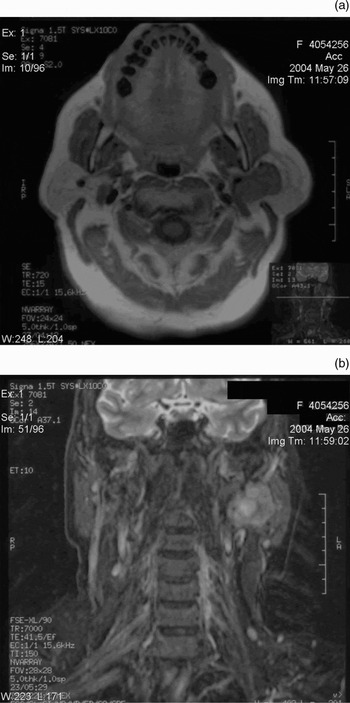
A fine needle aspiration (FNA) was performed but the specimen was non-diagnostic. A second FNA was also non-diagnostic, so imaging was requested prior to surgical excision. A magnetic resonance imaging scan demonstrated a mass within the superficial lobe of the left parotid gland (Figure 1).
(a) Axial, T1-weighted magnetic resonance image demonstrating a lobulated, intermediate-signal mass within the superficial part of the left parotid gland, displacing the retromandibular vein medially. (b) Coronal STIR image of the same high-signal mass.

At surgery, an unusual, firm, multinodular lesion was found within the superficial lobe of the left parotid gland. A lateral conservative parotidectomy was performed. The well circumscribed tumour was excised complete with the superficial lobe of the parotid.
There were no post-operative complications, and facial nerve function was unaffected. The patient was discharged home the following day.
At followup, the wound had healed well. After eight months, there was no sign of local recurrence or further lesions.
Histopathological findings
Macroscopically, a firm, whitish yellow, 42×28×19 mm, multinodular tumour was observed attached to the superficial parotid gland by a thin core of parotid tissue. Serial sectioning of the parotid gland revealed normal salivary gland tissue and a normal intraparotid lymph node.
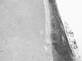
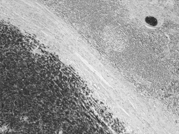
Microscopy of the tumour revealed blunt-ended, spindled nuclei with cytoplasmic vacuolation, arranged in bundles and fascicles. At the periphery of the tumour, there was well defined lymphoid tissue with all the constituents of a lymph node, including a peripheral sinus and capsule (Figure 2). There was no evidence of necrosis, and mitotic activity was absent. Typical of a neurilemmoma, the tumour showed moderate cellularity and was composed of spindled, focally ‘wavy’ nuclei (Figure 3) with areas of Antoni A and Antoni B morphology. A diffuse if modest chronic inflammatory cell infiltrate and areas of hyalinisation were seen within the tumour. These features and foci of nucleomegaly were deemed typical of ‘ancient neurilemmoma’.
Low power photomicrograph demonstrating a small amount of parotid parenchyma (right), the edge of a lymph node and the very well circumscribed tumour (left) (Hematoxylin-eosin; ×12.5).

High power photomicrograph of the tumour, demonstrating moderate cellularity with spindled and focally ‘wavy’ nuclei (Hematoxylin-eosin; ×100).

Immunohistochemical analysis demonstrated that the tumour was strongly positive for S100 (Figure 4) and glial fibrillary acidic protein, and negative for desmin and α smooth muscle actin. The tumour was also negative for epithelial markers, CD68, CD117, synaptophysin and HMB-45. This confirmed that the tumour was of nerve sheath origin, and a definitive diagnosis of a parotid intranodal neurilemmoma was made.
S100 immunohistochemical analysis, demonstrating strong positivity of the tumour and also positivity of a small peripheral nerve (top right) (× 50).

Discussion
Neurilemmomas arise from the nerve sheath and consist of Schwann cells in a collagenous matrix. The two main growth patterns are described as Antoni types A and B. Type A tissue has elongated spindle cells arranged in irregular streams and is compact in nature. Type B tissue has a looser organisation, often with cystic spaces intermixed within the tissue.Reference Piana, Gelli, Cavazza, Serra and Gardini3 The cystic spaces can result in high signal intensity on T2-weighted magnetic resonance imaging. Tumours originating in Schwann cells can be detected at immunohistochemical examination by virtue of their positive staining for S100 antigen.
Weiss et al. reviewed a number of cases previously thought to be neurilemmomas arising in lymph nodes.Reference Weiss, Gnepp and Bratthauer1 Further investigation revealed strong immunohistochemical reactivity to smooth muscle actin and desmin and negativity to S100. This, along with the presence of stellate areas of collagen deposition (‘amianthoid fibres’), suggested a diagnosis of palisaded myofibroblastoma, a distinct spindle cell tumour. The authors proposed that true S100-positive neurilemmoma was a distinct and much rarer histological entity.
Neurilemmomas arising within lymph nodes are very rare.Reference Weiss, Gold blum, Weiss and Goldblum4 A literature search of Medline revealed only two previous case reports of true intranodal neurilemmomas.Reference Griffiths, Ironside and Gray2, Reference Piana, Gelli, Cavazza, Serra and Gardini3 One report was of a two-year-old infant with a neurilemmoma arising in a subscapular lymph node. The other reported an elderly patient with a neurilemmoma arising within a mesenteric node, found incidentally following bowel resection for an adenocarcinoma.
Our case demonstrates the features typical of a true intranodal neurilemmoma, namely, a tumour of neural origin with ill-defined Antoni A and B areas consisting of spindle cells strongly positive for S100, encapsulated by normal lymphoid tissue. The tumour showed no evidence of amianthoid fibres, distinguishing it from the more common palisaded myofibroblastoma. A further intriguing feature in this case was that the tumour arose within a lymph node, itself within the parotid salivary gland, thus presenting as a parotid gland mass.
• We report a 67-year-old woman who underwent surgery for a parotid mass
• The mass was found to be a true benign intranodal neurilemmoma, an extremely rare tumour arising from a nerve sheath within a lymph node
• These tumours can be differentiated from other intranodal neoplasms by their characteristic microscopic appearance and positive staining for S100 antigen
• This case report is the first of its kind, and supports the proposal that intranodal neurilemmoma be considered as a distinct histological entity
Primary benign non-lymphoid neoplasms of lymph nodes are uncommon.Reference Warnke, Weiss, Chan, Cleary and Dorfman5 The differential diagnosis of intranodal spindle cell tumours includes proliferative lesions derived from smooth muscle cells, such as leiomyoma, angiomyolipoma, leiomyomatosis and lymphangiomyomatosis, all of which show immunoreactivity to smooth muscle actin. It is important to distinguish these benign tumours from lymph node metastases of carcinomas, melanomas and other malignant tumours, which usually show marked cellularity, mitotic activity and cytological pleomorphism, together with different immunohistochemical staining patterns. The treatment of a benign neurilemmoma therefore relies on accurate histological diagnosis. Complete surgical excision is adequate as recurrence is rare, and neurilemmomas rarely show malignant transformation.Reference Wright and Jackson6
Conclusion
We report a case of a neurilemmoma arising within a parotid lymph node. The lesion showed characteristic morphological and immunohistochemical features of an intranodal neurilemmoma. This finding supports the proposal of a histological entity distinct from the more common palisaded myofibroblastoma.
Acknowledgements
We thank Dr Jane Ferguson, Specialist Registrar in Radiology, Royal Devon and Exeter Hospital, for her assistance with the magnetic resonance images, and Mr Nigel Drury, University of Birmingham, for his advice on the manuscript.