Introduction
Epistaxis provides a challenge to all otorhinolaryngologists and is the most common ENT emergency. Sixty per cent of the general population experience epistaxis at some point in their lifetime and 6 per cent of these will seek medical attention.Reference Supriya, Shakeel, Veitch and Wong Ah-See1 Most cases are mild and self-limiting, and do not require hospital admission. However, a proportion of cases are severe and lead to significant morbidity and even mortality.Reference Melia and McGarry2 Bleeding point identification is the key to efficient epistaxis management because it allows for control by cauterisation and reduces the need for more complex procedures.
Traditionally, epistaxis has been divided into anterior and posterior, with anterior epistaxis accounting for 90–95 per cent of cases. Little’s area or Kiesselbach’s area, situated on the anteroinferior part of the cartilaginous septum, is well established as the source of most bleeding in anterior epistaxis. It is supplied by the anterior ethmoidal artery, sphenopalatine artery, greater palatine artery and superior labial artery.
There is no universally accepted definition of posterior epistaxis. One of the most commonly used definitions is bleeding not seen on anterior rhinoscopy. However, this is examiner-dependent and is not widely accepted.Reference Supriya, Shakeel, Veitch and Wong Ah-See1 In this study, however, we did not define epistaxis as anterior or posterior. Anterior bleeds are by and large treated with direct visualisation by anterior rhinoscopy and managed using chemical or electrocautery. In the vast majority of cases they are treated in the emergency department and do not require referral to an otolaryngologist or in-patient admission.
This study focuses on a subset of patients with intractable epistaxis requiring intervention by the otolaryngology team. There have been several articles that have identified sites of bleeding as part of their study but few have concentrated on the bleeding site alone.Reference Chiu and McGarry3 This study aimed to identify the most common sites of bleeding in patients with uncontrolled epistaxis.
Material and methods
We identified all patients with intractable epistaxis who underwent endoscopic examination of the nasal cavity under general anaesthesia between 1989 and 2024. All patients were those who continued to bleed despite usual first-line interventions. All procedures were performed by a consultant otolaryngologist. Patients were identified from hospital in-patient enquiry data. Operation notes were reviewed to ascertain the location of the bleeding point found.
Patients were excluded if they had secondary epistaxis, bleeding secondary to coagulopathy, recent nasal surgery, trauma, tumours or haemorrhagic hereditary telangiectasia. In our institution, the majority of patients with epistaxis are referred from the emergency department.
There were no consistently applied criteria regarding the timing of surgery, which was influenced by the severity of bleeding and availability of the operating theatre and surgeon. Thus, some patients with bleeding after a single packing were managed by a more senior doctor repacking the nose and/or with a more effective pack. Others were taken to the operating theatre at this point.
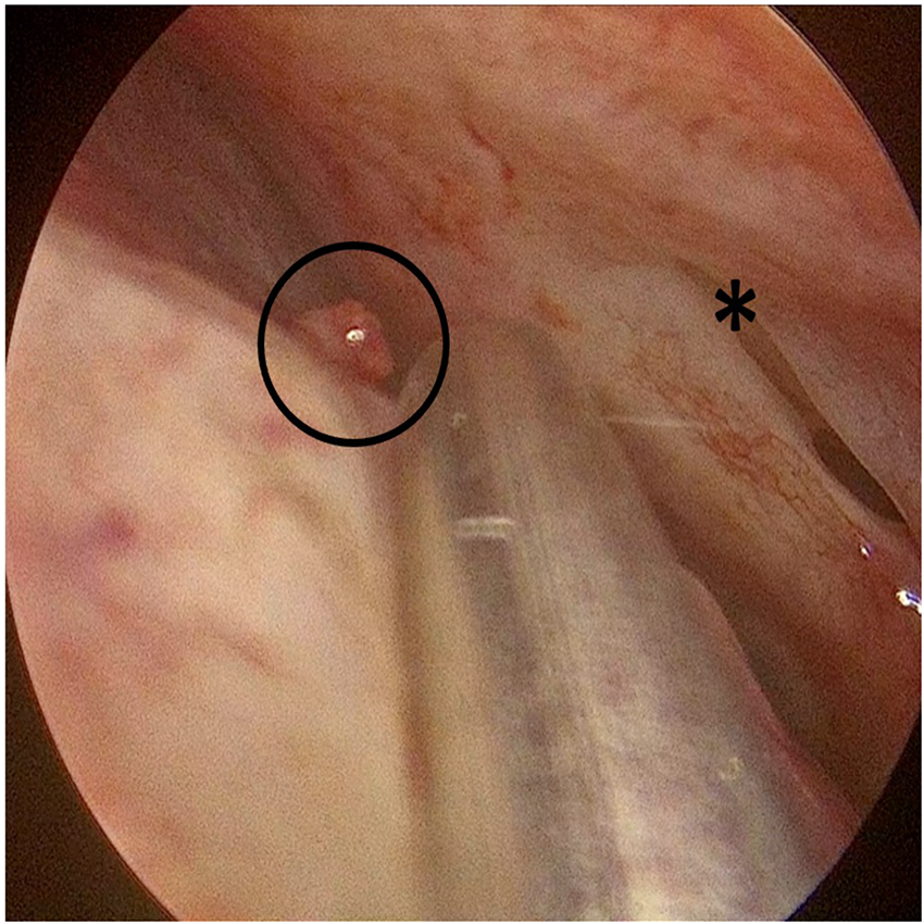
In general, patients underwent the same procedure consisting of removal of nasal packs if in situ and removal of clots from the nasal cavity. Neurosurgical patties soaked in 1:1000 adrenaline were generally placed in the nasal cavity. This is vital to ensure adequate vasoconstriction and visualisation. A 4-mm nasendosope was used in all cases. A systematic examination was carried out, starting at the septum, with a particular focus on the anterosuperior septum at and above the level of the axillar of the middle turbinate. The septum was generally manipulated using a Freer elevator to ensure good visualisation of this area (Figure 1, 4).
Examination of the anterosuperior septum (S-point) – axilla of the middle turbinate indicated by *.

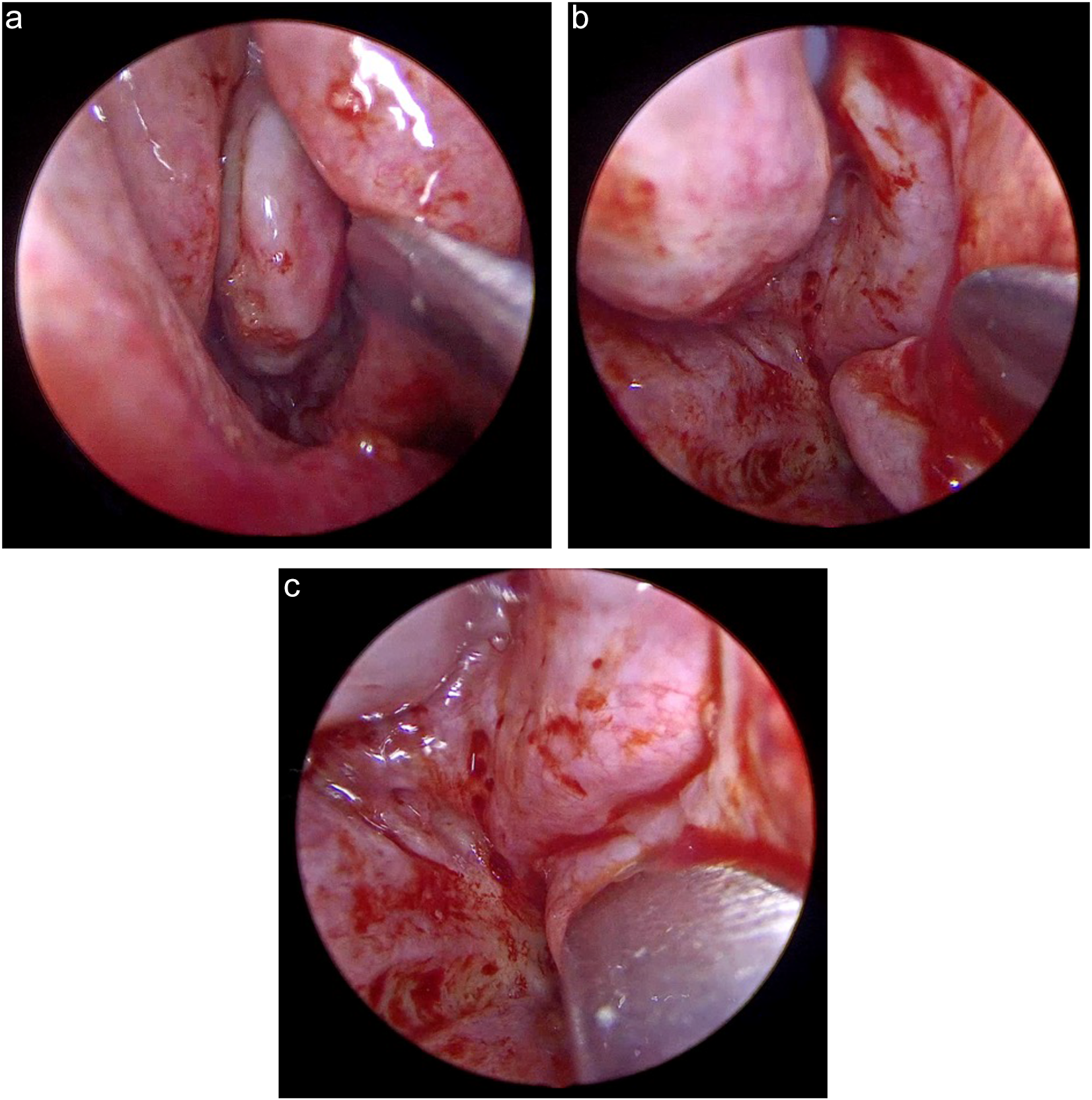
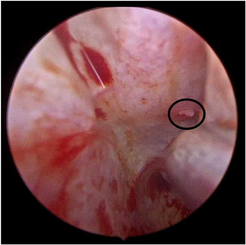
Examination incorporated medialisation of the middle and inferior turbinates to allow examination of the middle and inferior meatus (Figures 2a-c, 3). It is imperative to reveal the mucosal fold at the posterior end of each meatus to identify a potential vessel. Once the bleeding point was located, electrocautery was performed. Examination findings were documented. Patients in whom a bleeding point was identified were discharged on the same day. Where no bleeding point was identified, patients were transferred to the ward for 26 hours of observation without nasal packing to ensure no further bleeding.
(a-c) Examination of middle meatus.

Bleeding point at the posterior end of the inferior meatus, indicated by the black circle.

Bleeding point on anterosuperior septum (S-point) – bleeding point indicated by the black circle, axilla of the middle turbinate indicated by *.

Results and analysis
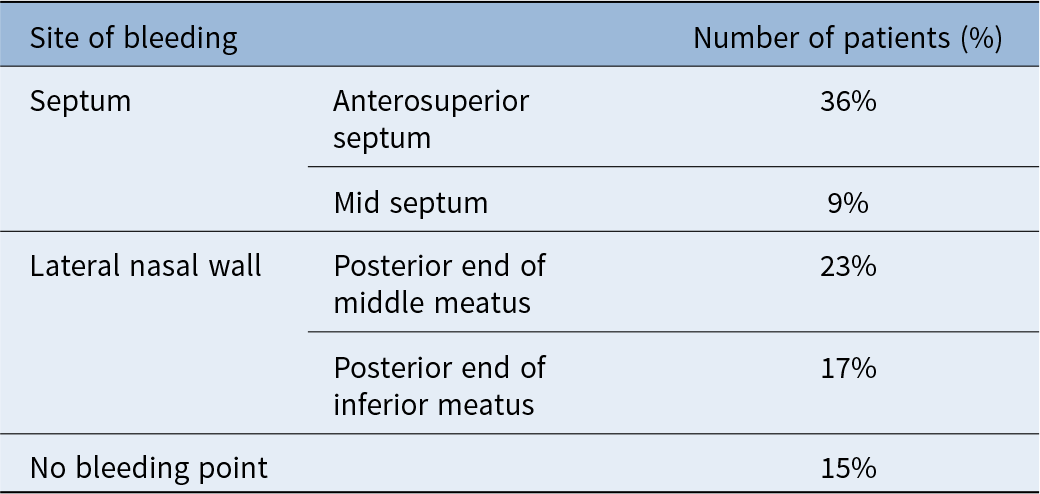
From hospital in-patient enquiry data 194 patients were identified, of which 74 per cent were male and 26 per cent were female. The ages ranged from 17 to 94 years, with a mean age of 61 years. Of the 194 patients, 89 (46 per cent) had a bleeding point identified on the septum and 70 (36 per cent) of these bleeding points were high on the anterosuperior septum above the axilla of the middle turbinate (Table 1). This territory is supplied by the anterior ethmoidal artery complex. The remainder of the points were identified on the mid-septum (19 patients, 9 per cent). A bleeding point was identified on the lateral nasal wall in 77 patients (40 per cent), 44 (23 per cent) of which were at the posterior end of the middle meatus and 33 (17 per cent) of which were at the posterior end of the inferior meatus. In these locations, the bleeding point is either on the lateral aspect of the turbinate or on the opposite lateral nasal wall. No bleeding point was identified in 29 patients (15 per cent).
Site of bleeding points

Hemostasis was achieved in all patients. Packs were not inserted even in those patients in whom a bleeding point was not identified. No patients required sphenopalatine and/or anterior ethmoidal artery ligation or embolisation over this 35-year period.
Discussion
Epistaxis is the most common ENT emergency, but at present no detailed consensus guidelines exist for its management. As a result, there is a wide variability in the management and outcomes for these patients.
Intractable epistaxis is a condition most often encountered in the elderly co-morbid population. Indeed the 2016 Integrate epistaxis national audit carried out in the UK reported a 3.4 per cent 30-day all-cause mortality rate in patients admitted with epistaxis. The audit acknowledged that while epistaxis may not be the direct cause of death in these cases, it does highlight the level of morbidity amongst this population of patients.4
Control of bleeding by cauterisation (direct therapy) has been somewhat overlooked in recent years. It is the most simple and uncomplicated method of controlling bleeding, and allows rapid treatment and prompt discharge from hospital. However, it requires the identification of precise bleeding points.Reference Melia and McGarry2 A national survey carried out in the UK in 1996 concluded that only one in five patients admitted to ENT departments were managed with direct control of the bleeding. This low proportion may be due to perceived difficulty in locating the bleeding point within the nose.Reference Kotecha, Fowler, Harkness, Walmsley, Brown and Topham5 In addition, the majority of cases of epistaxis are managed by the most junior members of the ENT department.Reference Melia and McGarry2, Reference Kotecha, Fowler, Harkness, Walmsley, Brown and Topham5 As a result, many of these patients are managed with anteroposterior nasal packing. In addition to a high failure rate, packing can be associated with prolonged hospital admissions, marked patient discomfort and complications such as pressure necrosis, hypoxia and toxic shock syndrome.Reference O'Donnell, Robertson and McGarry6
Recently there has been a shift in focus from cauterisation to techniques such as artery ligation and embolisation, in particular endoscopic sphenopalatine artery ligation. Authors have suggested the need for earlier endoscopic sphenopalatine artery ligation, with Dedhia et al. recommending it as an initial treatment option for medically stable patients with posterior epistaxis in preference to posterior packing.Reference Dedhia, Desai, Smith, Lee, Schaitkin and Snyderman7 However, while successful, these techniques all have the potential for significant complications. Our study also found that the most common site of bleeding was the anterosuperior septum, which is supplied by the anterior ethmoidal artery, not the sphenopalatine artery.
The 2016 Integrate epistaxis audit, which included 1152 patients, reported that only 36 patients required surgical intervention. Of these, 19 had sphenopalatine artery (SPA) ligation whereas only 9 had electrocautery of identified bleeding points. In addition, 22.6 per cent of these patients re-presented with recurrent epistaxis following surgery and required further procedures. These high rates of recurrent epistaxis are probably due to inadequate identification of the causative bleeding vessel.4
In a large review of bleeding sites in epistaxis, Jiang et al. found a site was identified in 61 per cent of cases, which is slightly lower than in our cohort.Reference Jiang, Yu, Zhang, Tu, Liu and Chen8 In the 395 cases where they did identify a bleeding point, the most common site was the inferior meatus in 32.9 per cent, followed by the septal surface of the olfactory region in 25.8 per cent, lower nasal septum in 20.3 per cent and middle meatus in 15.2 per cent. A common bleeding location on the anterosuperior septum, which Jiang et al. term the septal surface of the olfactory region, has been named Stamm’s S-point. It is located on the upper nasal septum, at the level of the axilla of the middle turbinate. It is an arterial pedicle of the septal branch of the anterior ethmoidal artery. This is an under-recognised source of intractable epistaxis despite being the most common site in our cohort.Reference Jeong, Choi, Lee, Kim, Min and Kim9–Reference Kosugi, Balsalobre and Stamm11
Our study is a continuation of Thornton et al.Reference Thornton, Mahesh and Lang12 Their study included 43 patients, who are included in this study. Although most bleeding points were clearly visible, some were concealed within a horizontal groove on the lateral aspect of the middle and inferior turbinate. This groove or mucosal fold was a fairly consistent anatomical finding. It is important to look into the depths of this groove to avoid missing a bleeding point concealed there. Visualisation of the depths of this groove can require the use of a 30° scope and a Freer’s elevator to retract the mucosa.
In a minority of cases, a microdebrider was used to remove the mucosa to assist in the visualisation and cauterisation of bleeding points in this area. Despite the fact that the majority of our patients had at least one nasal pack inserted, or in some cases multiple packs prior to endoscopic examination, the anterosuperior septum and the inferior and middle meatus remain virgin territories, allowing the identification of the bleeding point.
It can be difficult to carry out a thorough examination under local anaesthesia, particularly for examining the posterior end of the middle and inferior meatus. For this reason, all our cases were carried out under general anaesthetic. However, it should be possible to examine and cauterise bleeding points high on the anterior septum under local anaesthetic and this should be considered in patients who are high-risk surgical candidates. All bleeding points were accessible with electrocautery, which controlled bleeding without complication. Failure to identify a bleeding point after thorough examination under general anaesthesia does not require further intervention unless complicated by further bleeding.
• It is known that 60 per cent of people experience epistaxis and 6 per cent of people require medical treatment for epistaxis
• Localisation of epistaxis has not previously been well described
• Localising a bleeding point allows for effective cautery at the source, preventing more complex interventions and minimising hospital stay and morbidity
Conclusion
Endoscopic examination of the nose has been overlooked in recent times. Direct cauterisation avoids the morbidity associated with nasal packing, the cost of prolonged hospital admission and more invasive procedures such as SPA ligation. We believe the increasing adoption of early aggressive endoscopic ligation is not the best solution for managing such patients.
This study showed a remarkable consistency of the location of bleeding points in intractable epistaxis: high on the anterior septum at the axilla of the middle turbinate, which is supplied by the anterior ethmoidal artery (S-point), the posterior end of the middle meatus and the posterior end of the inferior meatus, which are supplied by the sphenopalatine artery. Examination with rigid nasendoscopes to identify the precise bleeding point with a particular focus on these three areas should be the first step in the management of this cohort of patients following usual first-line interventions.
Competing interests
None declared.