Introduction
Vestibular schwannomas (VS) constitute approximately 10% of all intracranial neoplasms and 90% of all cerebellopontine angle tumours.Reference Massager, Nissim and Delbrouck1 They are benign slow-growing tumours arising from schwann cells of the myelin sheath of the vestibular branch of eighth cranial nerve. They are commonly unilateral or can be rarely bilateral when associated with neurofibromatosis type 2 syndrome.Reference Mallory, Pollock, Foote, Carlson, Driscoll and Link2 Their location and proximity to critical structures such as brainstem, cerebellum and lower cranial nerves make them present with complex symptoms such as unilateral decreased hearing, tinnitus, vertigo, imbalance, decreased sensation over face, deviation of mouth, altered taste and raised intracranial tension symptoms. While tumours <3 cm in size can be managed with stereotactic radiosurgery (SRS) or microsurgery, tumours >3 cm require microsurgery that may be total or planned subtotal excision followed by SRS. SRS has been proposed as the treatment of choice as primary modality for VSs smaller than 3 cm in size.Reference Kondziolka, Lunsford, McLaughlin and Flickinger3 SRS is less invasive and is associated with a short hospital stay and with better quality of life long term. If the late side effects are minimal, SRS probably can replace microsurgery for tumours <3 cm in size and hence, we aimed to analyse the outcome—tumour control, adverse effects—facial function and hearing, and their correlation to patient and treatment-related factors in patients with VSs who received Linear Accelerator (LINAC)-based SRS.
Materials and Methods
Study population
One hundred and seven patients and 108 tumours (1 patient with bilateral tumours) were treated with LINAC-based SRS between June 2007 and June 2014 at our centre. A retrospective review of the hospital records of these patients was conducted after approval from the Institutional Review Board. There was no follow-up data for nine patients and hence these were not included for analysis.
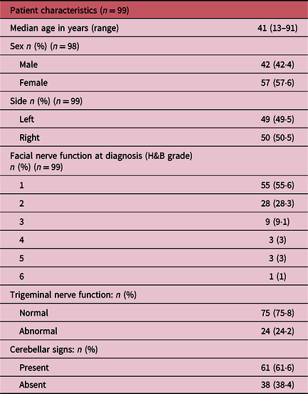
Median age of patients studied was 41 years (range: 13–91). Ten patients had neurofibromatosis type 2 and bilateral schwannomas and one patient received SRS for both lesions. The baseline characteristics are summarised in Table 1.
Patient characteristics

Treatment details
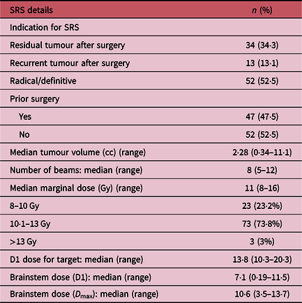
The stereotactic frame (Brown-Robert-Well) was fixed by the neurosurgeon on the day of SRS under local anaesthesia, and a contrast-enhanced computed tomography (CECT) scan of the head with 3 mm slice thickness was performed. The scan was transferred to the planning system (Brainscan, BrainLab, Germany); T1W post-gadolinium magnetic resonance images (MRI) (volumetric acquisition) were fused with CECT in the planning system and used to define the target volume, brainstem and optic apparatus. All plans had a single isocentre. Unilateral non-coplanar beams were used for all patients except for nine of them where one contralateral beam was added in order to achieve better target coverage, reduce hot spot and achieve a better conformity index. The patients were treated using 6 MV photons on a Primus Linear accelerator (Seimens, USA) fitted with a 3-mm micromultileaf collimator (mMLC, Brainlab, Germany) attached to it as a secondary collimator. The patients who had <3 cm in size tumours were offered SRS. The prescribed dose was 13 Gy to 80% isodose (tumour margin covering isodose) except in three patients in whom the dose was more than 13 Gy. Table 2 summarises the treatment parameters of SRS. SRS as primary treatment was delivered to 52·5% of the patients and 47·5% had prior surgery, and the residual tumour was treated with SRS.
SRS treatment parameters
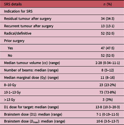
D1, – Dose to 1% brainstem volume; D max – Maximum brainstem dose.
The most common presenting symptom was impaired hearing. Pure tone audiometry evaluation was done at diagnosis for all patients. Hearing was impaired (moderate-to-severe hearing loss) in 93·9% of patients and out of whom complete hearing loss was found in 6·4%. Severe facial nerve paresis (House–Brackmann [H&B] scores 5&6) was found in 4·04%. Trigeminal nerve was found affected in 24·2%. Cerebellar dysfunction was present in 61·6%. Tinnitus was another common symptom and was found in 29 (29·2%) and headache was in 21 (21·2%).
Follow-up
All 98 patients and 99 tumours were followed up clinically and radiologically with contrast MRI/CT at 1 year and thereafter every 1–2 years. Those patients who had not visited the hospital for follow-up were contacted telephonically and the details were obtained. The MRI scans that were available were analysed for tumour control and signal changes in the brainstem, cerebellum and middle cerebellar peduncle. The patients with initial minimal (2–3 mm) increase in size of tumour with central necrosis but asymptomatic were recorded as asymptomatic minimal progression and considered as stable disease. The patients with increase in size (>3 mm) of tumour with symptoms were recorded as symptomatic progression.
At follow-up, the patients were assessed for facial nerve function and H&B grading was recorded and worsening of hearing was noted if the patient complained and hence it was subjective patient-reported outcome of hearing.
Outcome variables
The treatment parameters such as volume of tumour irradiated, radiation dose delivered to margin of tumour, conformity index and dose received by the brainstem were collected from the treatment plans. The indication for SRS, hearing and facial nerve functioning prior to SRS and at follow-up were noted from the patient’s medical records.
Statistical analysis
Data were screened for outliers and extreme values using Box–Cox plot and histogram (for shape of the distribution). Summary statistics were used for reporting demographic and clinical characteristics. T-test was used for the analysis of continuous data with normal distribution and Mann–Whitney U test for data with non-normal distribution with grade. Chi-square test was performed for categorical variables and the outcome variables. Differences were considered significant at p < 0·05. Estimation of radiological progression was performed using the Kaplan–Meier method. All the statistical analyses were performed using SPSS 16.0.
Results
Follow-up
Ninety-eight patients had a minimum follow-up of 1 year. Median follow-up period was 5·6 years (range: 1–12 years). A total of 59 (59·6%) patients had 5 years follow-up or more.
Only one patient expired of pulmonary embolism 2 years after SRS who was 91 years of age at the time of SRS. There was no disease-related or treatment-related mortality in this cohort of patients.
Tumour control
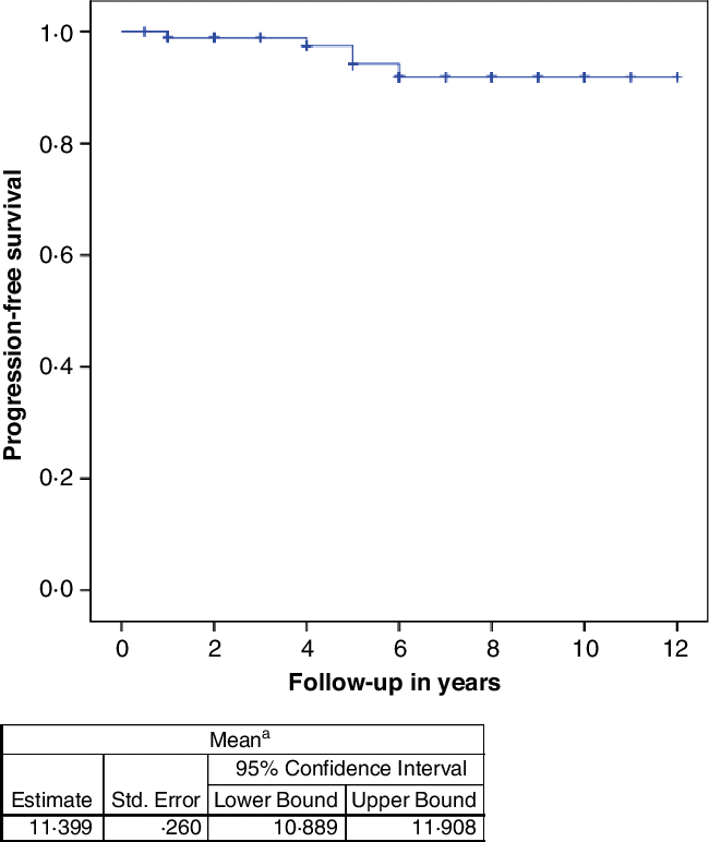
The response to treatment (clinical and radiological combined) was stable in 37 (37·4%), regression in 46 (46·5%), asymptomatic minimal progression in 9 (9·1%), and symptomatic progression in 5 (5%) and unknown in 2 (2%) patients. Among the 98 patients and 99 tumours for which the follow-up data are available, the local control rate was 95%. Figure 1 gives the Kaplain–Meier curve for progression-free survival of the cohort. The five patients progressed at 1st, 4th, 5th, 5th and 6th year of follow-up after SRS. One of them underwent surgery at 5th year and the other four patients were advised further follow-up as the symptoms and progression were minimal. One patient needed ventriculoperitoneal shunt and the other shunt revision.
Kaplain–Meier curve for progression-free survival of the cohort.

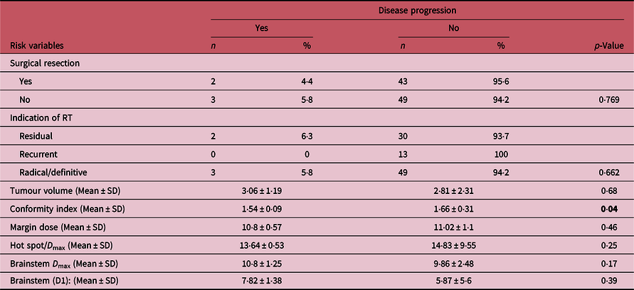
Table 3 demonstrates that none of the risk variables such as surgical resection, indication for radiation therapy (RT), treated tumour volume and tumour margin dose statistically significantly affected tumour control. Conformity index has p-value of 0·04 which is significant. This indicates that treating the tumour with irradiating less normal tissue around is sufficient for good tumour control.
Risk variables for disease progression.

Radiological assessment
Pre-SRS MRI studies were available for 99 tumours, while follow-up studies were available for 89 tumours. Residual tumour was present in all follow-up studies.
Median largest diameter of pre-SRS tumour was 21 mm (range: 11–39) and median volume was 2·63 cc (range: 0·3 −23·2). At last follow-up MRI scans, the median largest diameter of the tumour was 19 mm (range: 7–38) and median volume was 2·26 cc (range: 0·1–10·9).
Pre-SRS imaging demonstrated distortion of brainstem secondary to the mass effect of the schwannoma in 29 (29·3%) cases. Four (4%) cases also demonstrated associated T2W hyperintensity within the brainstem secondary to the mass effect. On the follow-up imaging, only one case demonstrated new development of T2-weighted hyperintensity involving the brainstem. This was attributed to the increase in the cystic component of the adjacent schwannoma rather than radiation-induced change.
Pre-SRS imaging demonstrated a mass effect on ipsilateral cerebellar hemispheres in 12 (12·1%) cases and on ipsilateral middle cerebellar peduncles in 50 (50·5%) cases. This was associated with signal changes in the form of T2-weighted hyperintensity in 25 (25·3%) and 22 (22·2%) cases, respectively. More cases demonstrated signal changes involving the cerebellum due to changes related to previous partial tumour excision. For the available follow-up scans in 86 tumours, a new development of-T2 weighted hyperintensity was seen in two cases (one case each for cerebellar hemisphere and middle cerebellar peduncle) and was related to increase in the cystic component of the residual tumour with resultant mass effect. No signal changes were seen that could be attributed to SRS.
Hearing function on follow-up
Hearing was assessed subjectively (patient-reported outcome) which is the limitation in this study. Six (6·06%) noted improvement in hearing, 82 (82·83%) noted no change in hearing of which 6 of them continued to have normal hearing, 8 (8·08%) noted worsening in their hearing post-SRS and unknown status in 3 (3·03%) patients. There was no new-onset hearing loss. Among 98 patients (99 tumours), the hearing preservation rate after SRS is 92%.
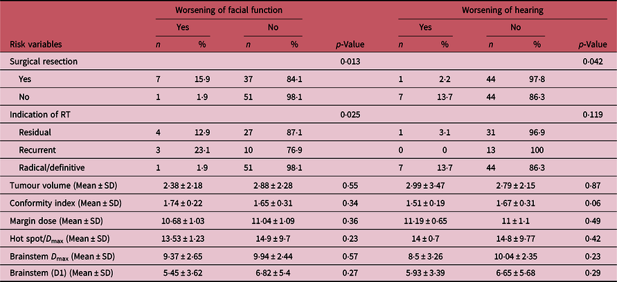
Table 4 demonstrates that none of the risk variables such as indication for RT, treated tumour volume, conformity index and tumour margin dose statistically significantly affected worsening of hearing. Worsening of hearing was seen predominantly in the population that received definitive SRS than those who had prior surgery and SRS.
Risk variables for worsening of facial function and hearing

Facial nerve function on follow-up
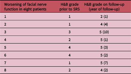
Facial nerve function was assessed clinically and 12 (12·1%) had improvement in H&B grade to 1 or 2, 76 (76·8%) had stable findings, 5 (5%) had worsening of H&B grade, 3 (3%) had new-onset facial palsy and unknown in 3 (3%) patients. Table 5 describes the H&B grades at worsening and the year of worsening from the time at SRS.
Worsening of facial nerve function

Table 4 demonstrates that none of the risk variables such as treated tumour volume, conformity index and tumour margin dose statistically significantly affected worsening of facial function. The p-value was significant for prior surgery and indication for RT. The patients who had prior surgery and then SRS were predisposed to developing a facial paresis after SRS than those who received definitive SRS alone. The patients who developed worsening of facial function were predominantly in the cohort that had prior surgery.
Discussion
Epidemiology
VSs are detected incidentally more often in recent times; probably, due to more use of MRI scans for the brain.Reference Ikonomidis, Pica, Bloch and Maire4 The causative factors for schwannomas are still unknown. Oren Berkowitz et al reported that there is no sex predilection for this tumour and median age of presentation is 50–60 years.Reference Berkowitz, Iyer, Kano, Talbott and Lunsford5 Median age in this cohort was 41 years.
Choice of modality of treatment
The treatment modalities include observation, microsurgery and radiation therapy. Smaller asymptomatic and incidentally found lesions (Koos grade 1–3) can be observed as schwannomas have a slow-growing nature.Reference Ikonomidis, Pica, Bloch and Maire4 Radiation therapy as SRS or hypofractionated SRS or stereotactic radiotherapy (SRT) have been equally effective as shown by Murphy et al.Reference Murphy and Suh6 For lesions <3 cm, radiosurgery is the preferred choice when compared to microsurgery.Reference Wolbers, Dallenga, Mendez Romero and van Linge7–Reference Lunsford, Niranjan, Flickinger, Maitz and Kondziolka11 For larger lesions (>3 cm), fractionated SRT is an option or facial nerve preserving microsurgery or both. As far as SRS is concerned, LINAC-based SRS has shown comparable results to that of Gamma knife-based SRS.Reference Spiegelmann, Lidar, Gofman, Alezra, Hadani and Pfeffer12
Radiation therapy
Schwannomas are considered to be late responding tissues to radiation and the surrounding brain tissue also is considered late responding both with α/β ratio of 2.Reference Santacroce, Kamp, Budach and Hänggi13 The optimal dose for SRS has been demonstrated to be 12–13 Gy to the margin. This dose has been effective in achieving optimal tumour control and with very minimal risk of cranial neuropathies.Reference Flickinger14 When resurgery is needed after SRS, Singh et al. reports that with doses <13 Gy, resurgery is feasible.Reference Singh15
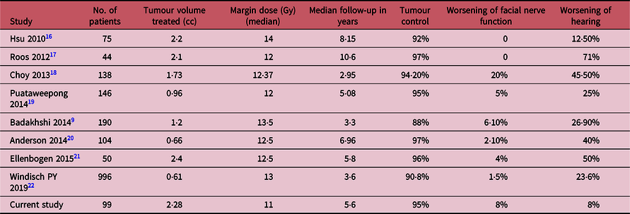
Tumour control: Table 6 shows published results of tumour control of schwannomas after the year 2010.Reference Hsu, Chang and Lee16–Reference Windisch, Tonn and Fürweger22 Tumour control radiologically varies from 88% to 97%. Our study also shows 95% tumour control for a median follow-up of 5·6 years. Recent meta-analysis has shown that SRS gives good tumour control for cystic schwannomas too.Reference Ding, Ng and Romiyo23 Initial transient tumour growth in these tumours up to 3 years duration is a known phenomenon.Reference Okunaga, Matsuo and Hayashi24,Reference van de Langenberg, Dohmen, de Bondt, Nelemans, Baumert and Stokroos25 The percentage of tumours that show such phenomenon varies from 11% to 55% in various published results and it was 9% in our series.
Local control and toxicity of LINAC SRS for VSs

In our series on the follow-up MRI scans, most patients had signal changes in brainstem, cerebellum and middle cerebellar peduncle secondary to surgery. Only three patients had signal changes that were not related to surgery and all were due to an increase in the size of cystic component of the residual tumour. No SRS-related signal changes were seen in the posterior fossa structures. In cases with signal changes in SRS field, it is important to differentiate between recurrence and radiation necrosis.Reference Walker, Ruzevick and Malayeri26
Facial nerve function worsening: Facial nerve function worsening has been seen in 2–20% of these tumours according to recent literature as shown in Table 6. Our study shows 8% of patients having worsening of facial function and five out of eight developed this complication within 3 years of treatment. Surgical resection was a covariate that was statistically significant for this complication and hence can be inferred that surgery prior to SRS increases the chances of worsening or new-onset facial nerve paresis. Murphy et al. found that treated tumour volume and tumour dose as factors affecting facial nerve worsening in their study.Reference Murphy and Suh6 Tumour <3 cm and tumour dose < 13 Gy reduces the chances of this complication. Cranial nerve length irradiated has also been thought of as a risk factor for facial nerve worsening.Reference Linskey, Flickinger and Lunsford27 This was not assessed in our series.
Worsening of hearing: Table 6 lists reported published literature for worsening of hearing that varies from 12·5% to 70%. Our study reports only 8% and this is probably due to subjective assessment at follow-up. Puataweepong et al. notes that early intervention in those patients who are observed without treatment for VSs preserve their hearing function better. Pretreatment hearing assessment into serviceable and non-serviceable using Gardner–Robertson classification or subjectively as ‘ability to hear and converse over telephone in the affected ear’ is useful to carefully plan treatment and preserve hearing. When we plan radiation therapy, delineating cochlea and restricting dose to ≤ 4 Gy is another option to try and preserve hearing. Pretreatment hearing function can also predict hearing outcome.Reference Ikonomidis, Pica, Bloch and Maire4
To consider in future patients: Although observation is an option, there is a risk of development of hearing dysfunction that needs to be considered.Reference Leon, Lehrer and Peterson28 SRT can be offered for larger lesions; Persson et al have reported good tumour control with SRT.Reference Persson, Bartek, Shalom, Wangerid, Jakola and Förander29 Cochlea and facial nerve to be delineated while planning SRS and doses to be minimised to within tolerable limits accordingly (4 Gy to cochlea and <13 Gy to cranial nerve) and length of cranial nerve irradiated to be minimised in order to reduce chances of hearing and facial function worsening.Reference Lerner, Lee and Naples30,Reference Deora and Tripathi31 Objective hearing assessment at follow-up will help in better assessment of worsening of hearing.
Limitation of this study
The hearing and facial functions were documented based on clinical examination during follow-up visits and no objective assessments were made for hearing on follow-up. Retrospective nature of the study is another limiting factor.
Conclusion
SRS is an effective modality to treat VSs <3 cm in size. Tumour control rate was 95% with a median follow-up period of 5·6 years. The complication rates were 8% each for facial function worsening and worsening of hearing. The p-value for radiation treatment factors such as treated tumour volume, tumour dose and brainstem dose was not statistically significant to affect the outcomes: tumour control or complications. Prior surgery was a significant factor that affected facial nerve function deterioration. Restricting dose to cochlea to attempt to preserve hearing function needs to be studied further. Cranial nerve length irradiated and the dose to it is another factor to be studied.
Acknowledgements
None.
Financial Support
Nil.
Conflicts of Interest
None.