Cardiac tumours are benign or malignant neoplasms that can be either primary or metastatic.Reference Nadas and Ellison 1 – Reference Gunther, Schreiber, Noebauer, Eicken and Lange 6 Most cardiac tumours are benign in all age groups. Malignant tumours account for only about 8% of cases in children.Reference Uzun, Wilson, Vujanic, Parsons and De Giovanni 7 , Reference Padalino, Vida and Boccuzzo 8 Nevertheless, the size and location of the masses can be clinically significant in terms of morbidity and mortality.Reference Padalino, Vida and Boccuzzo 8 – Reference Marx and Moran 12
An autopsy series in the general population reported an estimated incidence of primary cardiac tumours ranging between 0.0017% and 0.03%.Reference Nadas and Ellison 1 – Reference Beghetti, Gow, Haney, Mawson, Williams and Freedom 4 , Reference Marx and Moran 12 Paediatric necropsy studies have reported a frequency between 0.027% and 0.05%.Reference Nadas and Ellison 1 , Reference Basso, Valente and Thiene 11 , Reference Marx and Moran 12 – Reference Centofanti, Di Rosa and Deorsola 15 Echocardiography databases report an incidence of 0.17–0.2% among children referred for cardiac examination.Reference Beghetti, Gow, Haney, Mawson, Williams and Freedom 4 , Reference Gunther, Schreiber, Noebauer, Eicken and Lange 6 , Reference Basso, Valente and Thiene 11 , Reference Reynen 16 , Reference Freedom, Lee, MacDonald and Taylor 17
We report our 27-year experience on the diagnoses of primary cardiac tumours among children in a referring centre for Paediatric Cardiology in order to better analyse the clinical presentation, natural history, and treatment of this rare disease.
Methods
This is a retrospective review of all consecutive patients admitted to our centre after having been diagnosed prenatally or postnatally with a cardiac tumour between April, 1982 and April, 2009.
The diagnosis of cardiac tumours was made using echocardiography and/or magnetic resonance imaging.Reference Butany, Nair, Naseemuddin, Nair, Catton and Yau 2 , Reference Uzun, Wilson, Vujanic, Parsons and De Giovanni 7 , Reference Basso, Valente and Thiene 11 , Reference Marx and Moran 12 , Reference Fyke, Seward and Edwards 18 The records were examined for characteristics such as clinical history, familiarity of cardiac tumours, and associated symptoms or tuberous sclerosis complex. Similarly, the diagnosis of the benignity or malignity of the mass was based on epidemiological data, clinical course, and conditions at follow-up.Reference Butany, Nair, Naseemuddin, Nair, Catton and Yau 2 – Reference Elderkin and Radford 5
Histopathological analysis was carried out mainly in surgically managed patients. The association to tuberous sclerosis complex was evaluated according to the diagnostic criteria drawn at the tuberous sclerosis complex Consensus Conference in 1998.Reference Roach, Gomez and Northrup 19 Finally, intracardiac masses for which none of the previous criteria were applicable were excluded from this series because they were masses of other nature, such as a thrombus, endocarditis vegetation, and calcification. In contrast, when a cardiac tumour was not clearly defined in vivo as a histotype – fibroma versus rhabdomyoma – and when other nature of the mass was undeniably ruled out, the mass was defined as “other tumour”.
Demographic, prenatal, diagnostic, clinical, and surgical data were collected from patients’ medical records after searching the Congenital Echo Cardiac Database of the Department of Pediatrics. Morphological and histological results of the resected masses were included in the present revision. The clinical follow-up included the annual physical examination, two-dimensional and Doppler echocardiography, electrocardiography and electrocardiography Holter monitoring.
Partial tumour regression at follow-up was defined as a reduction of 15% or greater in tumour diameters when compared with the previous echocardiographic evaluation. A complete regression was diagnosed when the tumour was no longer detectable by echocardiography, or if it persisted only as a residual hyperechoic myocardium.Reference Di Liang, Ko and Huang 20
Fisher's exact probability test was used to compare proportions; statistical significance was reached if the p-value was <0.05.
Results
Over a span of 27 years, 52 patients were diagnosed with a primary cardiac tumour (M/F = 27/25) at our Children's Hospital in Padua, Italy, and were included in our study group.
During this period, a total of 120,085 echocardiographies were performed on 4285 children, accounting for a prevalence of cardiac tumours of about 0.012%. Associated congenital cardiac diseases were reported in three patients (6%), comprising ventricular septal defects in two patients and patent ductus arteriosus in one patient.
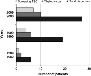
A total of 18 patients (35%) were diagnosed prenatally during a routine obstetric scanning with no other cardiovascular alterations, leading to a caesarean delivery in two cases due to foetal haemodynamic instability. Rhabdomyoma (67% of all foetal detections) was the most common type of cardiac tumour diagnosed prenatally; one teratoma was reported because of the onset of a pericardial effusion at 33 weeks of gestation. There were no pre-term births or foetal deaths in our series (Fig 1).
The number of patients diagnosed with cardiac tumours, prenatal detections, and diagnoses using the screening program for TSC, from 1982 to 2009, are shown. In this figure, it is shown graphically that, despite the increased number of total diagnoses of cardiac tumour, the rate of prenatal detection is stable over the decades, setting between 32% and 37%, with a global frequency of 34.6%. TCS = tuberous sclerosis complex.

In the remaining 34 patients, postnatal diagnosis was made at a median age of 3 months (range 1 day–18 years). In eight children (23.5%), the diagnosis was made immediately postpartum. In 83% of patients, the cardiac tumours were diagnosed before the age of 6.
Number and location of the masses
Heart tumours may occur as a single lesion or as multiple lesions usually located within the ventricle. We had 31 patients (60%) with single mass tumours and 21 patients (40%) with multiple mass tumours. The most common form of tumour was rhabdomyoma, which was multiple in 95% of patients, whereas single in only 39%. Children diagnosed with a single rhabdomyoma were older (mean age 3 years) compared with those with multiple tumours (mean age 18 days).
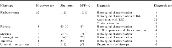
The tumour mass was found in the ventricles in 85% of patients (44 patients), whereas it involved the atria in 11% of patients (six patients) and the pericardium in 4% of patients (two patients). All fibromas and all but two rhabdomyomas were intraventricular, whereas myxomas always appeared as polypoid masses arising from the atrial wall. Bilateral ventricular involvement was observed in 46% of patients (24 patients). The mean diameters of the masses, measured by echocardiography or direct examination, are reported in Table 1.
Histopathological data and diagnostic modalities (n = 52 patients).

ECHO = echocardiography; F = female; M = male; TSC = tuberous sclerosis complex
Clinical presentation
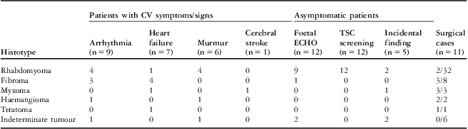
Cardiovascular symptoms or other clinical signs were present in 23 patients (44%).
Arrhythmias occurred in nine patients. Of four of these nine patients, who were diagnosed prenatally, two were born with an incessant supraventricular tachycardia – rhabdomyoma and haemangioma; one was born with Wolff–Parkinson–White syndrome – rhabdomyoma; and one had ventricular ectopic beats – rhabdomyoma. In the other five patients, postnatal diagnosis was prompted by the presence of ventricular ectopic beats in three patients – two had fibromas and one had rhabdomyoma; Wolff–Parkinson–White syndrome in one patient – indeterminate mass; and ventricular tachycardia in another – fibroma. There was no significant difference in the type of arrhythmia among the histological subtypes (p = 0.1282).
Congestive cardiac failure was found in seven patients soon after birth; two of these patients were diagnosed prenatally with a cardiac mass.
Of the remaining seven symptomatic patients, six were diagnosed with a cardiac tumour incidentally after finding a cardiac murmur during their physical examination and after the occurrence of a cerebral stroke in one case of a 7-year-old boy.
Among the 29 asymptomatic patients (56%), 12 were diagnosed prenatally, 12 had a cardiac evaluation in the context of the screening programme for tuberous sclerosis complex, and five were incidentally diagnosed during routine paediatric check-ups or evaluation for other illnesses. Clinical data of the patients at presentation and follow-up are summarised in Table 2.
Clinical data at presentation and management (n = 52 patients).

CV = cardiovascular; ECHO = echocardiography; TSC = tuberous sclerosis complex
Diagnostic techniques
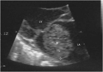
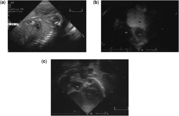
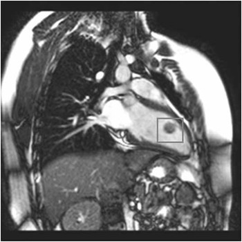
All patients were studied by means of two-dimensional echocardiography (Figs 2 and 3) and electrocardiography. A total of five patients (10%) had undergone additional magnetic resonance imaging (Fig 4) and two (4%) had undergone computed tomography in order to achieve a detailed diagnosis. Cardiac catheterisation was performed for six children (11.5%), preoperatively in three of them.
A two-dimensional echocardiogram showing a myxoma in the left atrium of a 6-year-old girl. LA: left atrium; LV: left ventricle; M: myxoma.

Echocardiograms showing a giant rhabdomyoma in the left ventricle: ( a ) on foetal two-dimensional echocardiography (3A), ( b ) on neonatal two-dimensional echocardiography, and ( c ) after 2 years, only a residual mass between Ao and PV was detectable. Ao = aortic valve; LV = left ventricle; PV = pulmonary valve; RB = rhabdomyoma; RV = right ventricle.

A steady-state free precession cine cardiac magnetic resonance image showing a cavernous haemangioma with myxomatous lacunae (within the red frame) in the left two-chamber view. LV = left ventricle.

All masses were primary benign cardiac tumours. Neither secondary nor malignant cardiac tumours were detected in our experience. The diagnosis was clinical in the majority of the patients; it was based on histological analysis in 12 patients only (23%). In our series, there were 32 cases of rhabdomyomas (61.5%), eight of fibromas (15.4%), three of myxomas (5.8%), two of haemangiomas (3.8%), and one of teratoma (1.9%). Finally, the precise in vivo diagnosis of the histotype of the cardiac tumour was uncertain for six patients in whom all other non-cardiac causes were excluded undeniably.
Simultaneous diagnosis of tuberous sclerosis complex was carried out in 23 patients (44%). This frequency was higher when compared with rhabdomyoma (72%). In two patients, there was a family history of cardiac tumours (Table 1).
In 12 patients, the instrumental diagnosis of the histotype was confirmed by histological examination after the surgery (11 patients) or after a surgical excisional biopsy of the tumour (one patient).
Management
A total of 11 patients (21%) underwent surgical treatment because of the presence of the following signs or symptoms: cardiac failure with intracardiac obstruction and cardiac dilatation in five patients, right ventricular outflow tract obstruction presenting as cardiac murmur in three patients, cardiac failure with severe pericardial effusion in one patient, cerebral stroke due to thromboembolism in one patient), and lastly paramyxomatous syndrome in one patient. All operations were successful. In nine patients, the tumour was completely resected, and in two patients the large size of the mass – fibroma in both patients – and the massive ventricular infiltration led to an orthotopic cardiac transplantation. There was no partial resection in this series. The age at operation ranged between 5 days and 18 years (median age 3.7 months). All patients who underwent surgical resection had no post-operative complications and did not need cardiovascular therapy. In contrast, the two transplanted patients required the usual immunosuppressant drug therapy, and hospitalisation stay was longer. Echocardiographic evaluation performed at discharge did not reveal any abnormalities in the cardiac function or any residual masses in the nine patients who had had undergone surgical resection. All resected masses were sent to pathology for histological examination and characterisation (Table 1).
A total of 41 patients (79%) were managed medically, with regular clinical follow-up at our outpatient clinic. Pharmacological treatment was deemed necessary in six out of the 41 patients (15%) at clinical presentation: antiarrhythmic drugs in five patients, and diuretics, digoxin, captopril, and warfarin in one patient.
Follow-up
At a mean follow-up of 7.2 ± 5.4 years (range 12 months–27 years, 96% completeness), two patients died 3.2 and 3.5 years after cardiac transplantion, due to the occurrence of a cerebral astrocytoma and H1N1 pneumonia, respectively. All the remaining patients are alive.
Among the surgically managed patients (nine patients), there was no recurrence of cardiac tumour, and they all show normal ventricular function and persistence of sinus rhythm. Among the medically managed patients (41 patients), 93% are asymptomatic and 90% are in sinus rhythm. All patients are free from any cardiac medications. Spontaneous regression, as defined above, occurred in 21 patients – partial in 15 and complete in six patients – all affected by rhabdomyoma, accounting for a regression rate of 66% in our series of rhabdomyomas (Fig 3a–d). In the remaining medically managed patients, the tumour mass remained unmodified throughout the follow-up period. Finally, 18 patients with rhabdomyoma (56%) presented with neurological symptoms related to tuberous sclerosis complex.
Discussion
We report one of the largest studies on paediatric cardiac tumours ever collected within a single centre, and as far as we know it is one of the two studies with the longest follow-up.Reference Beghetti, Gow, Haney, Mawson, Williams and Freedom 4
Historically, the first description of cardiac tumours dates back to 1559, by the pathologist Realdus Columbus,Reference Columbus 21 and it was only some centuries later (1934) that a cardiac tumour was diagnosed in a living patient.Reference Barnes, Beaver and Snell 22 The first successful total surgical excision was reported in 1954.Reference Marx and Moran 12 , Reference Acierno 23
Owing to their rarity and the lack of large study groups, the real prevalence of this disease is difficult to define. Beghetti et alReference Beghetti, Gow, Haney, Mawson, Williams and Freedom 4 reported an incidence of tumours rising from 0.06% during 1980–1984 to 0.22% during 1985–1989, and up to 0.32% during 1990–1995. This is probably related to improved diagnostic abilities and a more extensive screening for tuberous sclerosis complex.Reference Beghetti, Gow, Haney, Mawson, Williams and Freedom 4 , Reference Gunther, Schreiber, Noebauer, Eicken and Lange 6 , Reference Marx and Moran 12 , Reference Freedom, Lee, MacDonald and Taylor 17 , Reference Fyke, Seward and Edwards 18 , Reference Isaacs 24 – Reference Holley, Martin and Brenner 28
According to the literature, cardiac tumours are often an expression of a benign disease. Although they can have a very dramatic impact on the families and physicians, malignancy is very rare, especially in children.Reference Nadas and Ellison 1 , Reference Roberts 3 , Reference Beghetti, Gow, Haney, Mawson, Williams and Freedom 4 , Reference Padalino, Basso and Milanesi 25 In our study, no case of malignant tumour was identified. In addition, only 21% of the patients needed surgical treatment, which was successful in all patients undergoing resective surgery, whereas complications and post-operative late death occurred in cardiac transplanted patients only.
As previously reported elsewhere, the clinical presentation may vary from an incidental diagnosis in asymptomatic patients to a dramatic clinical scenario of a tamponade or severe congestive cardiac failure caused by outflow obstructionReference Padalino, Vida and Boccuzzo 8 , Reference Holley, Martin and Brenner 28 or even lethal arrhythmias.Reference Cho, Danielson and Puga 29 Usually, systemic embolisation, as much as constitutional symptoms such as fever, weight loss, and arthralgias, occurs long before diagnosis is established.Reference Gazit and Gandhi 30 – Reference Padalino, Basso, Moreolo, Thiene and Stellin 33 Nevertheless, it is noteworthy to underline that our experience confirms that the majority of cardiac tumours in children are usually detected in asymptomatic patients, often as an incidental finding.Reference Uzun, Wilson, Vujanic, Parsons and De Giovanni 7 – Reference Burke and Virmani 9 , Reference Acierno 23 , Reference Percell, Henning and Siddique Patel 34
In our study, 44% of the diagnoses occurred in association to tuberous sclerosis complex. According to the literature, suspected or known tuberous sclerosis complex is the main reason leading to cardiac evaluation, and multiple cardiac tumours during foetal life might be the signal for the diagnosis of tuberous sclerosis complex,Reference Uzun, Wilson, Vujanic, Parsons and De Giovanni 7 , Reference Basso, Valente, Poletti, Casarotto and Thiene 35 – Reference Drose 38 as confirmed by our own data.
We found that prenatal diagnosis was possible in almost one-third of our cases. Rhabdomyoma was the most commonly diagnosed tumour (67% of our foetal detections). In addition, in about 50% of prenatal diagnoses, multiple ventricular masses were visualised, and in all but one patient with double fibroma the multiple masses were rhabdomyomas. Thus, according to Drose,Reference Drose 38 we believe that detection at foetal echocardiographic screening of multiple intraventricular masses is highly suspicious for the presence of rhabdomyomas.
According to our experience, we agree with many authors in considering the association between tuberous sclerosis complex and the echo appearance and the spontaneous regression sufficient criteria for diagnosing rhabdomyoma. As reported elsewhere and by several authors,Reference Gunther, Schreiber, Noebauer, Eicken and Lange 6 , Reference Burke and Virmani 9 , Reference Reynen 16 , Reference Tworetzky, McElhinney and Margossian 39 , Reference Sallee, Spector, van Heeckeren and Patel 40 the accuracy of this in vivo echocardiographic diagnosis was usually confirmed by the histopathological diagnosis, whenever surgical resection was deemed necessary. Tumour biopsy and histological examinations continue to maintain a definitive role in confirming the diagnosis,Reference Padalino, Basso and Milanesi 25 but they are seldom necessary and even more rarely performed in asymptomatic patients. Presently, our policy is to request histological examination of the specimen after surgical resection.
In the last 20 years, two-dimensional echocardiography has become the imaging modality of choice for evaluating cardiac masses in infants and children. As our experience confirms, in addition to its non-invasive nature and intrinsic handiness, it can accurately define the number, size, localisation, mobility, and haemodynamic effects of the mass; it can help to define the clinical management, providing information on the subtypes, analysis of echodensity and echostructure differences.Reference Di Liang, Ko and Huang 20 , Reference Motto, Ballo, Bocelli, Gramenzi and de Martino 41 , Reference Zhou, Fan, Peng, Zhang, Fu and Wang 42
Despite the well-known fact that cardiac magnetic resonance imaging is by no doubt the gold standard technique to study cardiac tumours,Reference Sparrow, Kurian, Jones and Sivananthan 43 – Reference van Beek, Stolpen, Khanna and Thompson 47 it can be cumbersome and time-consuming and is not feasible in all patients, especially in neonates, infants or unstable patients. Thus, we consider cardiac magnetic resonance imaging as a second-level diagnostic instrument.
The spontaneous regression of rhabdomyomas is well documented in the literature, ranging from 50% to 80% of the diagnoses.Reference Beghetti, Gow, Haney, Mawson, Williams and Freedom 4 , Reference Gunther, Schreiber, Noebauer, Eicken and Lange 6 , Reference Bosi, Lintermans, Pellegrino, Svaluto-Moreolo and Vliers 48 We confirmed a tendency for regression in 65.6% of our patients with rhabdomyomas, particularly in those who were diagnosed early, as observed by Choi et al.Reference Choi, Bae, Noh, Yoon and Hwang 49
Interestingly, in our experience, the tumour mass regression occurred even after 5 years of age, despite the present literature stating that regression is more frequent during early infancy.Reference Bosi, Lintermans, Pellegrino, Svaluto-Moreolo and Vliers 48 Although in our study all tumours were completely resected so that they can regress spontaneously, we believe, as reported elsewhere, that partial surgical resection is often less risky and highly effective, especially in a subset of histotypes such as rhabdomyomas and fibromas.Reference Gunther, Schreiber, Noebauer, Eicken and Lange 6 , Reference Padalino, Vida and Boccuzzo 8 , Reference Gazit and Gandhi 30
Finally, the clinical follow-up did not show any recurrence of either symptoms or tumour lesions in surgically treated patients. In addition, patients who were managed medically did not develop new episodes of onset of tumor-related symptoms. This seems to confirm the benign nature and clinical course of this rare disease.
Conclusions
Overall, primary cardiac tumours are rare in infants and children. Over the years, the widespread use of prenatal and postnatal echocardiography is increasing the frequency of diagnosis along with tuberous sclerosis complex screening.
In our clinical experience, cardiac tumours in children have always been benign in nature and in the majority of the cases asymptomatic. Surgery is required only in a minority of cases.
Patients who are asymptomatic at diagnosis are likely to remain so. Cardiac tumours are usually detected within the first few years of life and exhibit a tendency towards a spontaneous regression, indirectly correlating with the age of appearance. Only a few patients died during our study from a cardiac tumour-related surgery (transplant); therefore, long-term prognosis is excellent in non-syndromic patients. The tuberous sclerosis complex-related neurologic prognosis represents, for these families, the most difficult feature to deal with. All these considerations should be taken into account while giving counselling on foetal cardiac tumours.
Acknowledgement
The authors thank Ms Barbara Hildebrand for revision of the paper and English editing.